By providing my email, I consent to receive Clarius webinar invitations, case studies, whitepapers, and more. I can unsubscribe anytime. Privacy Policy.
FREE WEBINAR
Practical Small Animal Ultrasound: 4 Urinary Tract Disease Cases Where POCUS Improved Patient Management
Watch On-Demand
Watch Now
RACE Approved for 1 CE/CPD Credit
Join ultrasound educator Dr. Camilla Edwards, DVM, CertAVP, MRCVS as she demonstrates POCUS techniques to assess the urinary tract and shares cases of feline and canine kidney and bladder abnormalities uncovered with wireless ultrasound.
During this 1-hour webinar, Dr. Edwards presents 4 actual patient case studies illustrating her ultrasound techniques and skills for obtaining detailed ultrasound images of the kidneys and bladder in dogs and cats. She teaches:
- How to perform a thorough ultrasound exam of the kidneys and bladder
- Ultrasound appearance and size of the normal vs. pathological kidneys
- How to diagnose nephroliths and uroliths versus sediment
- Ultrasound signs of sediment or masses in the bladder
- Ultrasound-guided cystocentesis techniques for safe urine collection

Ultrasound is a commonly used, non-invasive imaging tool for assessing and diagnosing urinary tract issues. In this webinar, Dr. Camilla Edwards showcases her systematic hands-on scanning techniques for the detection and diagnosis of urinary tract diseases and disorders in veterinary patients.
Small animal veterinary practices frequently encounter a wide spectrum of urinary tract diseases. Dr. Edwards shares four case studies, offering valuable insights and strategies necessary to identify, locate, and characterize various renal and bladder abnormalities using handheld ultrasound.
She instructs on recognizing the renal cortex, medulla, and pelvis, and discusses what to look for when assessing the urinary bladder contents and walls. This is followed by real-time ultrasound studies of 4 cases, which will aid in determining the appropriate course of action in each of these patients.
Case 1 is a dog who was imported from Greece a number of years ago and had a history of Leishmeniasis and bladder worm, which is rare in her region. POCUS, particularly dynamic imaging, identified some striking findings in both the kidneys and the bladder.
In Case 2, she examines a 10-year-old female domestic short haired cat with a history of weight loss and hyperthyroidism. Dr Edwards performs an ultrasound scan and discovers renal cortices with hyperechoic wedges and an irregular renal margin – highly suggestive of multiple infarcts.
Case 3 is a female German Shepherd dog with a history of haematuria and inappropriate urination. Using handheld ultrasound, a mass is discovered in the trigone area of the bladder extending into the urethra.
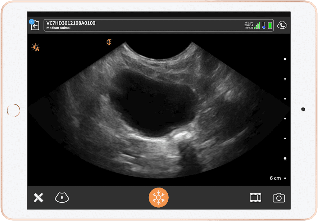
In case 4 of a male domestic short-haired cat with a history of urinary tract disease, Dr. Edwards was asked to perform an ultrasound with particular attention to the bladder. The ultrasound clearly demonstrates mineralization, which appears as mobile echoes with posterior shadowing, and an ultrasound guided cystocentesis was performed to confirm the diagnosis.
As a bonus, Dr. Edwards describes the value of cystocentesis in veterinary practice, and how ultrasound guidance can improve the accuracy of the procedure, thus improving the overall success. She demonstrates her technique using a bladder phantom and offers tips to hone your procedural skills.
It’s always helpful to review ultrasound of the normal kidneys and bladder, so as an adjunct to each of the 4 cases, Dr. Edwards will demonstrate her abdominal scanning techniques using videos taken with her own Nova Scotia Duck Tolling Retriever, Pippi. This helps not only hone your ultrasound scanning skills but also provides a baseline for distinguishing normal from abnormal anatomy.
Dr. Edwards is joined by your host and emergency physician Dr. Oron Frenkel and sonographer Shelley Guenther who showcases live scanning with the new Clarius C7 Vet HD3! Don’t miss this educational webinar – register today to take your kidney and bladder ultrasound scanning skills to the next level.

Peripatetic Veterinary Ultrasonographer | Educator | First Opinion Veterinary Ultrasound
Dr. Camilla Edwards, DVM, CertAVP, MRCVS
Dr. Camilla Edwards is passionate about first opinion level small animal veterinary ultrasound. She travels with her dog Pippi (a Nova Scotia Duck Tolling Retriever) within 50 miles of Cambridge as a peripatetic veterinary ultrasonographer, Camilla teaches ultrasound through FOVU and has built a thriving Facebook community for First Opinion Small Animal Vets. Through her website, www.fovu.co.uk, she reviews ultrasound machines with general practice small animal vets in mind. Camilla qualified as a vet in 2006 and has worked all over East Anglia, UK. Camilla is experienced in emergency and critical care, having gained her CertAVP in 2018.

Emergency Physician
Oron Frenkel, M.D., M.S.
Dr. Oron Frenkel completed his MS and MD simultaneously at the University of California Joint Medical Program in Berkeley and San Francisco, completing his residency in Emergency Medicine followed by a fellowship in Point-of-Care Ultrasound at Alameda County Medical Center in Oakland, California. He moved to British Columbia with the goal of increasing use of point-of-care ultrasound across the province, especially among rural practitioners. An avid educator, Dr. Frenkel is constantly evaluating the best teaching methods for disseminating this technology, how to measure competency in its practice, and its effects on outcomes for individual patients. Dr. Frenkel serves as Chairman of the Clarius Medical Advisory Board.

Medical Sonographer
Shelley Guenther, CRGS, CRCS
Shelley Guenther worked as a Nuclear Medicine Technologist for 2 years before entering into the ultrasound program at the Royal Alexandra Hospital in Edmonton. After graduating with specialties in general ultrasound as well as echocardiography, she worked as a clinical expert in the commercial world of ultrasound for over 25 years. As Clinical Manager at Clarius, Shelley Guenther is dedicated to providing the highest quality educational content for clinicians looking to add wireless ultrasound to their practice, including practical webinars and Clarius Classroom video tutorials.

