By providing my email, I consent to receive Clarius webinar invitations, case studies, whitepapers, and more. I can unsubscribe anytime. Privacy Policy.
FREE WEBINAR
Practical Small Animal Ultrasound: Kidney and Urinary Bladder Point-of-Care Scanning Techniques
Watch On-Demand
Watch Now
RACE-Approved: 1 CE/CPD Credit
Join Veterinary ultrasound educator Dr. Camilla Edwards, DVM, CertAVP, MRCVS as she shares her techniques for scanning abdominal lymph nodes in a point-of-care ultrasound examination.
In this 1-hour webinar, you’ll discover how easy and affordable it is to add wireless ultrasound to your veterinary clinic or animal hospital. We’ll explore:
- The value of ultrasound to improve patient care on the first visit
- Pro tips, tricks, and landmarks helpful in locating the bladder and kidneys
- The ultrasound appearance of normal anatomical structures
- Image interpretation – how to recognise what urinary tract pathology looks like
It’s important to include the evaluation of abdominal lymph nodes in an ultrasound examination. Normal nodes can be challenging to locate due to their size and location, but when affected by disease, their ultrasound appearance can change in a variety of ways. In this webinar, you’ll learn POCUS techniques to identify specific lymph nodes in the abdomen using landmarks that are easily identifiable with ultrasound.
Dr. Camilla Edwards has been performing veterinary ultrasound for many years and is a passionate ultrasound educator, providing veterinary professionals with the ultrasound skills they need to improve the care of their patients. In this webinar, she provides a hands-on approach to scanning abdominal lymph nodes and identifying pathology when it exists.
Observing live scanning sessions is extremely helpful when learning ultrasound techniques, and thanks to Dr. Edwards’ dog Pippi, you’ll see pre-recorded scanning sessions to provide you with tips and tricks needed to perfect this POCUS exam. You’ll learn how to evaluate the shape, size, contour, echogenicity, and blood flow of abdominal nodes using the latest advancements in handheld wireless ultrasound.
Dr. Edwards provides images that demonstrate the difference between normal and abnormal lymph nodes, to help you make a more accurate assessment and move on to treatment or ultrasound-guided intervention faster.
Ultrasound is the most used imaging tool in veterinary medicine because it’s fast, easy to perform, and non-invasive, which makes it well tolerated by small animal patients in a variety of settings. Advances in technology have made wireless ultrasound more affordable and accessible to more veterinary practitioners. Now, with the miniaturization of high-definition ultrasound and advances in artificial intelligence, learning and using handheld ultrasound is fast, affordable, and easy.
In this webinar, Dr. Camilla Edwards is joined by your webinar host Dr. Oron Frenkel and sonographer Shelley Guenther who will be presenting live canine scanning, so that you can see the new Clarius HD3 Vet scanner in action and hone your ultrasound skills. Don’t miss this webinar – watch today!

Peripatetic Veterinary Ultrasonographer | Educator | First Opinion Veterinary Ultrasound
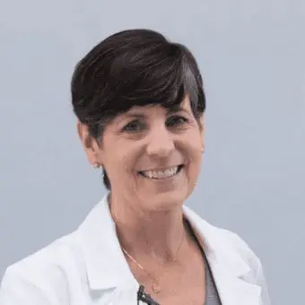
Dr. Camilla Edwards, DVM, CertAVP, MRCVS
Dr. Camilla Edwards, DVM, CertAVP, MRCVS, is passionate about first opinion level small animal veterinary ultrasound. She travels with her dog Pippi (a Nova Scotia Duck Tolling Retriever) within 50 miles of Cambridge as a peripatetic veterinary ultrasonographer, Camilla teaches ultrasound with Celtic SMR, Photon Surgical Systems and FOVU and has built a thriving Facebook community for First Opinion Small Animal Vets. Through her website, www.fovu.co.uk, she reviews ultrasound machines with general practice small animal vets in mind. Camilla qualified as a vet in 2006 and has worked all over East Anglia, UK. Camilla is experienced in emergency and critical care, having gained her CertAVP in 2018.

Medical Sonographer
Shelley Guenther, CRGS, CRCS
Shelley Guenther worked as a Nuclear Medicine Technologist for 2 years before entering into the ultrasound program at the Royal Alexandra Hospital in Edmonton. After graduating with specialties in general ultrasound as well as echocardiography, she worked as a clinical expert in the commercial world of ultrasound for over 25 years. As Clinical Marketing Manager at Clarius, Shelley Guenther is dedicated to providing the highest quality educational content for clinicians looking to add wireless ultrasound to their practice, including practical webinars and Clarius Classroom video tutorials.