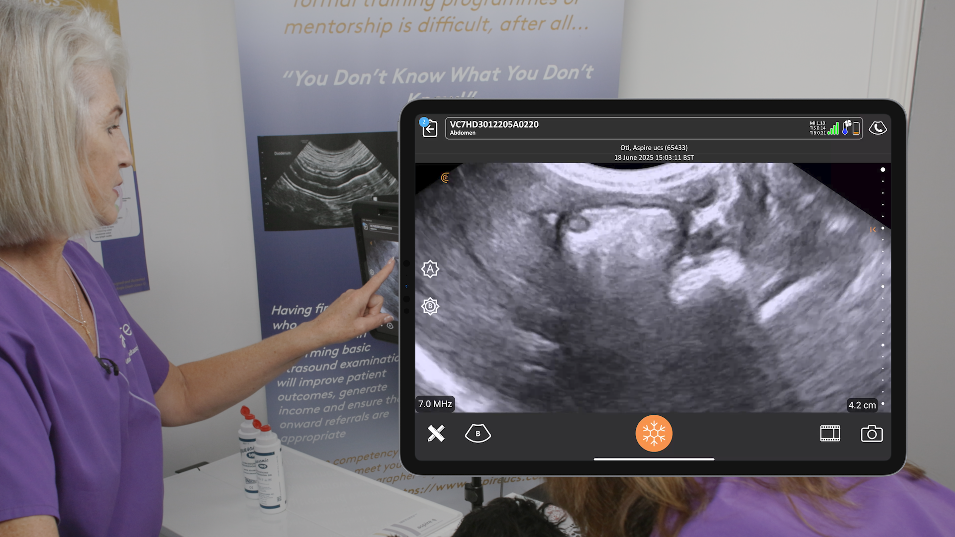
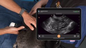
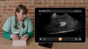
The liver and gallbladder, vital organs in our animal patients, often present diagnostic challenges. As Dr. Camilla Edwards highlighted in a recent webinar, ultrasound is an invaluable tool to help us unravel these mysteries.
The webinar, Practical Small Animal Ultrasound: Guiding Diagnosis and Management of Liver and Gallbladder Disease, is available now for you to watch at your convenience. It’s RACE approved for 1 CE/CPD Credit if you access it through the Vet Show.
Following are key clinical takeaways from Dr. Camilla’s insightful session:
1. The Importance of a Systematic Approach
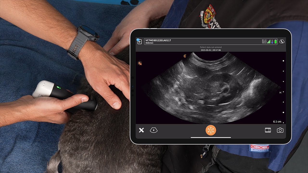
- Liver: The liver’s size and position can be deceptive. Ensure you visualize the entire organ, from the cranial edge nestled near the diaphragm to the caudal border. Fanning through both longitudinal and transverse planes is crucial for a comprehensive assessment.
- Gallbladder: Optimize your image for the gallbladder, focusing on wall thickness, luminal content, and the elusive common bile duct. Remember, the patient’s recent meal history significantly impacts gallbladder size.
2. Recognizing the Subtle Signs
- Liver:
- Size Matters: While there’s no strict reference range, be wary of a liver extending beyond the costal arch or displaying rounded edges, suggestive of hepatomegaly.
- Echogenicity: The liver should fall mid-range in echogenicity. Increased echogenicity can indicate steroid hepatopathy or lipid infiltration, especially in cats.
- Lesions: Characterize any lesions meticulously, noting size, number, and flow patterns. Even without sampling, this information is invaluable for monitoring progression.
- Gallbladder:
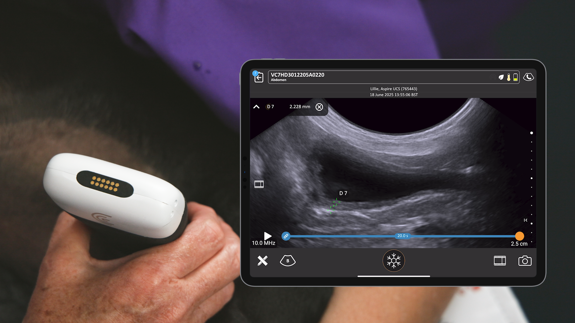
- Sludge vs. Mucocele: Incidental sludge is common, especially in dogs. However, be vigilant for slow-moving, heterogeneous sludge, a potential precursor to a mucocele.
- Wall Thickness: A thickened gallbladder wall can indicate cholecystitis or other systemic issues like right-sided heart failure.
3. The Power of Ultrasound-Guided Sampling
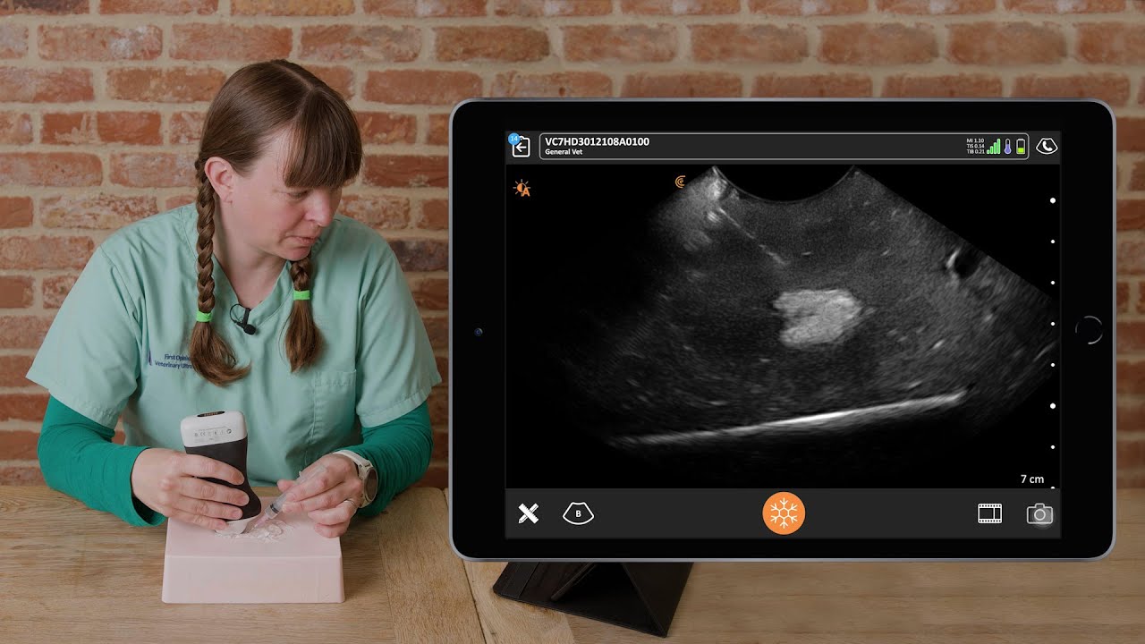
- Fine Needle Aspirates: When faced with a liver lesion, ultrasound-guided sampling can be the key to a definitive diagnosis. Practice your technique on phantoms or readily available materials like tofu to ensure accuracy and minimize patient discomfort.
- Gallbladder Aspirates: While potentially risky, aspirating gallbladder content can provide diagnostic information for culture and sensitivity testing, guiding targeted treatment.
4. The Patient’s Story Matters
- Clinical History: Always correlate your ultrasound findings with the patient’s clinical history and blood work. Vague symptoms like increased thirst or mildly elevated liver enzymes can be the first clues to significant liver or gallbladder disease.
- Owner Communication: Explain your findings clearly and discuss the pros and cons of further diagnostics like sampling or even surgical options like liver lobectomy. Empowered owners make informed decisions.
5. Never Stop Learning
- Continuing Education: Ultrasound is a dynamic field. Stay abreast of the latest research and techniques through courses and webinars. Dr. Edwards’ FOVU platform offers a wealth of resources tailored for general practice veterinarians.
Ultrasound is more than just a diagnostic tool; it’s a window into the inner workings of our patients. By mastering the techniques and interpreting the subtle signs, we can provide more targeted care, improving outcomes and enhancing the lives of the animals we serve.
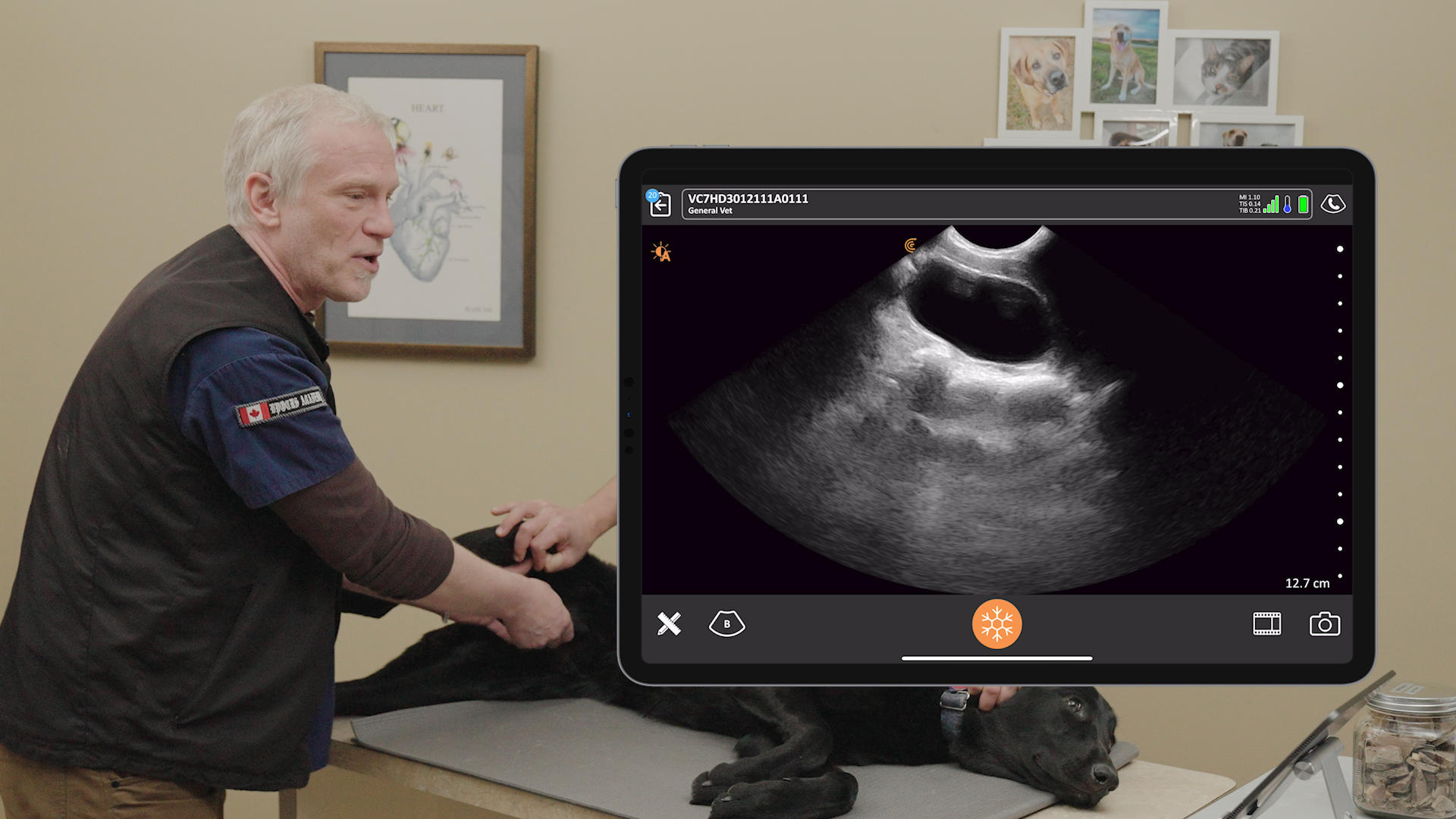
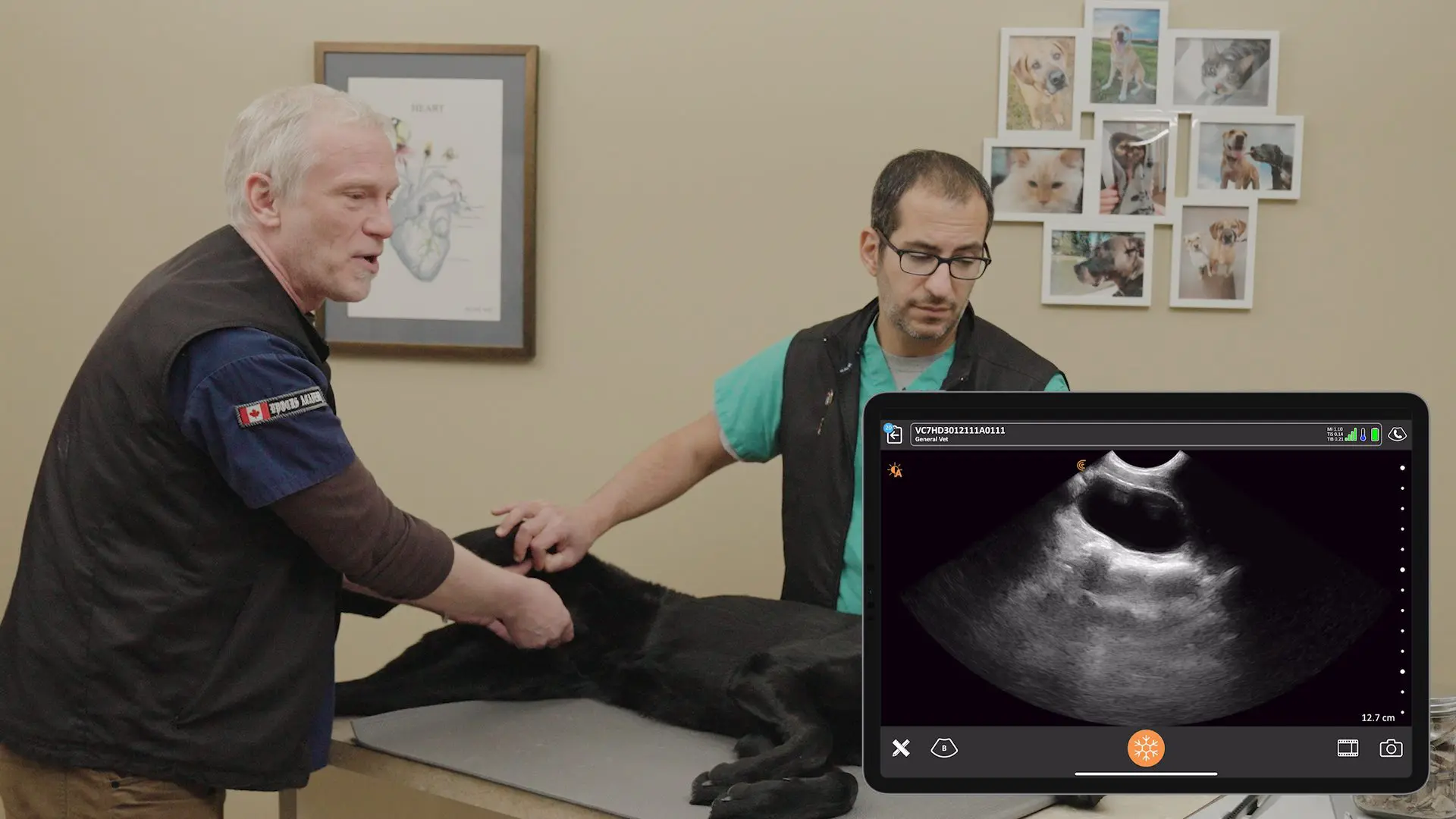
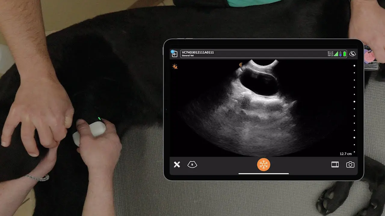
Video Demonstration: How to Scan the Canine Bladder
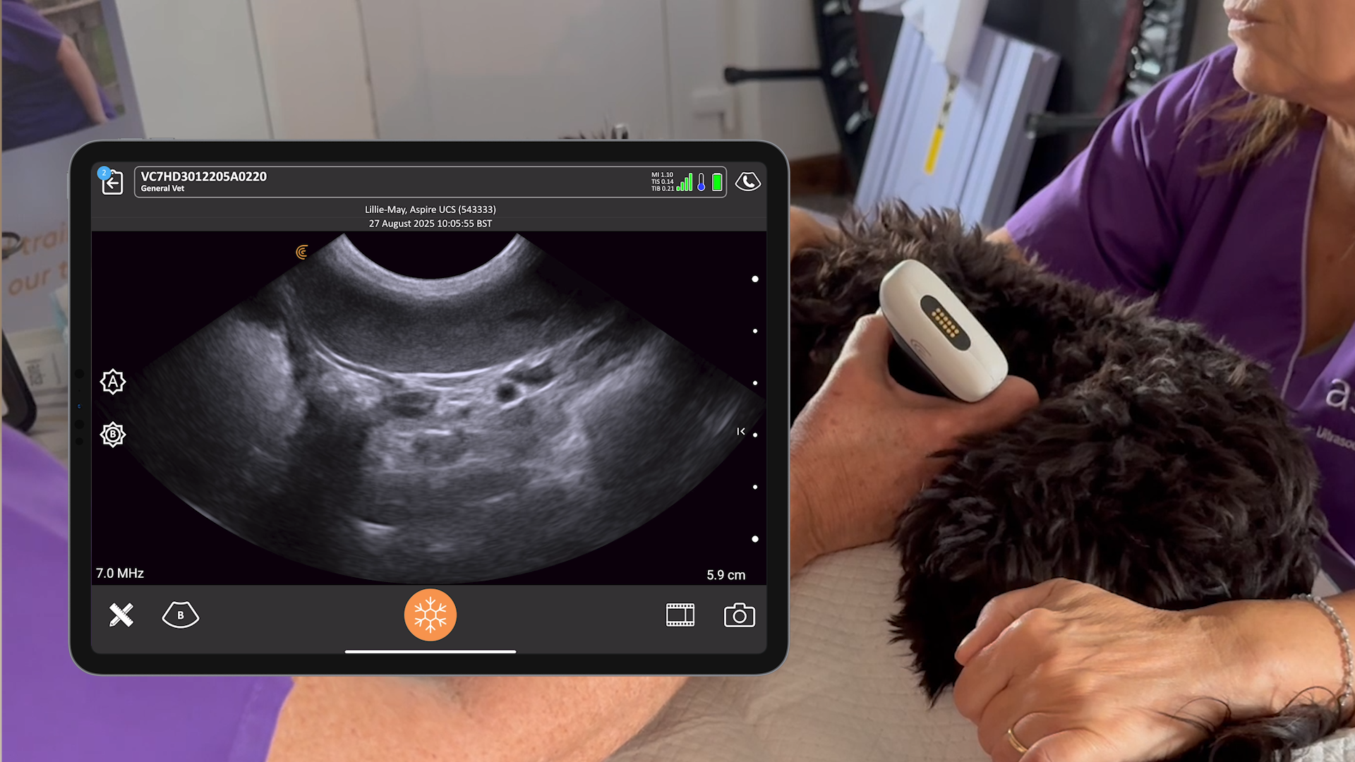
Watch this 1-minute video to see Dr. Edwards’ technique for locating and scanning through the gallbladder to check for stones, sludge, and mucocele using the Clarius C7 HD3 Vet wireless ultrasound scanner.
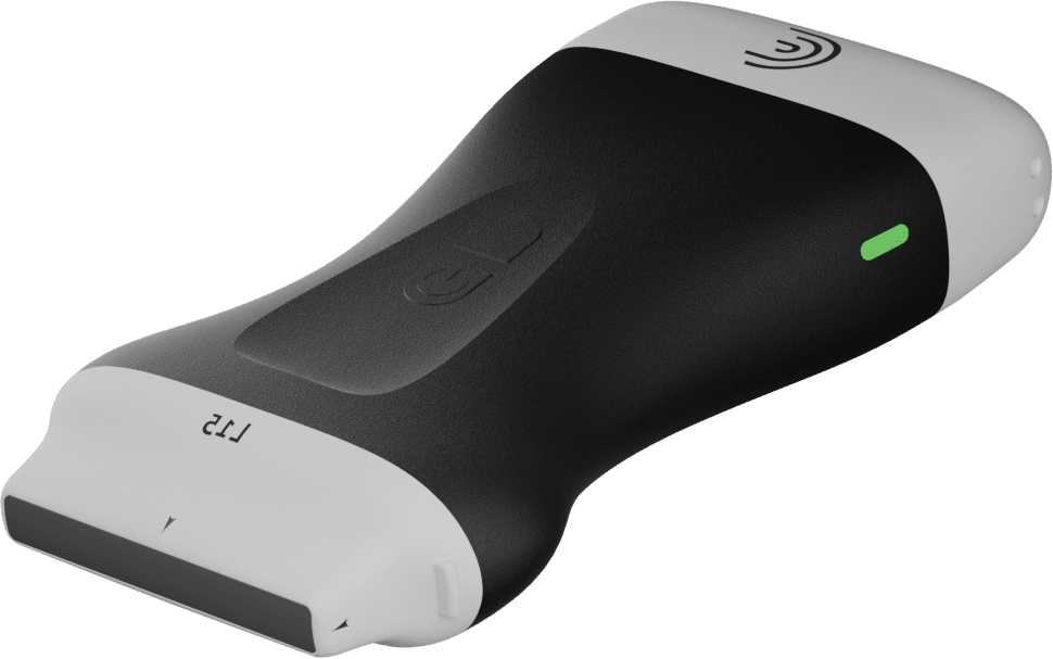
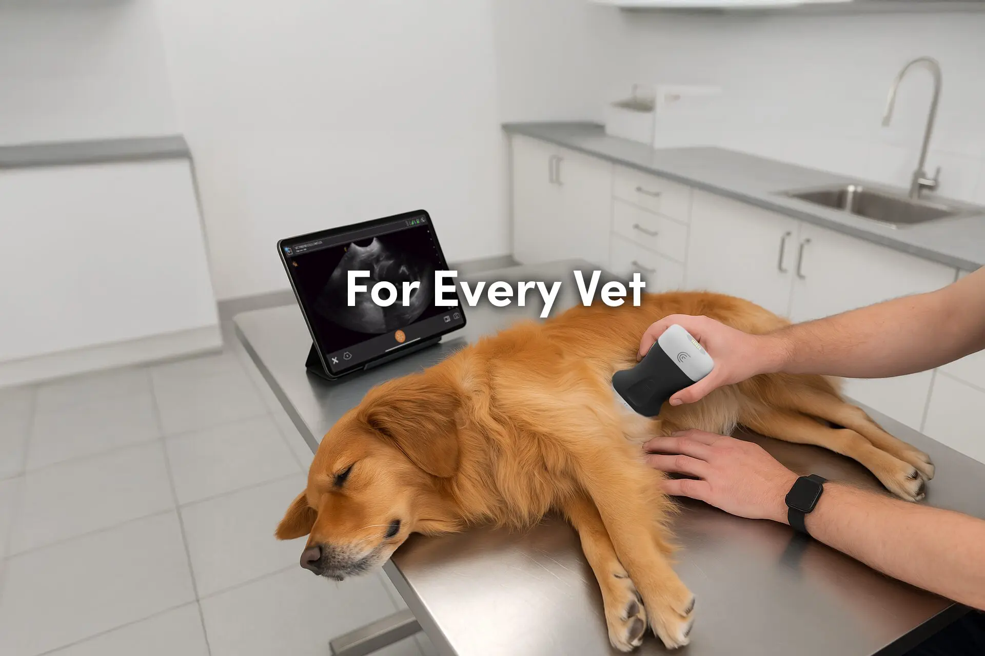
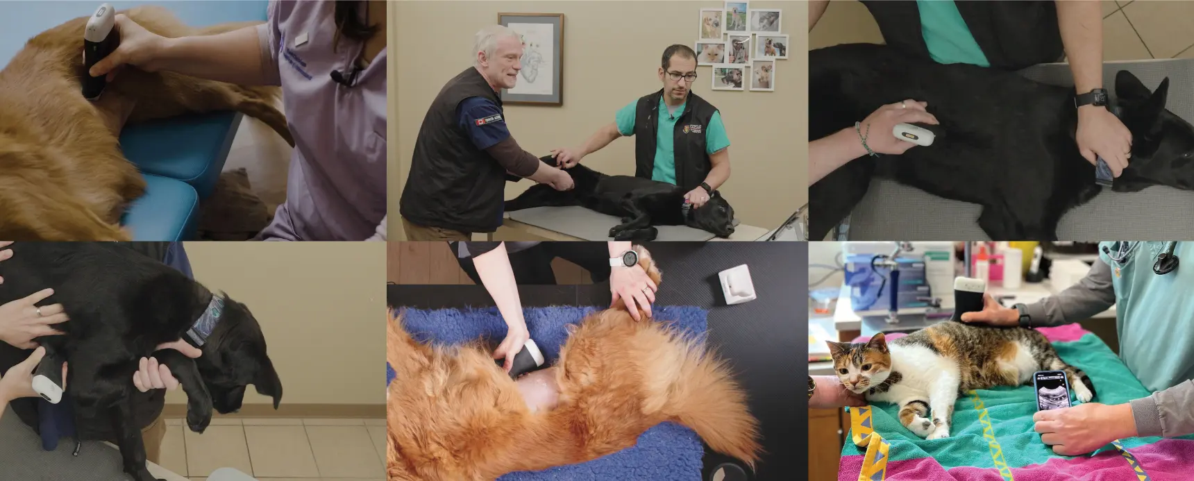
Please visit our Veterinary Specialty Page to learn how wireless ultrasound can benefit your practice. For a personal demonstration of our new smaller and lighter Clarius HD3 Vet scanners contact us today or request a virtual ultrasound demo.
