Clarius Classroom

Severe Hydronephrosis

Oron Frenkel, MD

Moderate-to-severe hydronephrosis can be identified as contiguous fluid extending from the pelvis into the calyces, suggesting a downstream obstruction. Both kidneys should always be scanned to distinguish unilateral from bilateral obstructions which would suggest a more distal etiology.
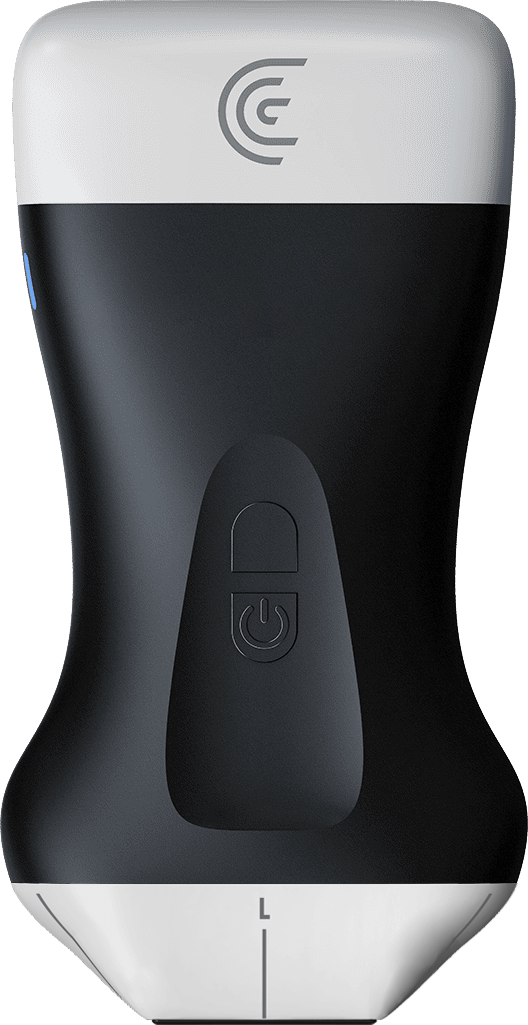
Specialties: Emergency Medicine, EMS, Primary Care, Rural
Applications: Abdomen
this video we see the phased array scanner on an abdominal preset here placing the coronal view on the flank and we're immediately caught by significant fluid collection in the kidney itself and as we scan through we can see that these actually represent the calyces of the kidney and not the pyramids which you would see normally and that these calyces are contiguous with the pelvis which is also significantly dilated we can also note that the cortex of the kidney here which is separated from the renal fat here that is hyperechoic has been thinned out by the significant fluid collection this all suggests severe hydronephrosis in this case and always to note the other kidney when it is available should be scanned to distinguish between a proximal or more distal obstruction that could cause bilateral hydronephrosis

Products Used

More Classes

Request a Quote

With 8 scanners available, we offer a wireless ultrasound solution tailored to your needs. Request a quote to discover which scanner can deliver the best ultrasound imaging for your practice.

⚠️ Clarius ultrasound is for medical professionals only.

Mit der Angabe meiner E-Mail-Adresse erkläre ich mich damit einverstanden, Einladungen zu Clarius-Webinaren, Fallstudien, Whitepapers und mehr zu erhalten, und ich stimme den Clarius-Datenschutzbestimmungen zu. Ich kann mich jederzeit wieder abmelden.