


Septic peritonitis (SP) is a severe, life-threatening condition in dogs and cats that requires early intervention for the best outcomes. Abdominal ultrasonography is widely known to be more accurate than radiography in detecting the presence of free abdominal fluid.
To brush up on point-of-care ultrasound protocols and techniques that put septic peritonitis high on the list of differential diagnoses, watch this webinar with Drs. Soren Boysen and Serge Chalhoub, leading experts in the field of veterinary POCUS. They will guide you through the essential aspects of SP diagnosis, including the key clinical indicators, pathophysiology, and the crucial role of ultrasound in early detection. Scroll down for quick highlights.
Webinar Case Study: Septic Peritonitis
During the webinar, Drs. Boysen and Chalhoub present a case study of septic peritonitis. The patient, Dyson, a 7-year-old Boxer cross, presented with a 3-day history of progressive vomiting and anorexia.
Physical Examination Findings
Physical examination revealed:
- Temperature of 40.7°C
- Heart rate of 168
- Respiratory rate of 24
- Bounding pulses
- Hyperemic mucous membranes with rapid capillary refill time
- Lethargy and nausea
- Abdominal pain
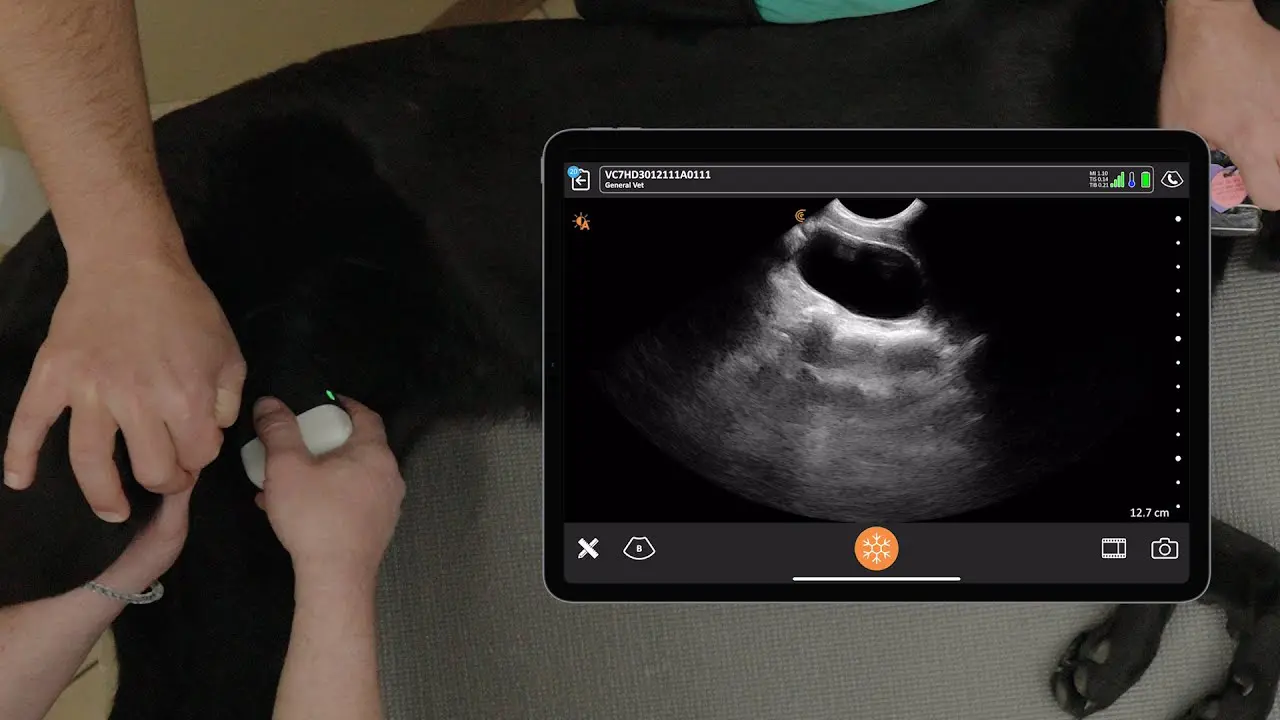
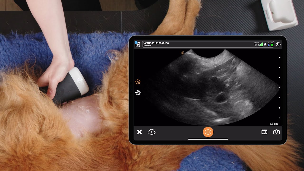
Ultrasound Findings
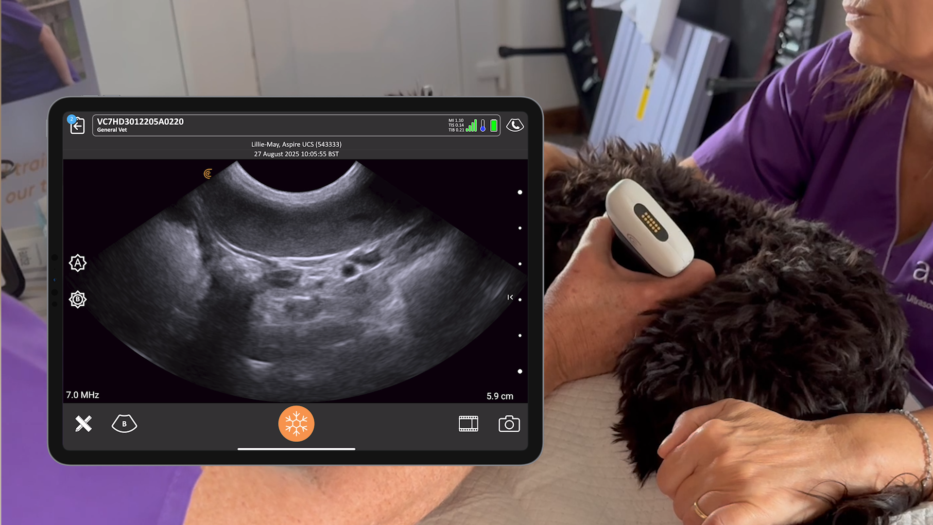
POCUS revealed free abdominal fluid, free abdominal air, and gallbladder wall edema with a halo sign.
Clinical Highlights
- POCUS is a valuable tool for detecting early signs of leakage from intestinal sutures following gastrointestinal surgery.
- Abdominal POCUS in the standing position can substitute for traditional right lateral scanning.
- The primary focus of POCUS in patients with acute abdomen and hemodynamic instability should be on the cardiovascular system and abdomen.
- Air is an enemy of ultrasound and appears as a hyperechoic interface with soft tissues.
- Three key steps to diagnose pneumoperitoneum:
- Find the peritoneal lining.
- Look for an enhanced peritoneal stripe sign.
- Look for reverberation artifacts.
- A normal caudal vena cava should exhibit at least a 25% change between the widest point at expiration and the narrowest point at inspiration.
Questions and Answers from the Webinar
Is free air in the abdomen ever normal?
Other than post-surgery, free air in the abdomen is not normal.”
How many scans does someone need to feel comfortable with POCUS?
For abdominal POCUS, about 20 scans are needed to gain a degree of confidence in identifying free fluid.”
If you suspect GI perforation and septic abdomen, are you comfortable going to surgery based on POCUS findings alone?
If you see free abdominal air and free abdominal fluid in a patient and cytology supports a septic abdomen, then it is appropriate to go to surgery.”
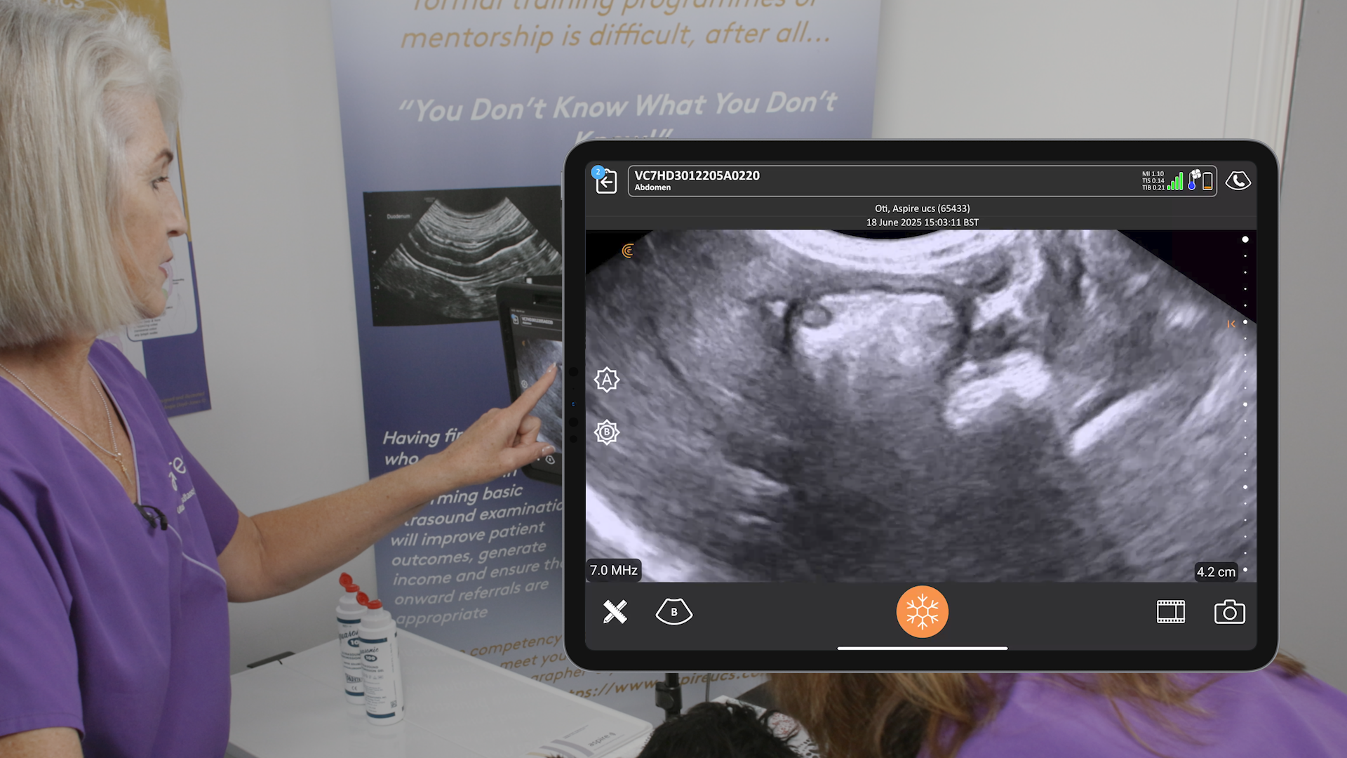
Do you ever use ultrasound to confirm pregnancy?
Ultrasound is more sensitive at picking up pregnancies earlier than radiographs and is useful for assessing fetal viability.”
If ultrasound is as good for diagnosing peritonitis in dogs, is it the same in cats?
It would be the same in cats. The same findings of free air and fluid will be present.”
Watch the full webinar for free: Veterinary POCUS and Septic Peritonitis: Ultrasound Findings in the Septic Abdomen
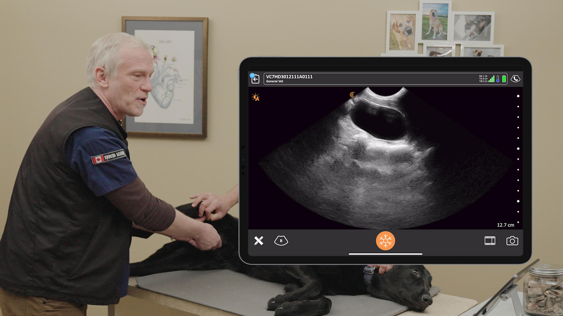
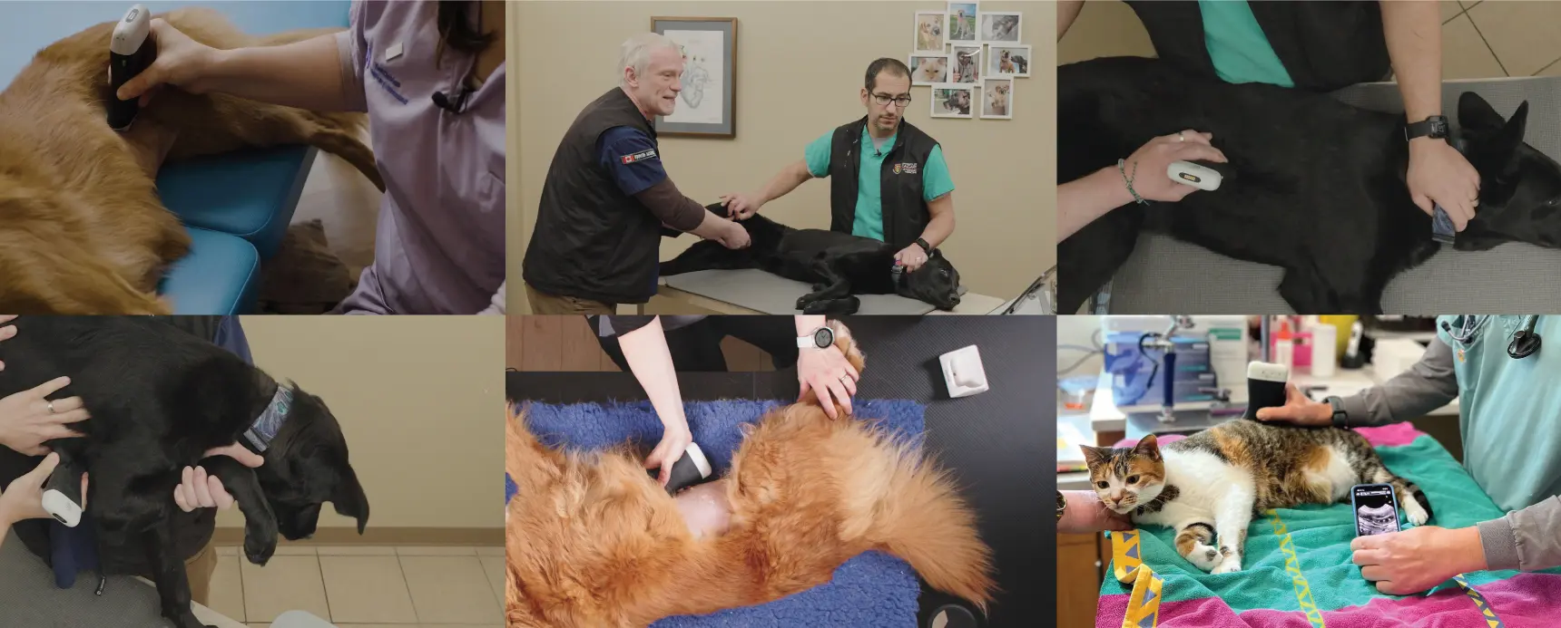
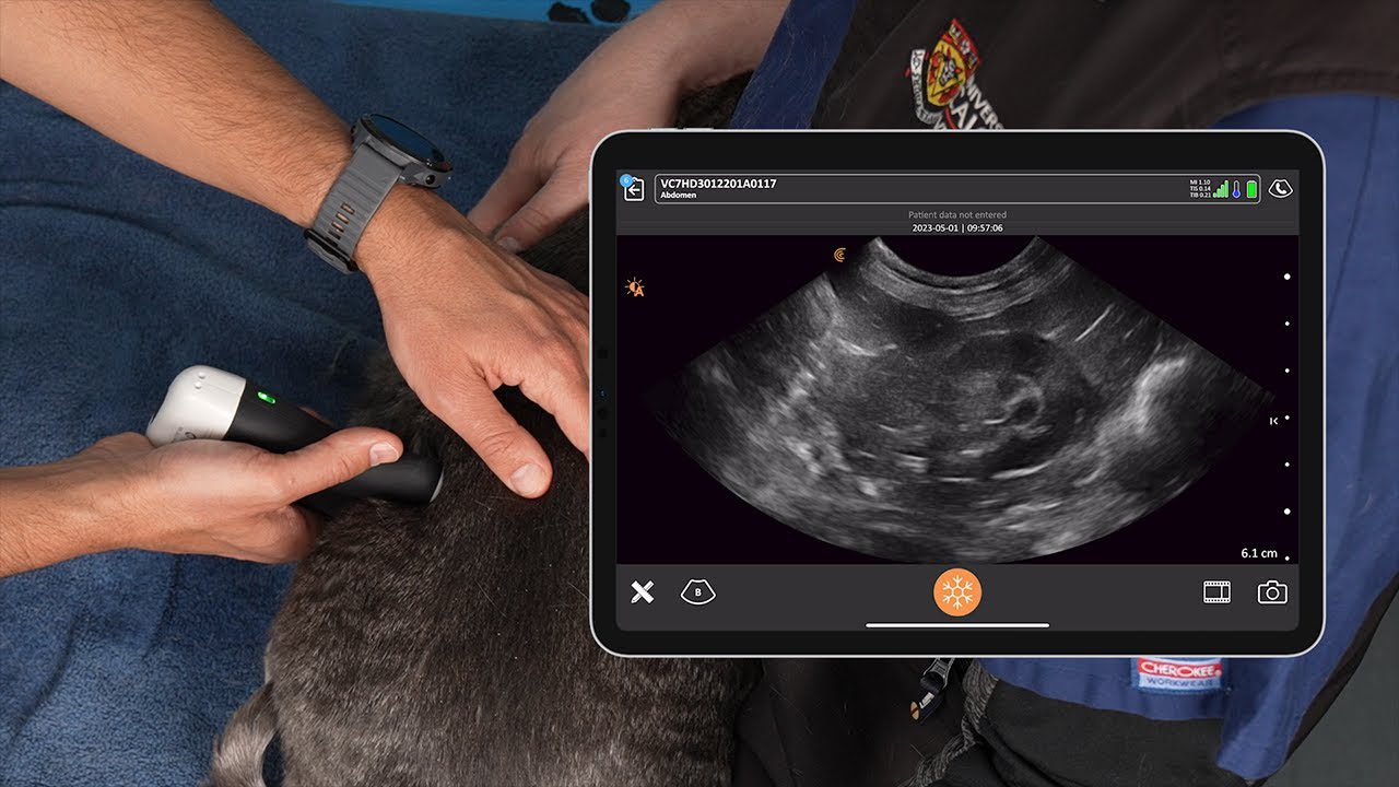
VIDEO: Scanning the Right Paralumbar Site
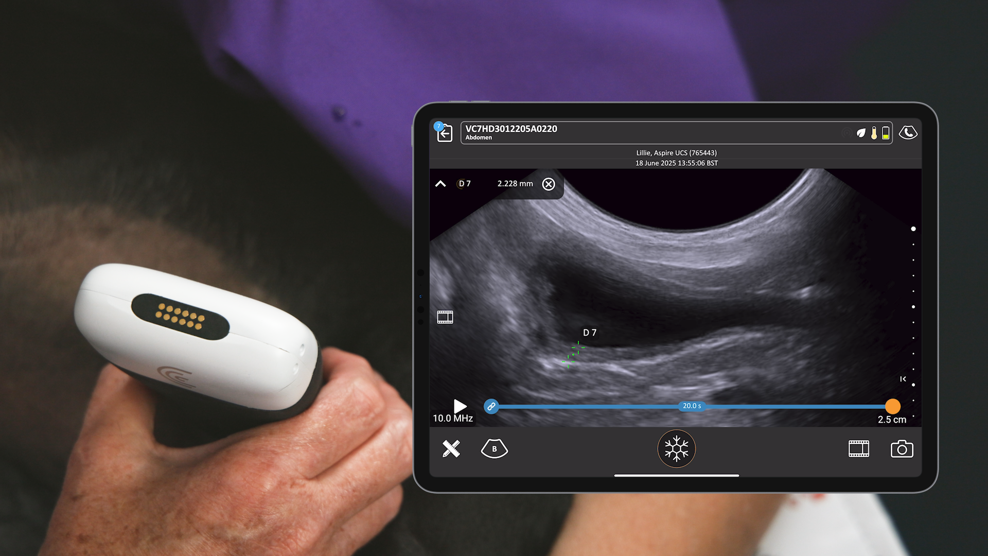
Watch this 6-minute video to see Drs. Boysen and Chalhoub demonstrate where to place the scanner to best evaluate this region for free intra-abdominal air or fluid. The right kidney and duodenum can also be assessed for obstruction. They’re using the Clarius C7 Vet HD3, which is designed for small animals.
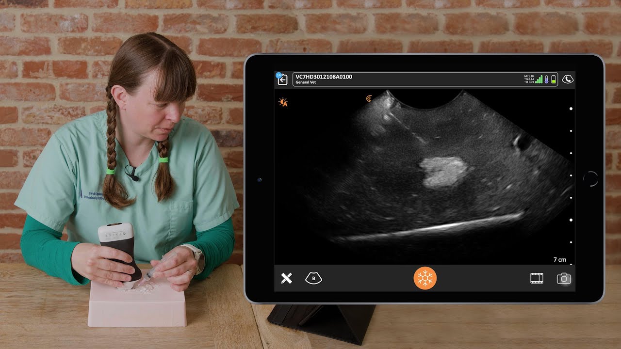
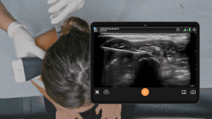
Clarius Ultrasound for New Users
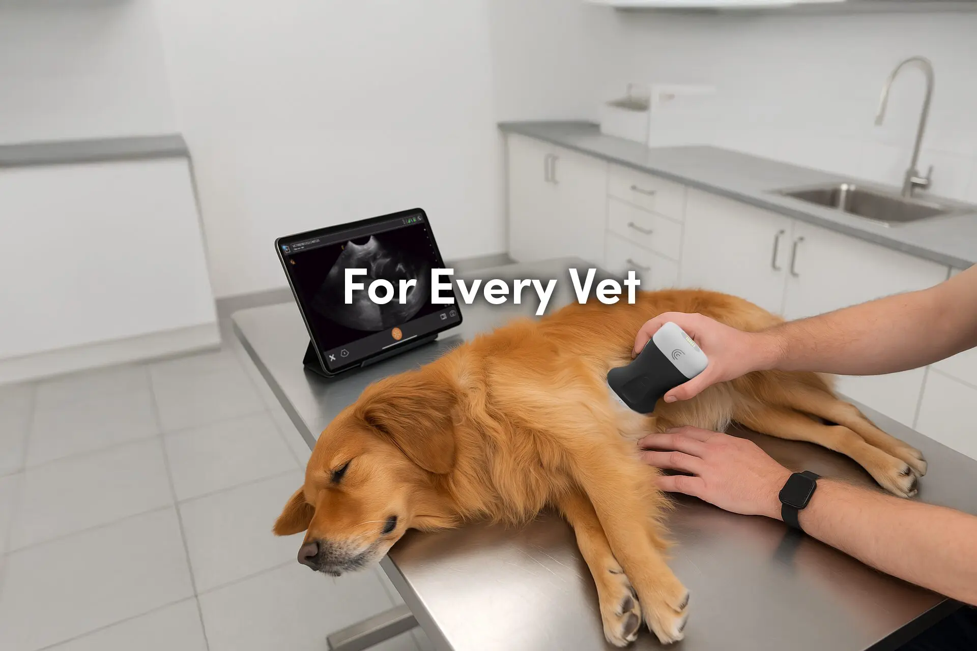
Clarius ultrasound is a good choice for new users because it is easy to use and provides high-quality images. The Clarius HD3 vet scanners are wireless and portable, making them ideal for use in a variety of settings. The scanners are also more affordable than larger cart-based systems. Additionally, Clarius offers a variety of educational resources to help new users get started with ultrasound, including in-app Clarius classroom videos and onboarding with Clarius clinicians.
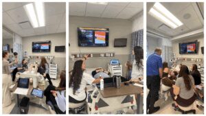
Clarius Wireless Ultrasound for Veterinary Practice
To learn more about how you can add wireless ultrasound to your practice, visit our Veterinary Specialty Page. There you’ll have access to additional webinars and classroom videos.
To find out which scanner is best for your practice, contact us today or request a personalized virtual ultrasound demo.
