

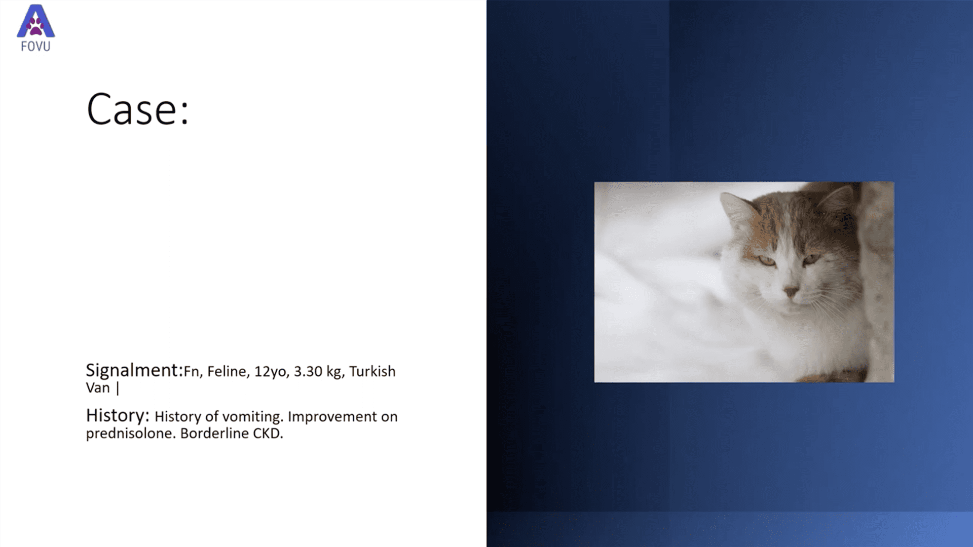
Dr. Camilla Edwards, DVM and founder of First Opinion Veterinary Ultrasound (FOVU) recently shared another great case study where ultrasound was used to identify a moderate amount of abdominal ascites, as well as a thickened stomach wall.
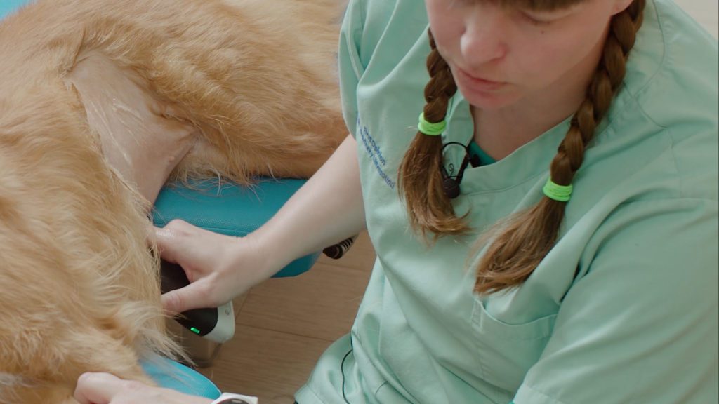
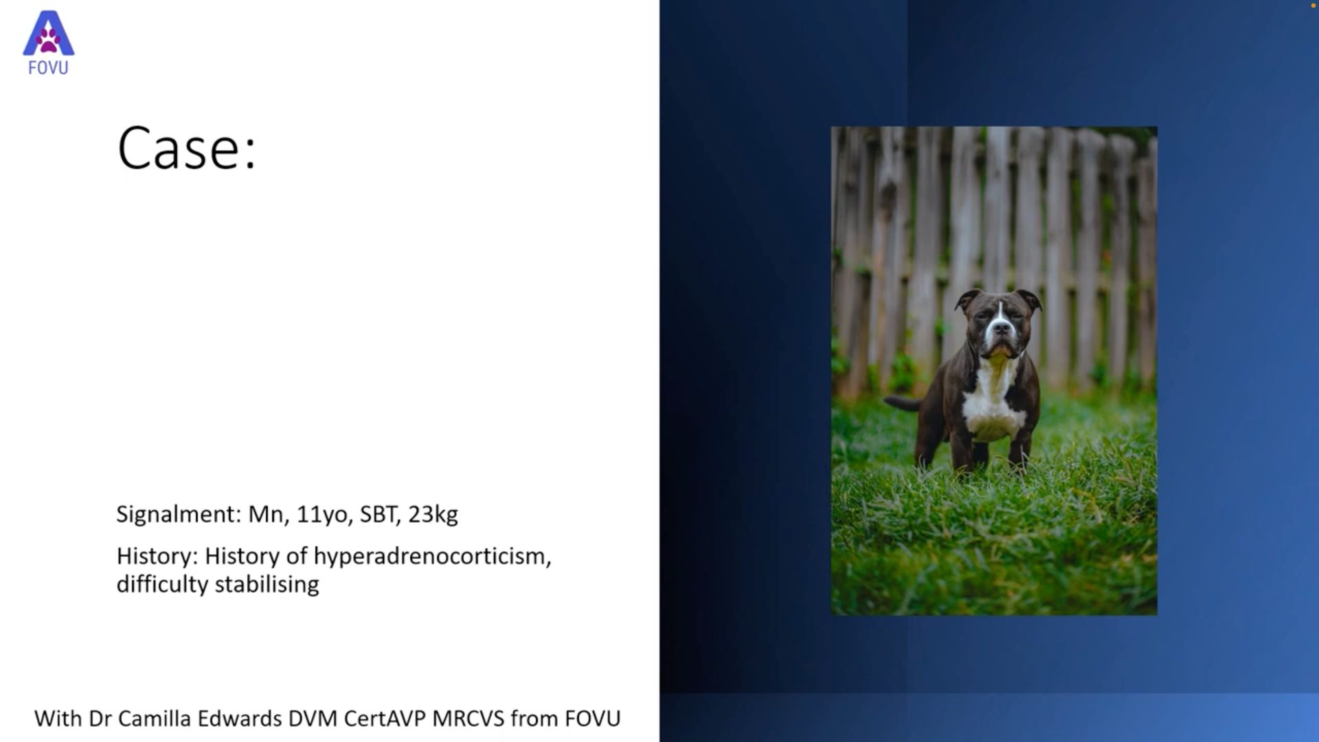
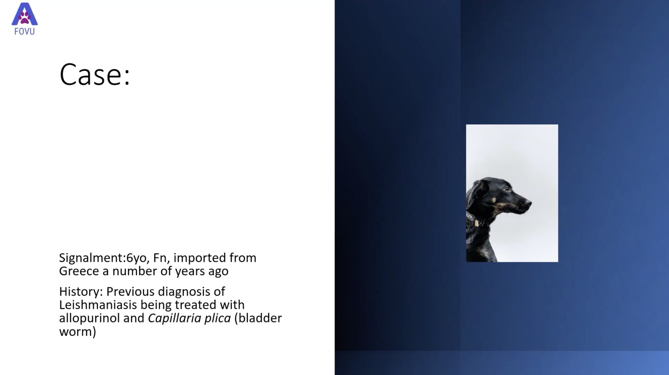
History: A 7-year-old m/n miniature Schnauzer presented with a history of diarrhea. Dr. Edwards proceeded, as always, to perform a very thorough abdominal ultrasound to investigate.
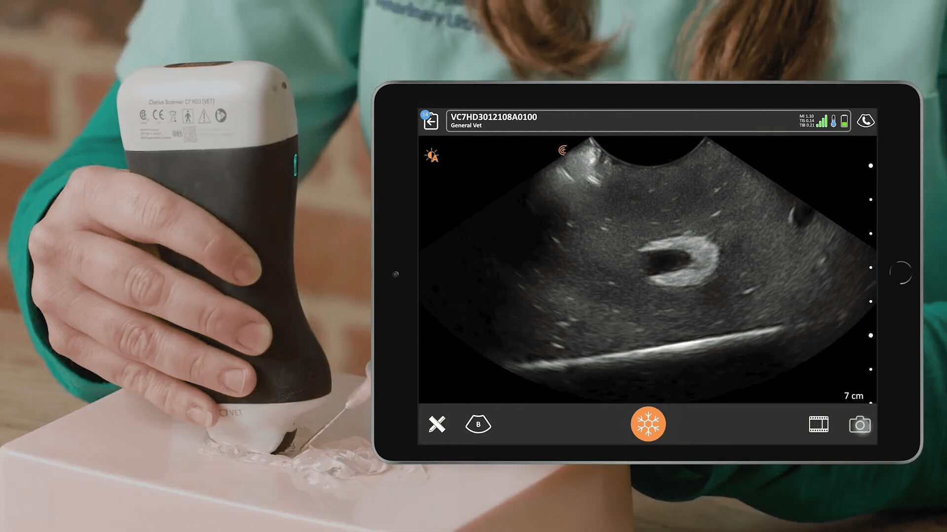
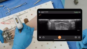
Ultrasound Findings
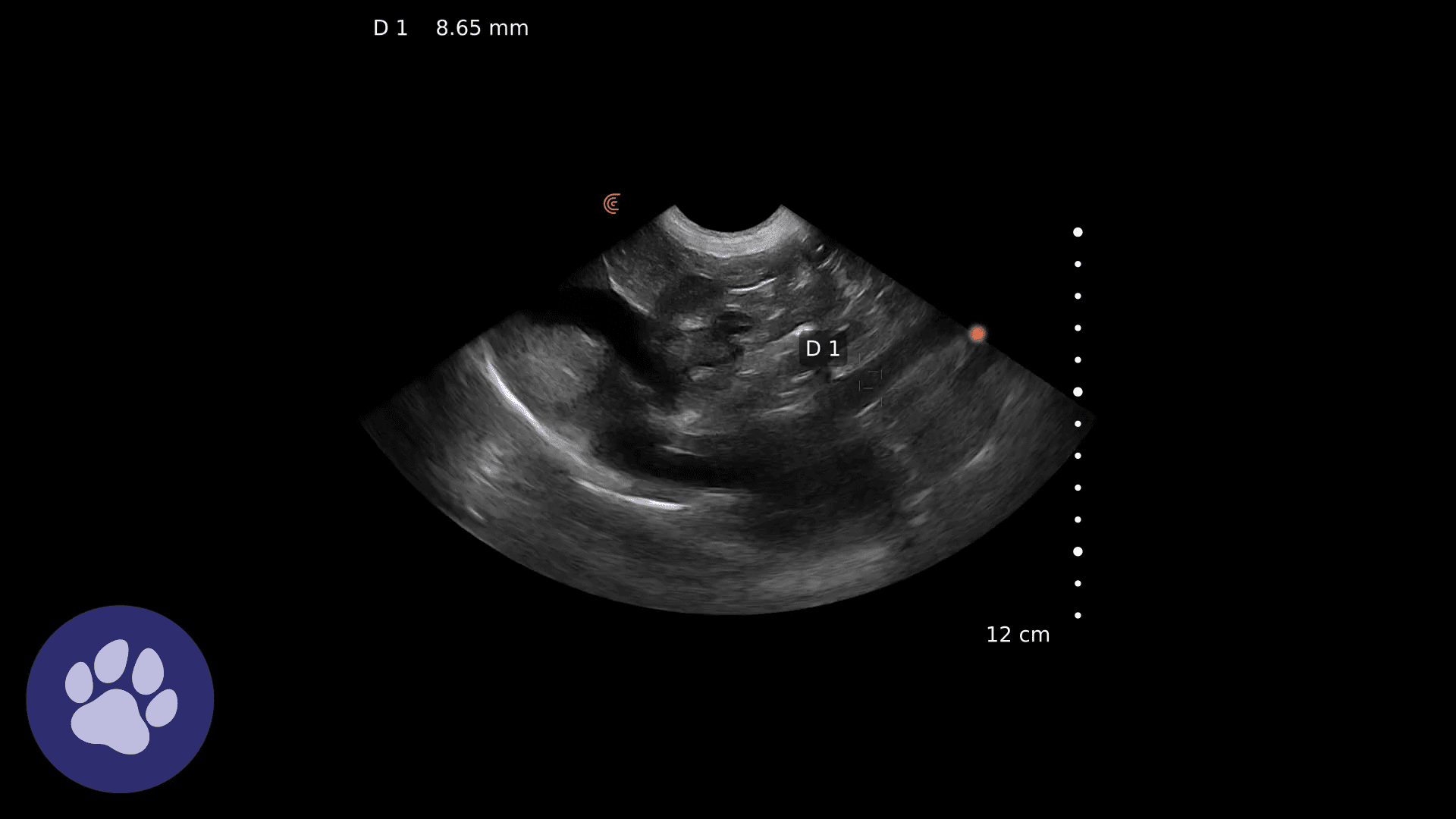
Almost immediately, at the cranial abdomen site, free fluid is visualized surrounding the liver and throughout the abdominal cavity. The liver appears more hypoechoic than normal, and the mesentery appears quite echogenic.
The stomach wall appears thickened, and in fact measures well above the 3 mm threshold at 7.4 mm. There is no focal mass seen.
The spleen, kidneys, pancreas, adrenals, jejunum, duodenum, bladder, and abdominal lymph nodes are all visualized well and appear normal.
In the region of the ascending colon there is dilated, distended bowel with very little peristalsis.
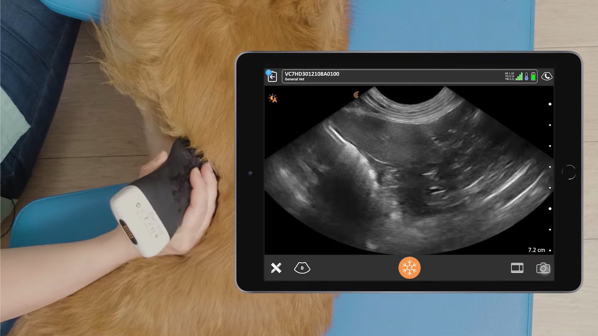
Video: Dynamic ultrasound of the canine abdomen demonstrates these findings beautifully.
Outcome & Discussion
There was no obvious cause of the abdominal ascites and thickened stomach wall in this dog, so an exploratory laparotomy was performed and samples of the small intestine were taken.
Small intestine histology: Chronic moderate to marked lymphoplasmacytic and neutrophilic enteritis with villous stunting and lymphangiectasia
Lymphoplasmacytic enteritis (LPE) is the most prevalent form of inflammatory bowel disease in dogs. The inflammation is caused by the infiltrations of lymphocytes, plasma cells, and in this case neutrophils, into the lamina propria of the stomach and/or small intestine. More severe cases involve villous stunting/clubbing and atrophy. There doesn’t appear to be a definitive cause, and treatment includes immunosuppressive therapy, as well as antibiotic therapy, and pre-and pro-biotics to help improve gut flora.
Free Webinar About Small Animal Ultrasound with Dr. Edwards
Dr. Edwards will teach how to rapidly identify palpable abdominal masses, characterize tumors, investigate spread, and offer prognosis during an upcoming one-hour webinar. Join us for Practical Small Animal Ultrasound: Guiding Diagnosis and Management of Palpable Abdominal Masses. You’ll learn to:
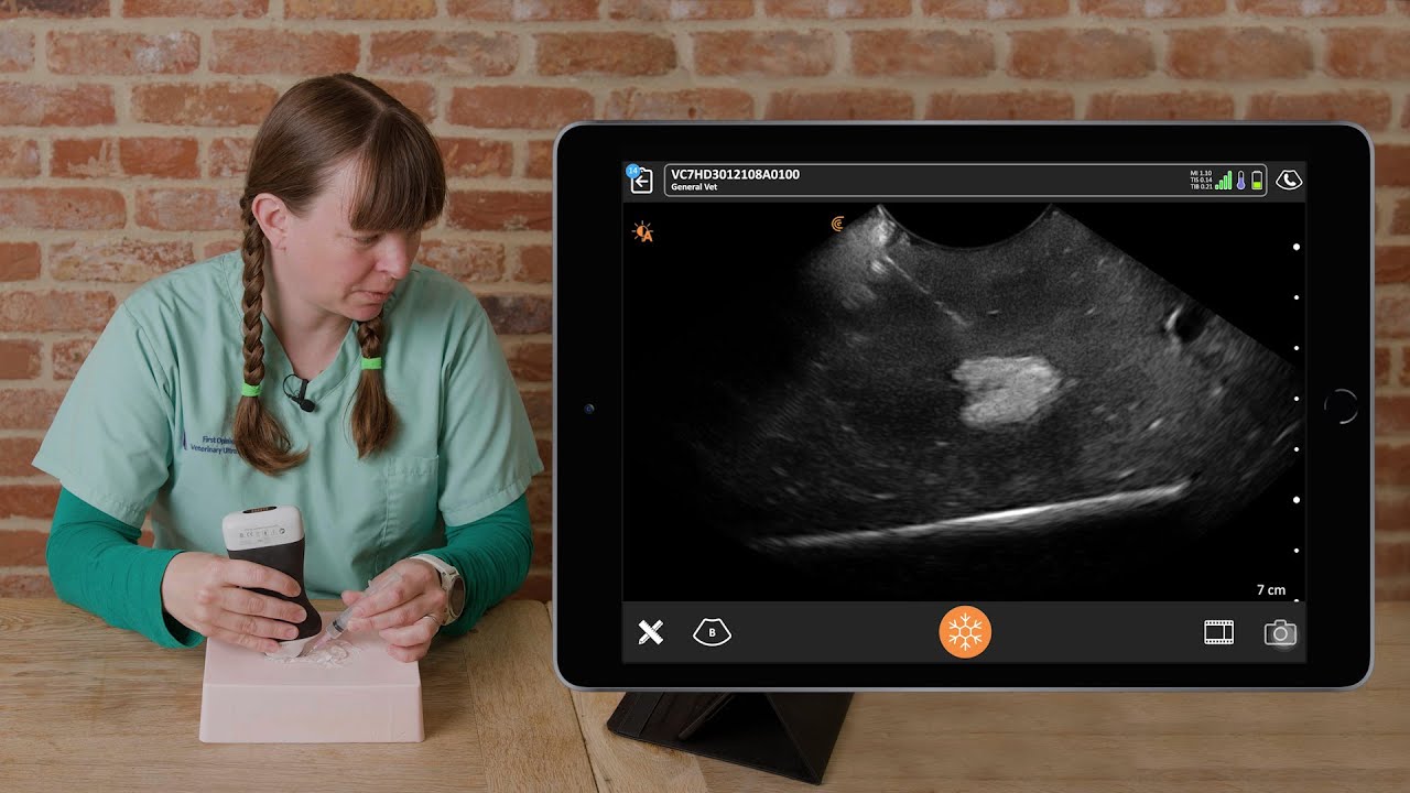
Clarius Ultrasound for Veterinary Medicine
Visit our Clarius Classroom where you’ll find several short tutorials with tips and tricks for scanning the abdominal organs in small animals.
Dr. Edwards uses the Clarius C7 HD3 Vet, which is specifically designed for clinical imaging of small and medium-sized animals. Our Advanced Veterinary Package offers more flexibility for users who need additional workflows for various animal exams.
To learn about how easy and affordable it is to add Clarius handheld ultrasound to your veterinary practice, visit our Clarius veterinary ultrasound page for product details. Or contact us today to discuss which scanner is right for your veterinarian practice.

