

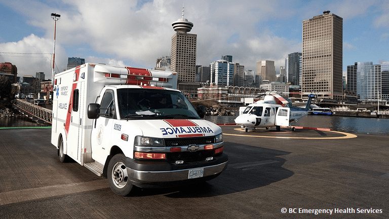
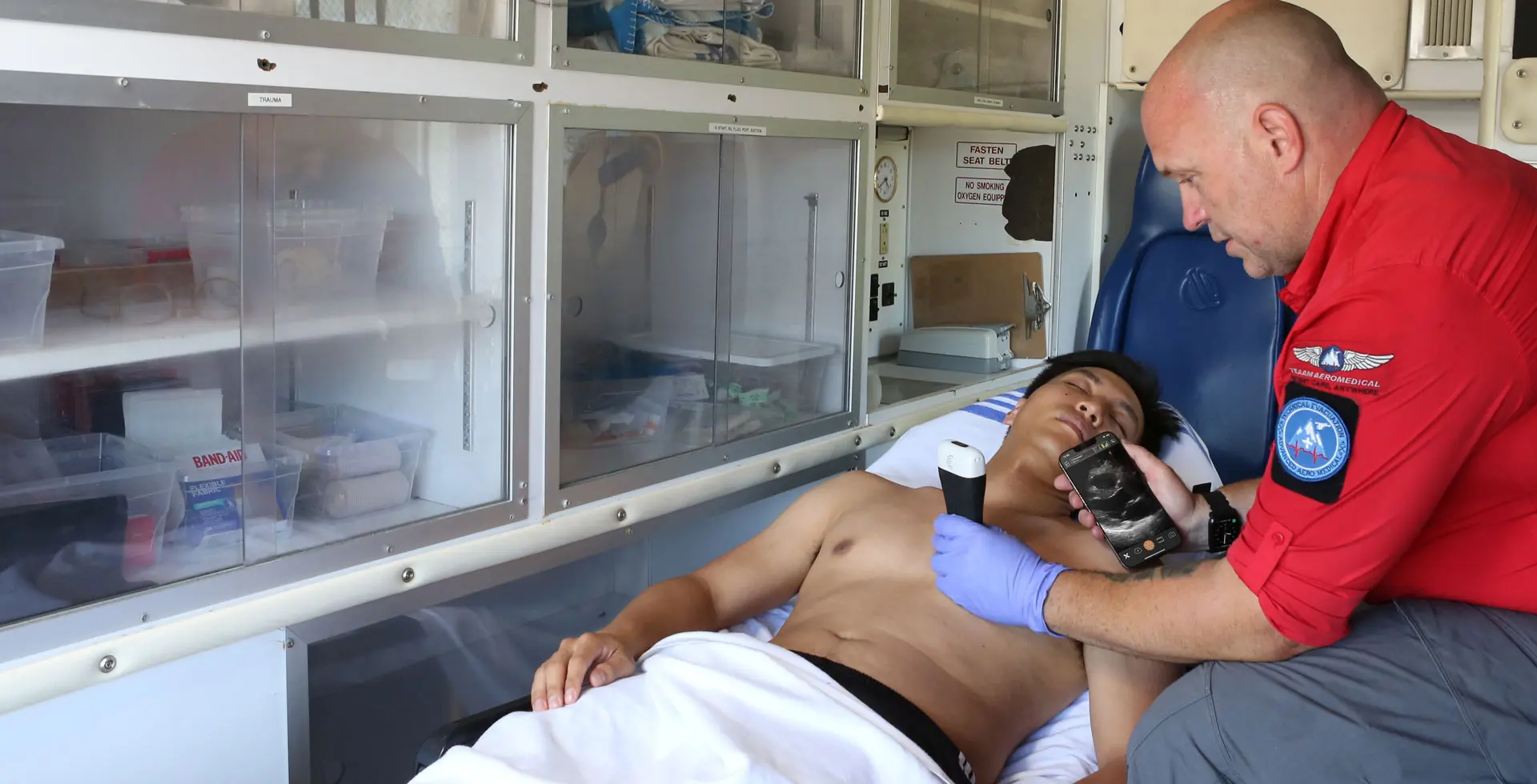
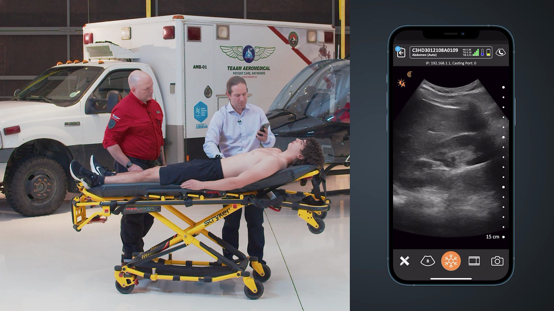
One of the most promising uses for Clarius is pre-hospital ultrasound, or ultrasound in Emergency Medical Services (EMS), before the patient reaches the emergency ward. In these situations, early diagnosis of conditions can make a significant difference to patient outcomes. Being wireless, ultra-portable and easy to use, Clarius will be ideally suited for such applications.
Dean Meenach, director of EMS Education at Mineral Area College, has written an excellent article at EMS1.com describing 9 potential uses of pre-hospital ultrasound.
Here is an excerpt:
- Causes of dyspnea
Field ultrasound increases the accuracy of diagnosing pulmonary edema versus chronic obstructive pulmonary disease as the cause of acute dyspnea [1,18,]. It is effective in patients with unexplained hemodynamic instability to help differentiate between cardiac and non-cardiac causes of shock [18,19]. In some limited cases the potential of field ultrasound to detect massive pulmonary emboli in patients has been demonstrated [15,20].
- Recognizing OB emergencies
Although advanced training is necessary, some prehospital providers have shown that ectopic pregnancy, placenta previa, and placenta abruption can be identified with about 95 percent reliability [4,21,22].Although advanced training is necessary, some prehospital providers have shown that ectopic pregnancy, placenta previa, and placenta abruption can be identified with about 95 percent reliability [4,21,22].
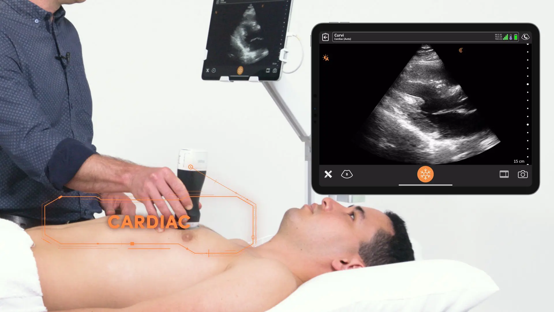
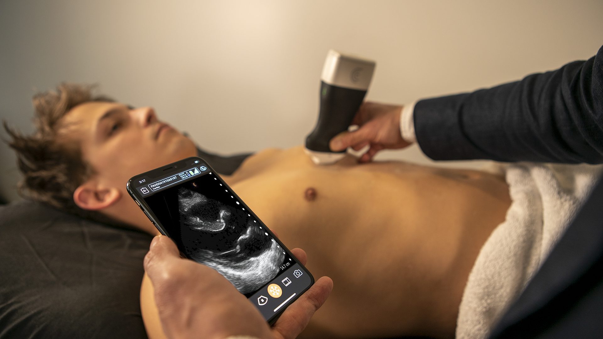
- Cardiac evaluation and resuscitation
Specialized prehospital resuscitation protocols using ultrasound have shown that in patients undergoing CPR, ultrasound helped prehospital providers determine cardiac wall motion when the initial ECG diagnosis was identified as asystole [23]. This was associated with an increased survival to hospital admission [24].
In addition to cardiac motion, ultrasound helped differentiate between true PEA — electromechanical dissociation — and pseudo-PEA — coordinated electrical activity with no palpable pulse [23,24]. Pseudo-PEA was also associated with increased survival to hospital admission when compared with true-PEA [23,24].
In patients in a peri-resuscitation state, ultrasound improved the diagnostic accuracy for potential diagnoses of tamponade, profound hypovolemia, myocardial insufficiency (severe left and/or right ventricular dysfunction), or thromboembolism (pulmonary or cardiac) [25,26]. EMS systems with prehospital protocols that use asystole or PEA as criteria for field resuscitation termination can benefit from adding ultrasound to such protocols [23,25,26].
- Airway placement confirmation and monitoring
Another diagnostic application of field ultrasound includes confirming endotracheal tube placement through high-resolution detection [27]. Although waveform capnography is considered the gold standard for successful ETT verification, this method has some limitations in specific situations such as cardiac arrest, low cardiac output, acute pulmonary embolism, and hypothermia [28].
Ultrasound offers prehospital professionals an alternative method for ETT confirmation for recognizing tube displacement or differentiating between main tracheal intubation and right mainstem intubation [27,28,29,30,31]. For example, Adi et al’s (2013) study showed an impressive result of 98.1 percent accuracy in initial verification [30].
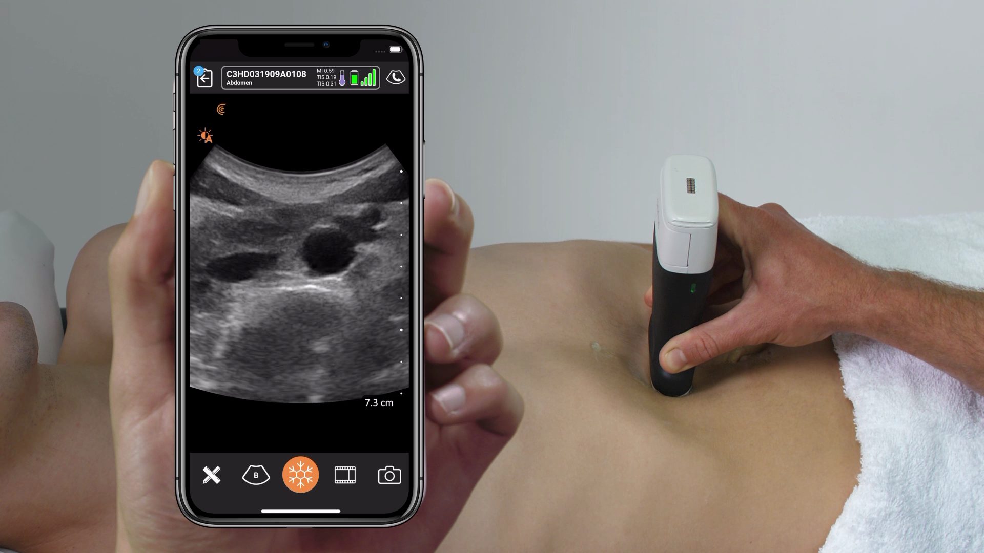
- Gastric tube placement confirmation
Gastric tube placement — nasogastric or orogastic — remains a recommended critical care intervention for all intubated patients as it decreases the risk of aspiration and improves tidal volume. Some EMS systems that place gastric tubes report that placement is easily confirmed using field ultrasound [32].
- Fracture determination
Many types of suspected long-bone fractures are managed in the prehospital setting. Growing evidence suggests that use of field ultrasound can successfully identify several types of long bone fractures [33,49,50].
- Pre-hospital needle thoracostomy placement
Pre-hospital ultrasound use in trauma patients with suspected pneumothorax can be effective in preventing unnecessary field needle thoracostomys [34,35]. One study showed that when thoracic ultrasound was used to detect pneumothorax, only 26 percent of the patients were actually found to not have a pneumothorax [34,35,36,37]. Using field ultrasound could help decrease potentially unnecessary needle thoracostomys and other invasive procedures en route to hospital [38].
- Peripheral intravenous access
Establishing vascular access is one of the most common procedures performed in the prehospital setting and on occasion is a high priority for the critically ill and unstable patient. The condition of the patient often presents challenges in attaining intravenous access. Conditions associated with difficult vascular access include very young age, obesity, chronic illness, IV drug abuse, and hypovolemia to list a few [39,40,41].
Patients with difficult IV access are often subjected to repeated attempts as in some cases time to IV placement can affect optimal resuscitation of the critically ill patient. Ultrasound guided IV access has shown to increase the success rate and decrease complications [42,43,44,45,46,47,48].
- Stroke diagnosis
It is well discussed in the literature that improving the outcome of stroke patients requires early and rapid time-sensitive diagnosis and treatment as well as transport to an accredited stroke center. Early diagnosis using telestroke protocols with field transcranial ultrasound for stroke diagnosis has shown to decrease diagnosis-to-fibrinolytic therapy times and expand the use of special interventional radiology procedures [51,52,53].
Read the full article at http://www.ems1.com/ems-products/technology/articles/72040048-Prehospital-Ultrasound-Emerging-technology-for-EMS/


