

Ultrasound imaging has evolved rapidly over the past few decades. It started with a single element A-mode scan (amplitude mode), which gave radiologists a way to analyze raw radio frequency signals for specialized meaning from the waveforms, similar to an electrocardiogram or EKG. Conversion of A-mode scans to greyscale was the invention of what we know today as M mode, or motion mode. Moving that single element back and forth using precision mechanics gives us B mode (brightness mode) or 2D ultrasound.
Ultrasound scientists and engineers are continually in pursuit of improved image quality, working tirelessly to extract additional diagnostic data from the sound waves that generate every single ultrasound image.
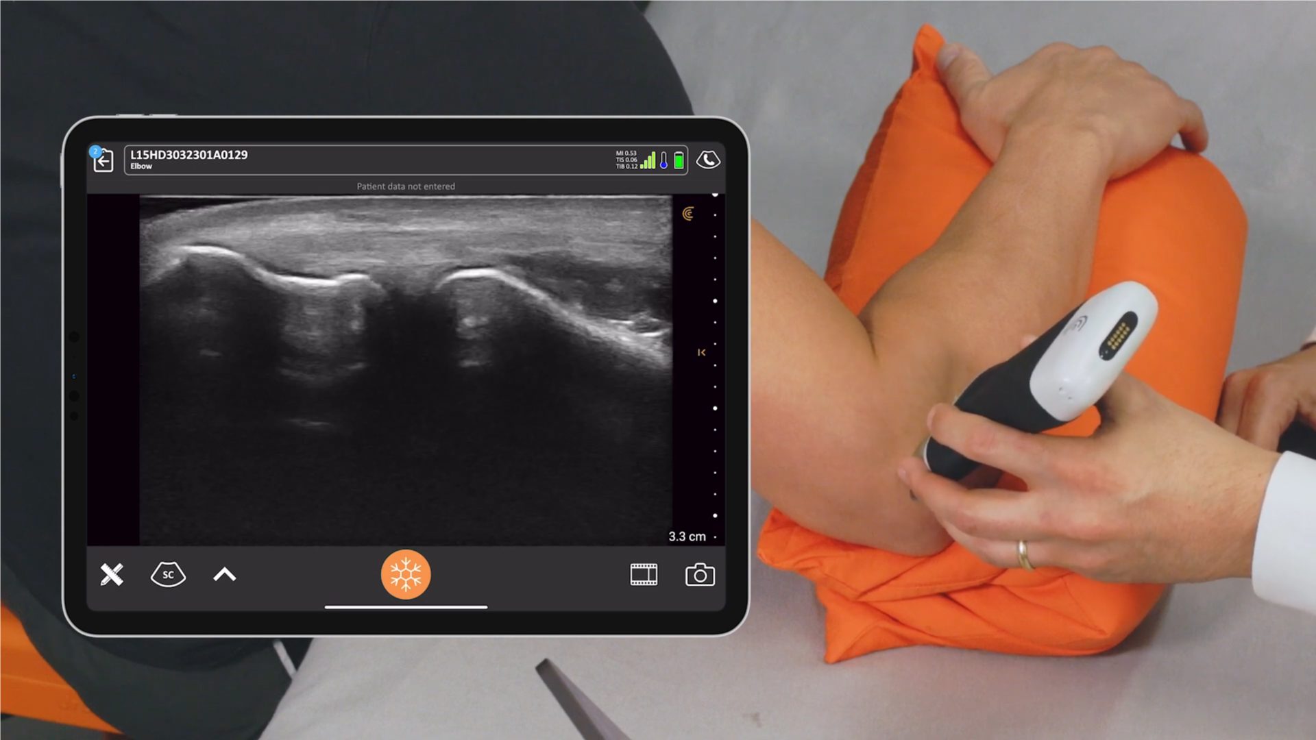
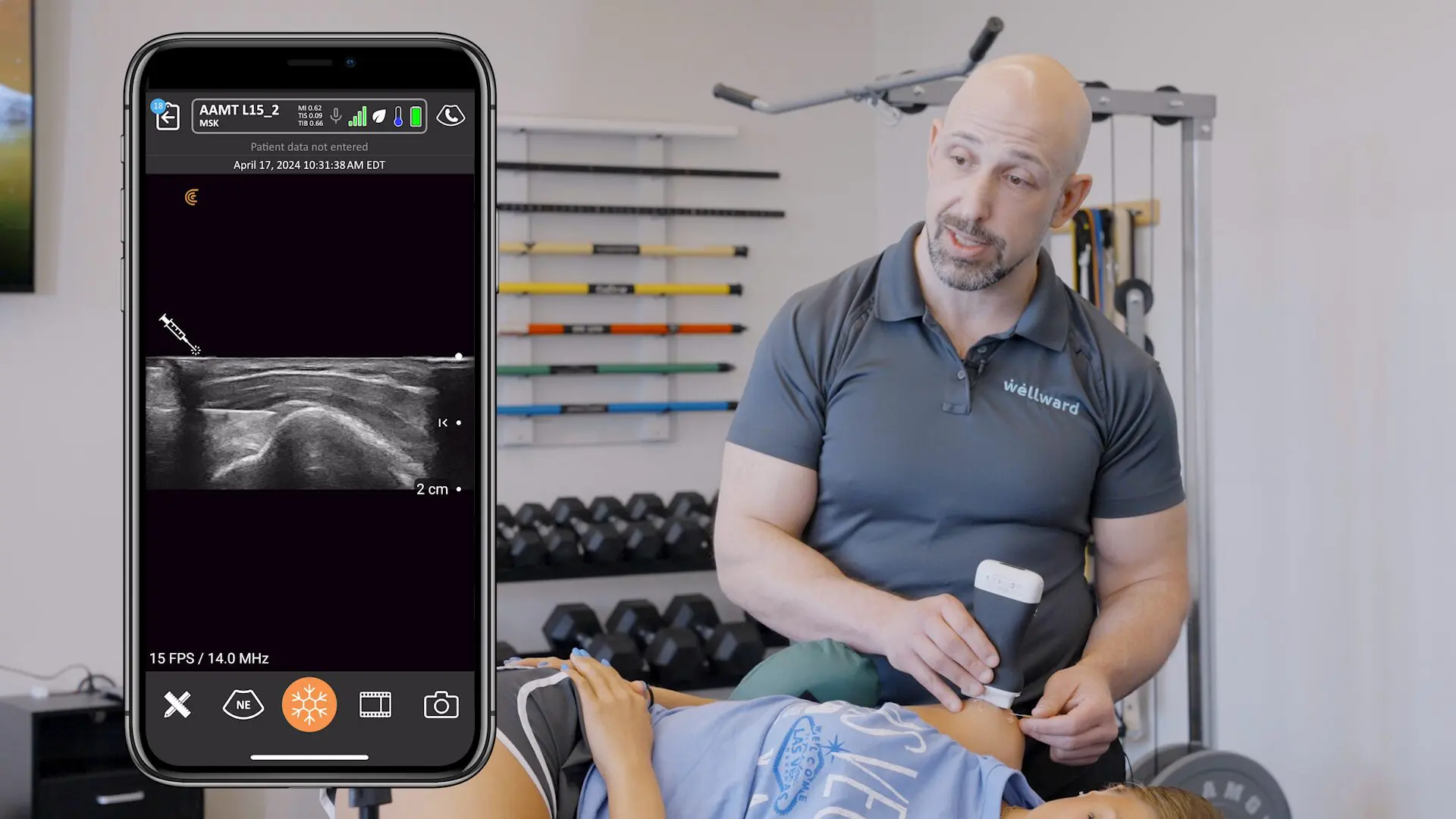
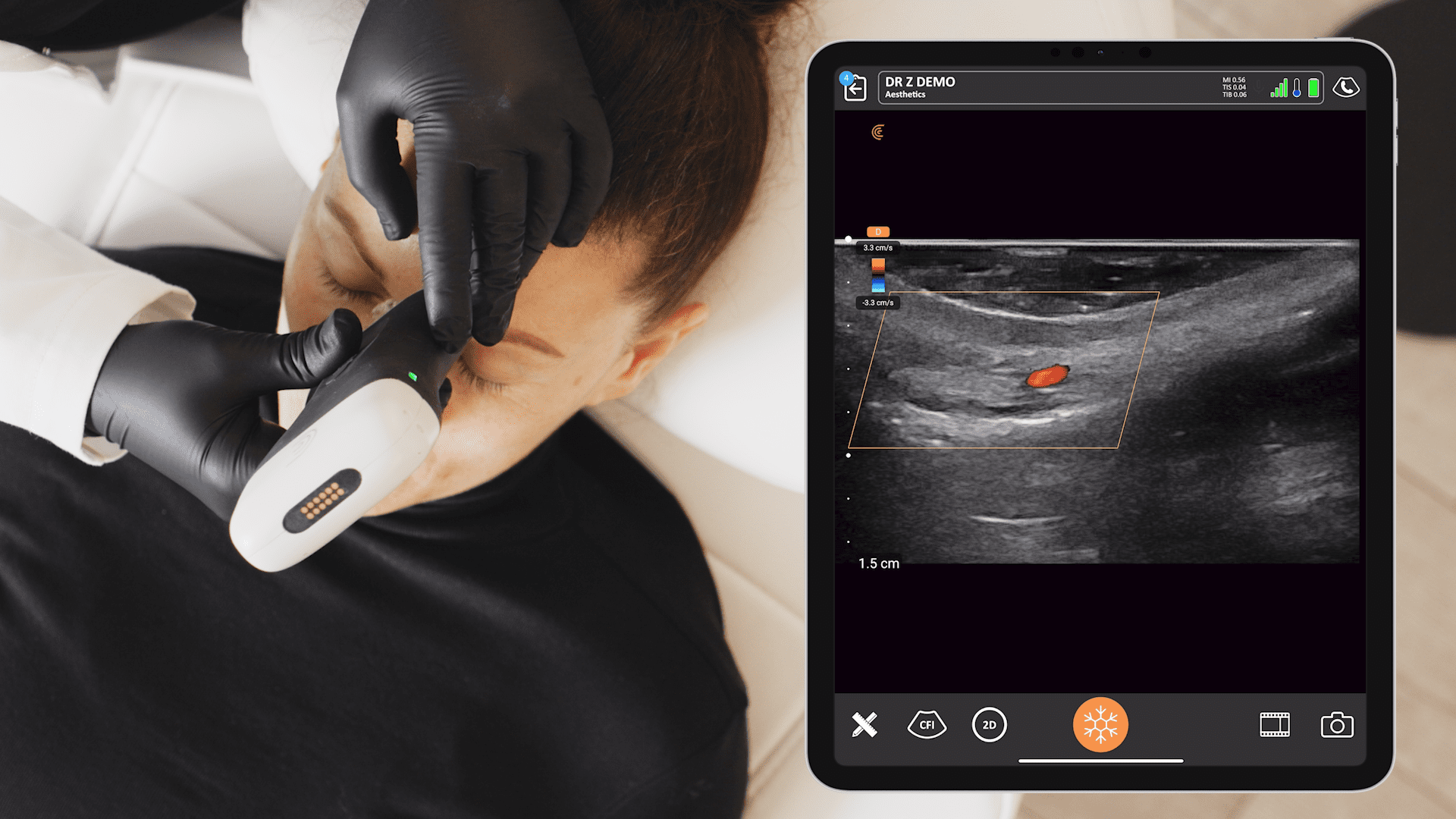
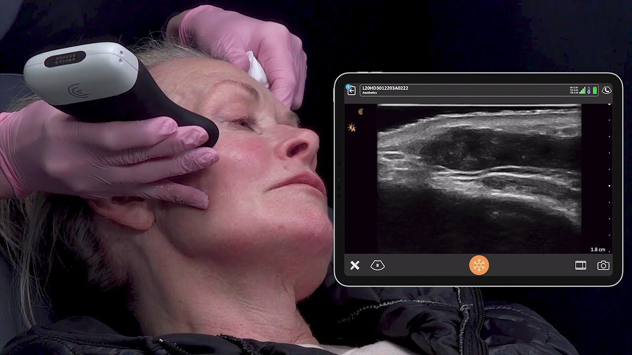
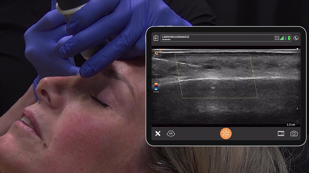
Over the last two months, we’ve been demonstrating the Clarius ultrasound scanner to clinicians from various medical specialties. The comment we hear most frequently is, “Wow! The image quality is great. Much better than I expected.”
In this post we talk to Narges Afsham, Sr. Research Scientist at Clarius to provide insights on how our software team has made great strides by focusing on delivering superior image quality on our handheld wireless ultrasound device.
High performance ultrasound systems typically have numerous real-time processors and high channel count to achieve great imaging,” Narges explained. “Given the size constraints of our wireless handheld ultrasound scanner, we were not able to use typical components to achieve the very high image quality goals we set.”
The Clarius team had to develop a new paradigm for designing ultra-compact ultrasound devices. Their “Ultrasound-on-a-Chip” design leverages the flexibility and performance of state-of-the-art miniature components that have been designed into the product.
With the limited resources of a technology start-up, we had to optimize our image processing routines to ensure they would fit inside the architecture and run in real-time without compromising frame rates. For example, one of our algorithms processed in the typical way would take about five seconds. By restructuring the algorithm and tailoring it for the Clarius architecture, we now achieve the same level of quality in less than 20 milliseconds,” Narges said.
Through sheer perseverance, a solid understanding of ultrasound technology and boldly pursuing new techniques for applying highly specialized compression algorithms, Narges and her software colleagues have engineered remarkably high frame rates. Now, Clarius easily achieves latency-free frame rates of more than 30 frames per second that run wirelessly to virtually any mobile smart device, without compromising image quality.


