

By James Ferrie, B.Pod, M.A. Pod.A
A sought-after Podiatrist based in Melbourne, Australia, James Ferrie first started using ultrasound in his practice to guide regenerative injection therapy. Now he says it has become an invaluable tool for diagnostic purposes as well. Read his article to learn more.
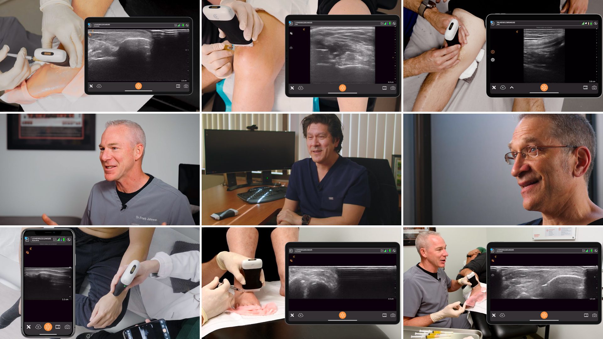
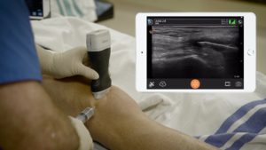
„As a Podiatrist, I’m very interested in the biomechanics and function of the foot. Unlike modalities like MRI, ultrasound lets me visualize anatomy dynamically and provides answers to clinical questions fast. Being able to ultrasound a patient on the spot helps improve the accuracy of my diagnosis and start treatment without delay. It saves my patients time, and it’s usually more cost-effective. It builds a patient’s trust and your confidence when you can point out pathology during the exam. The integration of a clinician’s detailed history and physical examination before an ultrasound scan is also extremely valuable.
As clinicians, we listen and take a comprehensive history. Before physically assessing the patient, we have already comprised a provisional diagnosis and several differential diagnoses. The integration of point-of-care diagnostic ultrasound during a clinical examination adds a sixth sense, the ability to see below the skin, it helps to confirm or disconfirm my provisional diagnosis quickly and accurately.
Clarius is also helpful with my training activities. I’m able to upload and save images securely on the cloud platform so I can easily share interesting cases with groups of podiatry colleagues who are new to ultrasound. I also use Clarius Live telemedicine to provide coaching sessions.
It’s a steep learning curve, so practising is essential. Here are some tips for building your ultrasound skills.
I started using ultrasound in my practice about five years ago when I was working in a rural location, with poor access to MSK imaging. I became interested in using ultrasound to guide regenerative medicine injection therapy. Now, I use ultrasound daily for enhanced diagnosis, and it has helped me provide better targeted patient care.
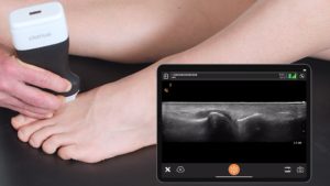
For clinicians new to ultrasound, I recommend getting started by focusing on the larger tendons and ligaments. When I’m teaching, I begin with the plantar fascia and Achilles tendons. There is a steep learning curve, and learning to master POCUS ultrasound takes practice. I recommend taking an introductory MSK course early on and then practicing extensively to develop probe skills and patterns recognition.
Developing a systematic approach to scanning is vital; learning to scan with your non dominate hand and using the ergonomic grip on the probe will go a long way to building your skillset.
Practice by scanning all the patients that you see. When requesting an MRI, ultrasound or X-Ray correlate it to your clinical ultrasound findings, this will fast track your pattern recognition ability.
I still refer for certain things: for example, thoroughly scanning the forefoot can be complicated and time-consuming. Sometimes it’s essential from a medical-legal point of view to have a second opinion on a complicated case.
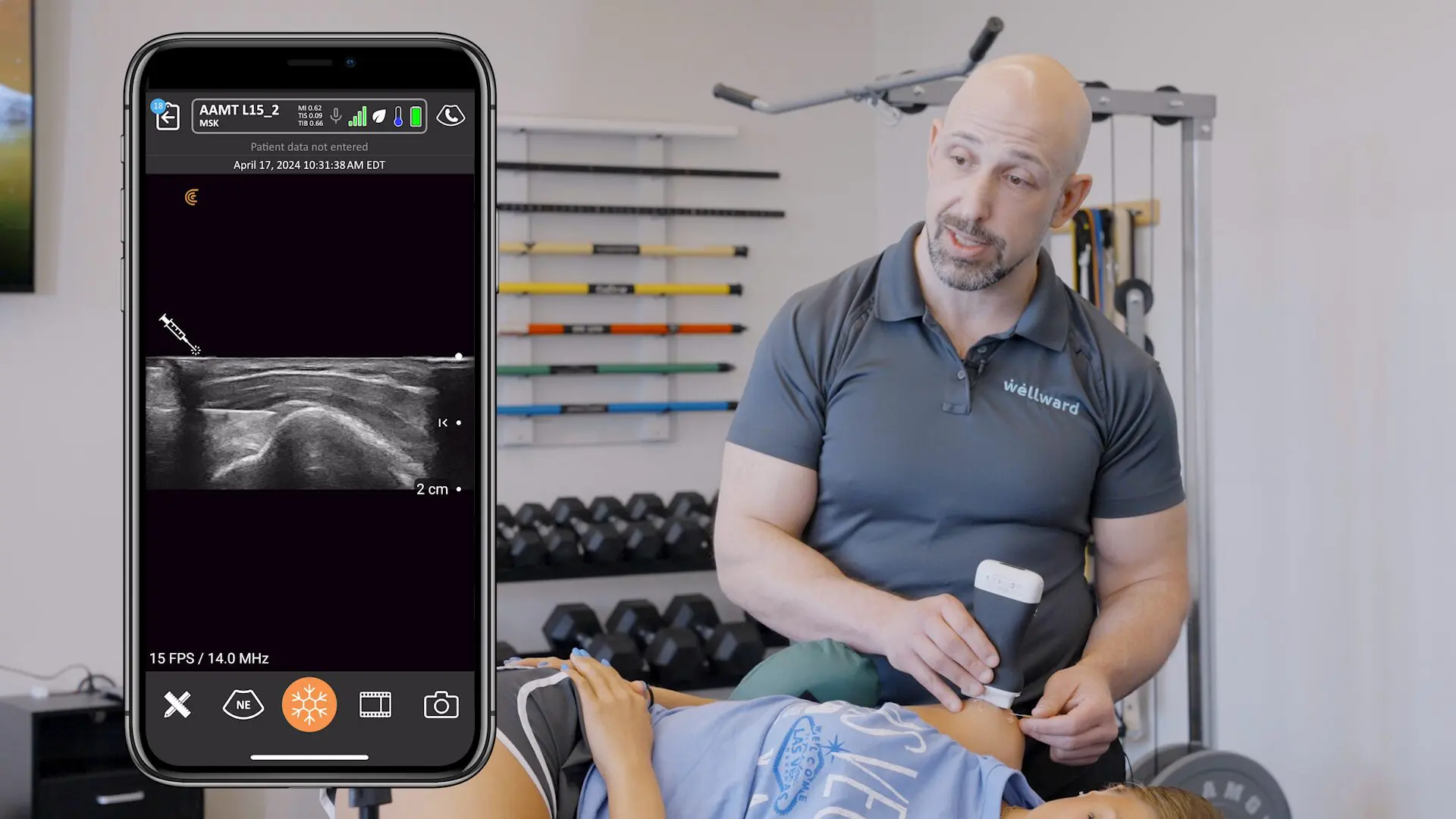
I refer to an excellent radiology department. I know the sonographers and the radiologists are very good at what they do. It’s not uncommon for them to find multiple issues. As clinicians, we need to add the clinical context to pinpoint the problem that is causing the primary clinical concern and decide how to treat that effectively. That’s where I find my Clarius L15 HD helps to visualize and sono-palpate the areas highlighted.
Proficiency with ultrasound builds confidence and trust, while dynamic imaging helps with difficult MSK cases.
Utilizing my sonography skills, I’ve been able to set up a diagnostic and intervention clinic with a prominent Foot and Ankle Surgeon. We work with patients who typically have complex cases. Our goal is to pinpoint what precisely is causing the clinical problem to target intervention and surgical planning.
Several weeks ago, we saw a patient with posterior ankle pain. Her MRI revealed accessory soleus, insertional calcinosis and tendinosis of the Achilles tendon. We needed to understand better the influence each pathology was having on her current symptoms.
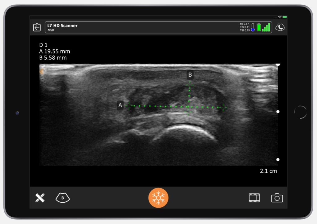
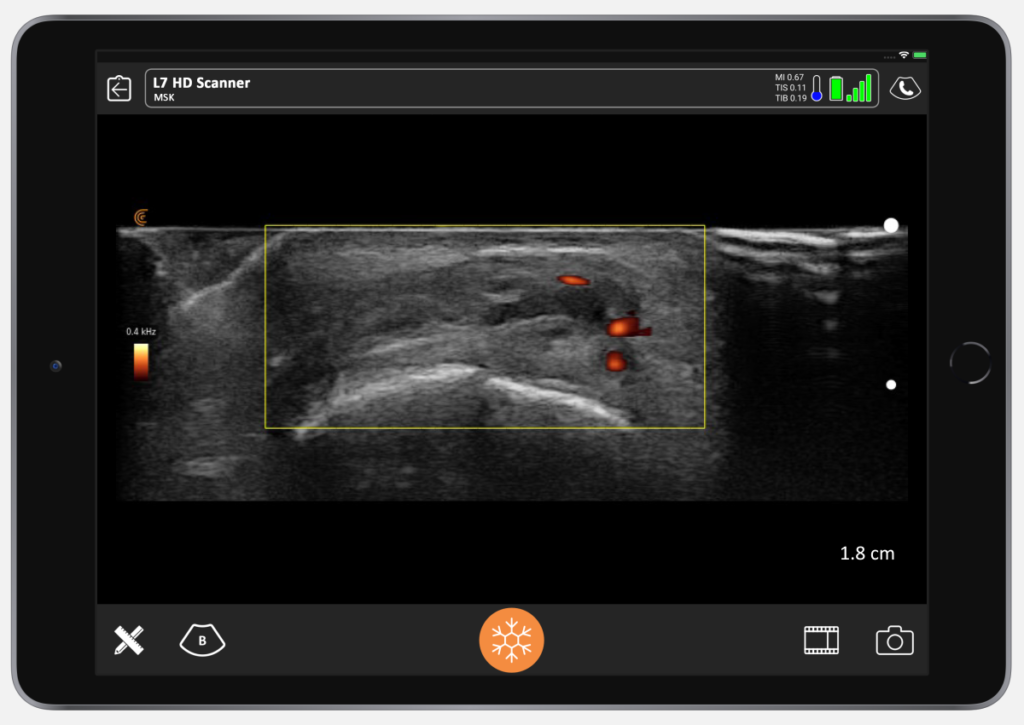
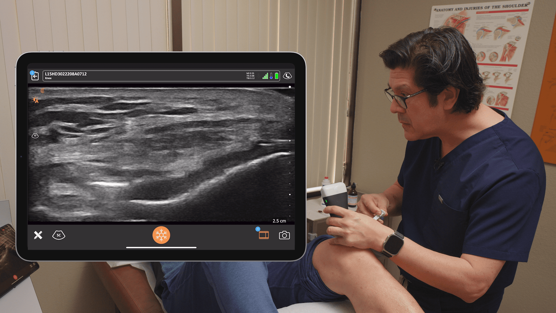
I was able to measure the accessory soleus before and after exertion and dynamically assessed the movement to evaluate for an impingement around the retrocalcaneal bursa. Using the Clarius wireless scanner, I was able to perform a dynamic ultrasound exam — both non-weight-bearing and also weight-bearing. The scan revealed an intrasubstance tear of the Achilles tendon, which became more apparent when the patient placed weight on the Achilles. This level of dynamic evaluation is not possible with MRI.
Our clinical, sonographic findings helped better understand the patient condition and guide conservative and surgical treatment planning. This level of insight provides clinicians with vital information to better inform our patients.“
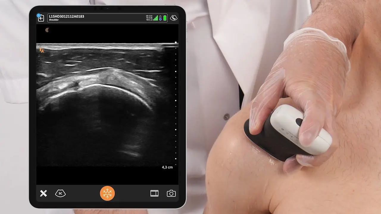
James Ferrie uses the Clarius L15 HD handheld scanner. He is based in Melbourne, Australia and practices at two clinics: My Sports Podiatrist and Ultrasound Training Australia.
Thank you James for sharing your success story! We also enjoyed learning how ultrasound helped deliver better care in the four specific cases you shared with us, highlighted in a previous blog article.
Watch three new video tutorials in Clarius Classroom to hone your podiatry ultrasound scanning skills!
At Clarius, we work with ultrasound scanning experts to produce educational videos for clinicians who are new to ultrasound. We have over 100 tutorials in Clarius Classroom, including the following three podiatry-focused videos by Dr. Oron Frenkle.
How to Scan the Calf
How to Scan the Heel
How to Scan Small Joints
To learn more about how easy and affordable it is to add Clarius HD to your podiatry or MSK practice, contact us today or request an ultrasound demo. We’d be happy to help!
Or come by to see us at APMA 2021 in Colorado for a live demonstration!

