

A specialist in the management of dermal filler complications, Dr. MJ Rowland-Warmann sees complication cases from other injectors nearly daily and has become a vocal advocate for using ultrasound to improve the safety and effectiveness of aesthetic procedures.
We recently had the pleasure of hosting a webinar with Dr. MJ, now available for replay at your convenience: Ultrasound-Guided Treatment for Extreme Aesthetic Complications: Part 2 – Permanent Facial Fillers. Scroll below for highlights.
Filler complications are out of control,” says Dr. MJ at the start of the webinar. “Not a week goes by when we don’t see a horror story in the news or on social media about someone’s awful experience with fillers. And these cases are difficult when patients are injured and upset and a resolution is needed quickly. I couldn’t be without ultrasound. My Clarius L20 is a staple in my practice, and I’ve been using it for around two years. It’s an absolute marvel.”
Types of Dermal Fillers and What They Look Like with Ultrasound
There are hundreds of different filler products out there, different types, different brands. Some countries have more products on their market. Some like the US with stricter licensing laws, they have less. On the UK market, there’s an estimated 500 plus filler products. Some are good, some not so much. To be approved medically, the ideal cosmetic filler should be non-toxic, non-carcinogenic, non-immunogenic, and to achieve excellent aesthetic results.
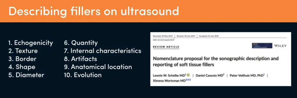
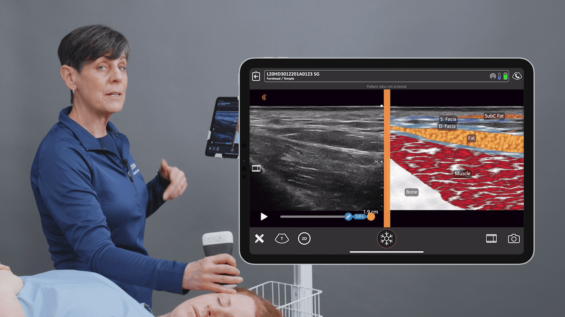
This paper by Schelke and others from 2019 is really helpful because it describes the ultrasound characteristics of dermal fillers using 10 parameters.”
Hyaluronic Acid Filler
Hyaluronic acid filler is undoubtedly the most commonly used type of filler due to its safety and efficacy. Net accounts for about 80% of fillers use worldwide. It’s what you see in your patient’s faces most of the time.
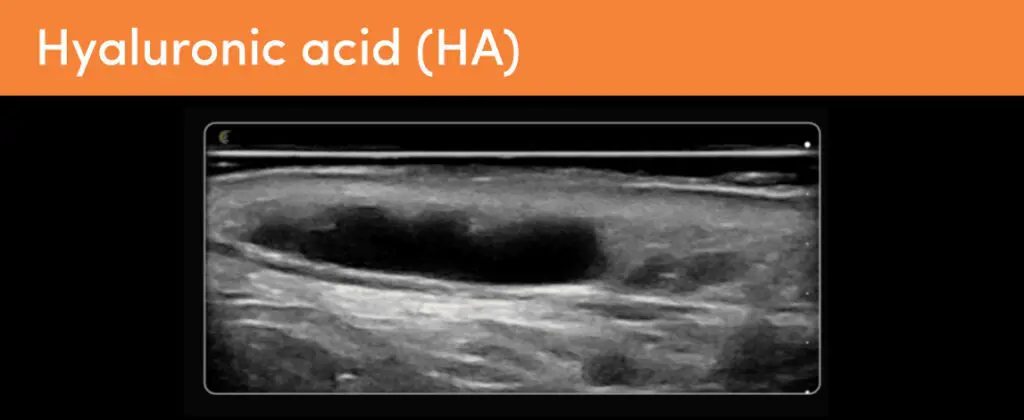
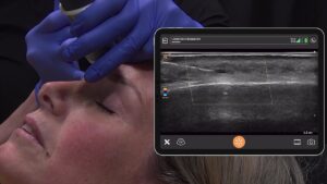
This is HA, it’s anechoic and homogeneous with a well-defined border. HA is usually round or oval in shape. The diameter varies according to the size of the deposits, but generally, per deposit we expect less than 0.5 to a ml, given the size of syringes on the markets. There are no internal reflections. And this tells us the product is liquid or gel-like.
There is a strong posterior enhancement due to the liquid contents, and this amplifies sound waves passing through the filler to the tissues underneath, making them appear whiter. And that’s what you could see there by the white band underneath the filler deposit. This is the classic presentation of HA in tissues. HA filler is extremely consistent over a long period in tissues even years.”
Poly-caprolactone Filler
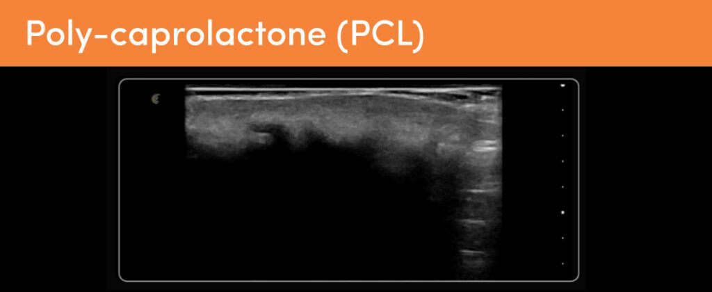
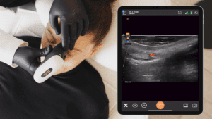
This is polycaprolactone, often marketed under the trade name Ellansé. It’s a hydrophobic filler that is hypoechoic on ultrasound. It appears homogenous. It’s got a poorly defined border and a band-like shape. PCL creates a significant posterior shadow. It blocks out the tissues underneath. This is the jaw line, and the tissues including the bone under the filler have been completely shadowed. Ellansé is a popular bio stimulator in South America and in Europe, so you will likely see this if you work there. It’s injected in a CMC carrier gel, which resorbs. And the results of PCL last about two to four years. It’s not reversible, but it does degrade over time.”
Poly-L-lactic acid (PLLA)
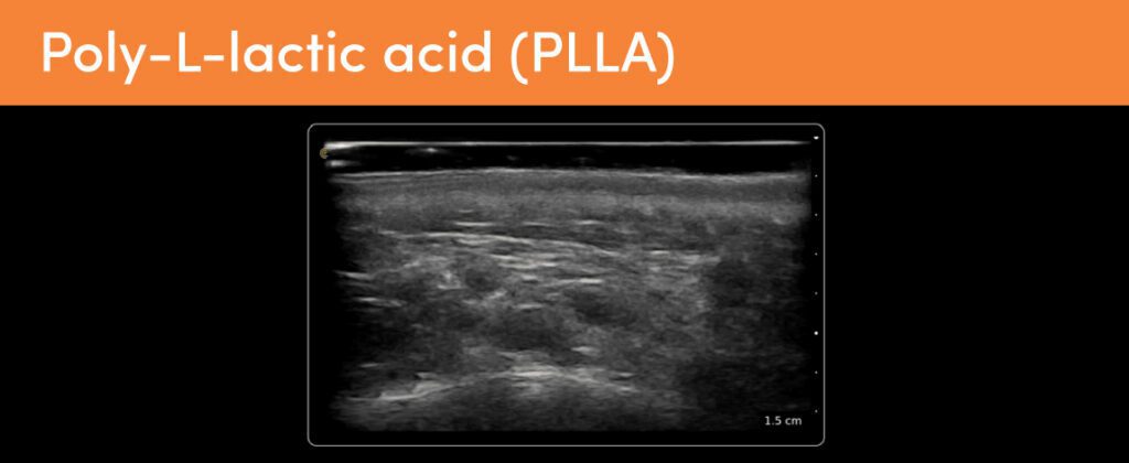
Here we see poly-L-lactic acid. We know it as Sculptra. It’s a hydrophobic bio stimulator. It causes fibrosis, and on ultrasound it’s characterized by widespread hyperechoic patches with slight posterior shadowing, making this diffuse sort of dappled appearance. PLLA is not retained within the tissues at the filler deposit, but instead it causes this fibrotic tissue response. And these kinds of hyperechoic patches are only often really evident when you are comparing it to normal isoechoic fat tissue.”
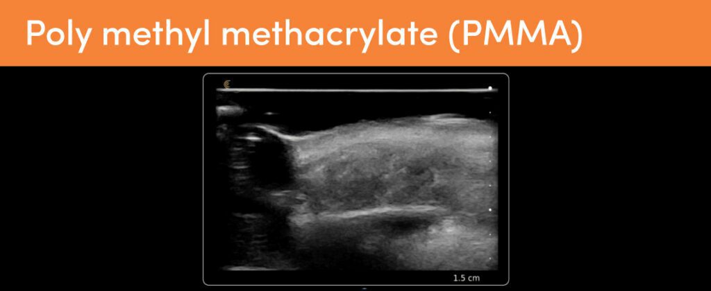
Polymethyl methacrylate (PMMA)
An uncommon synesthetic filler, this is PMMA or poly methyl methacrylate.This patient had cheek filler about 10 years prior, which over time displaced infraorbitally. And here you can see it underneath the orbicularis oculi as an ill-defined hyperechoic mass-like deposit with mini comet-tail artifacts. Those are those small hyperechoic dots that you can see within the filler deposit and which are typical of PMMA. PMMA microspheres are injected in a carrier gel like hyaluronic acid, which is why we’ve got that anechoic deposit right there. And it’s largely fallen out of favor as it’s permanent and it hardens with time. It is still used in South America, and this is where this patient had it placed. So, make sure you take a good history if you suspect PMMA.”
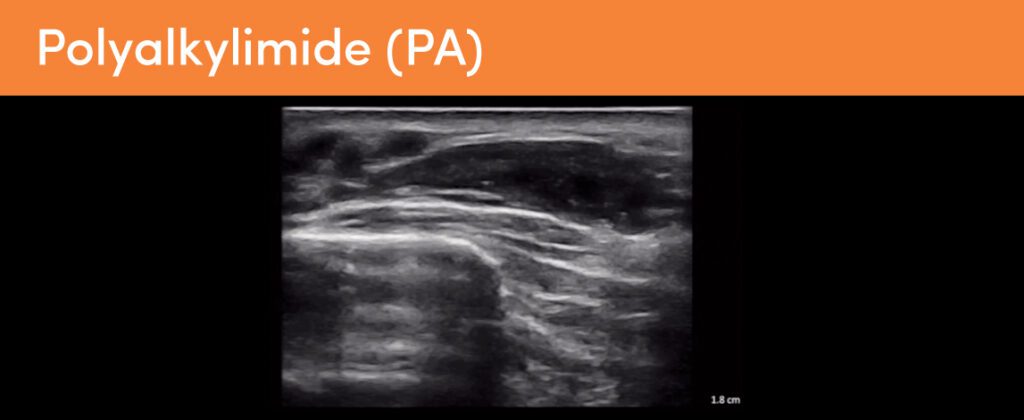
Video Case Review: Woman with Permanent Filler Complication
In this 5-minute video, Dr. MJ describes the case of a 57-year-old lady suffering from significant aesthetic disfigurement due to historic filler treatment. The midface is overfilled and lumpy. Dr. MJ demonstrates the ultrasound appearance of polyalkylimide permanent filler and discusses options for treatment. Watch the webinar for more details.
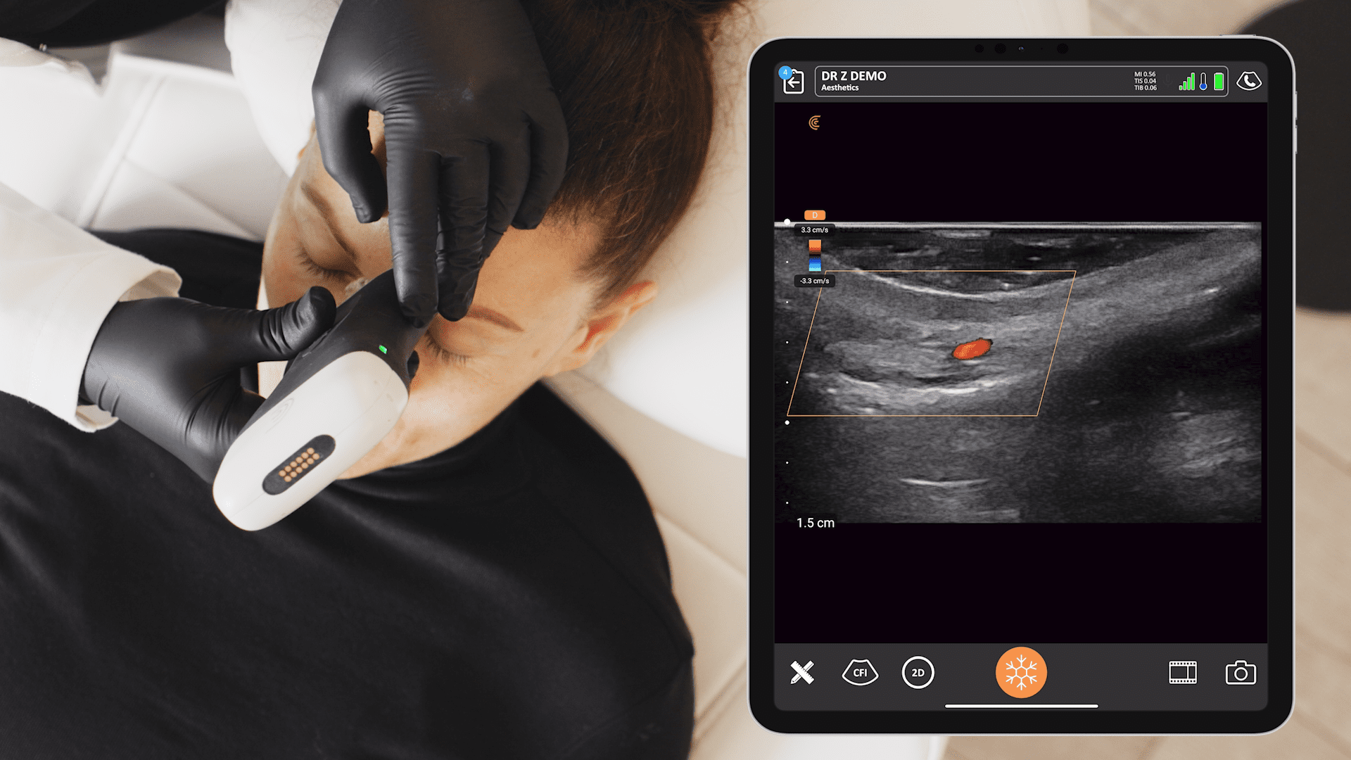
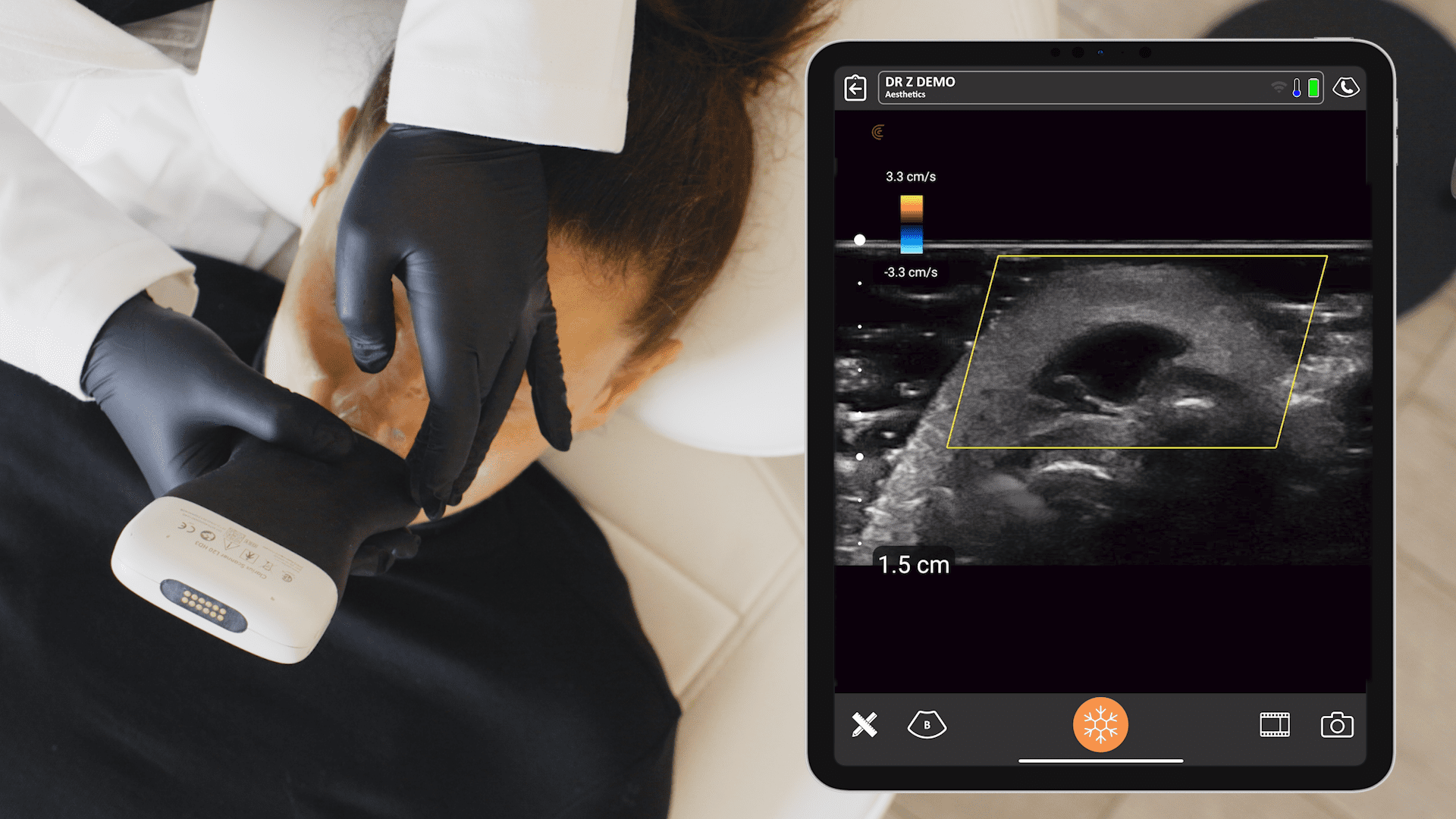
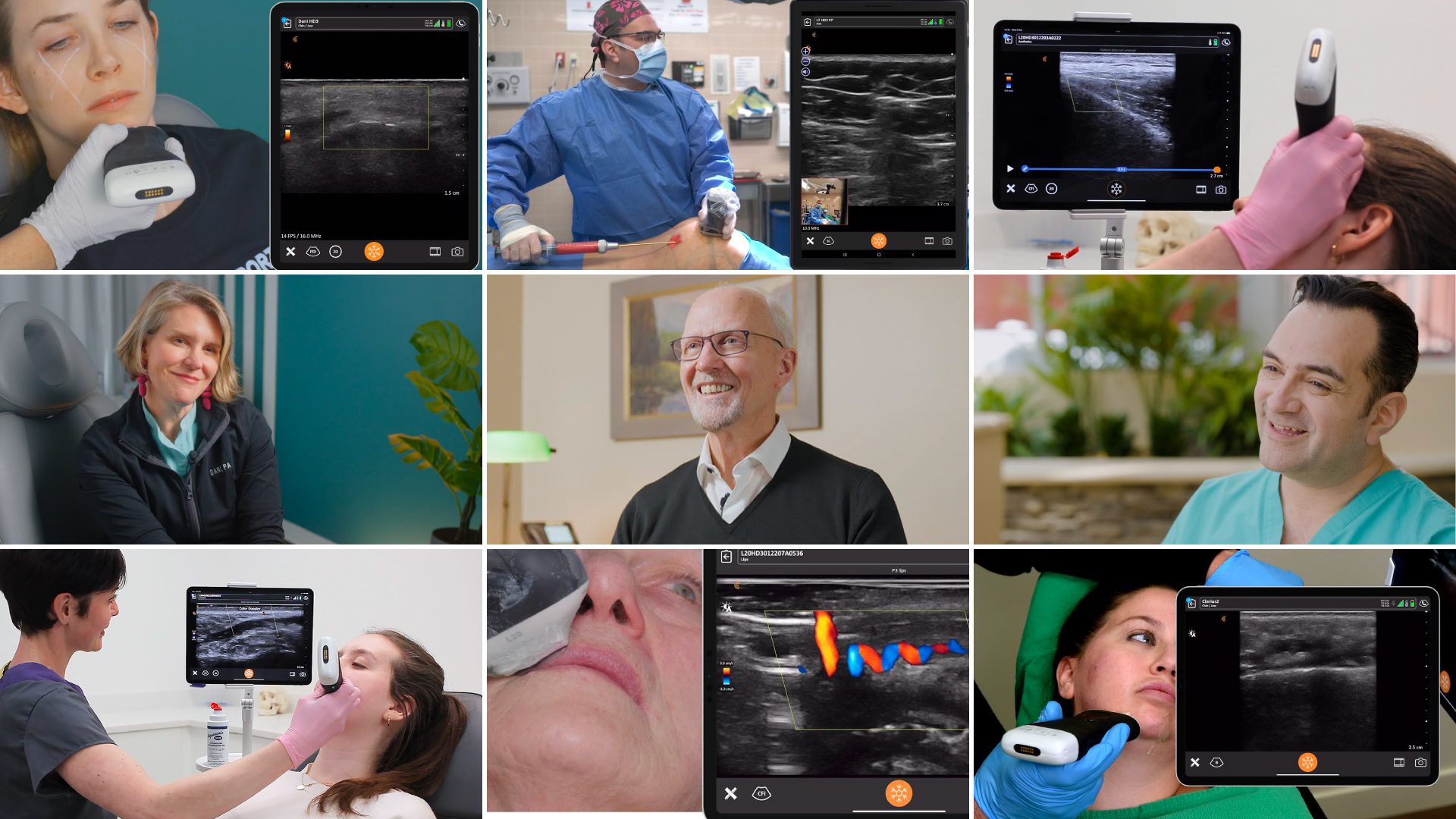
Affordable, High-Definition Ultrasound for Aesthetics
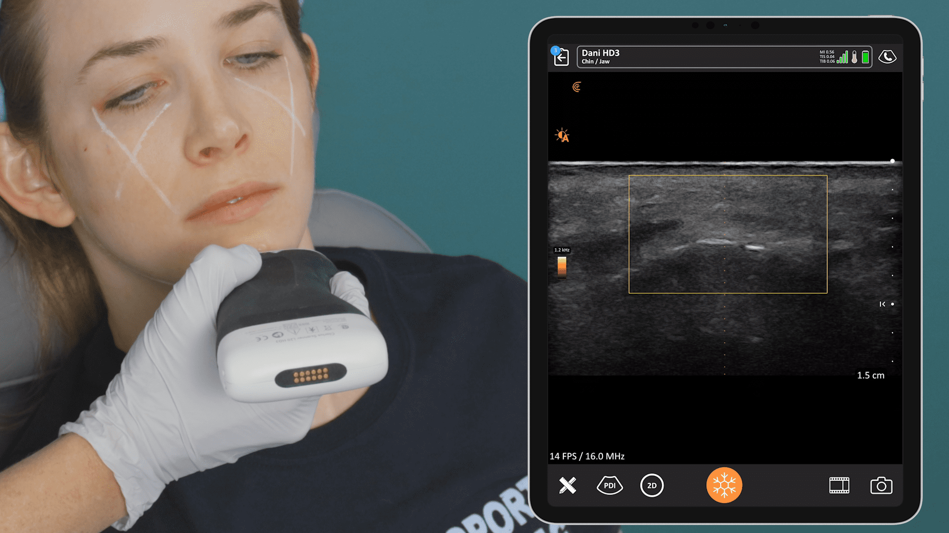
Wireless and pocket-sized, Clarius handheld ultrasound scanners deliver the high-definition imaging and performance of traditional ultrasound systems for a small fraction of the cost. They are the leading choice for plastic surgeons and aesthetics practitioners performing ultrasound-guided procedures to ensure patient safety.
New T-Mode™ AI by Clarius is a groundbreaking educational technology to help clinicians new to ultrasound advance their image interpretation skills using Clarius handheld scanners.
Visit our aesthetics page to learn more or request a virtual ultrasound demo today to learn how high-definition ultrasound imaging with voice controls can improve safety and deliver consistent patient outcomes at your aesthetic practice!


