

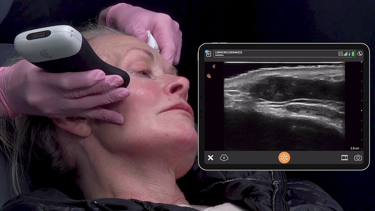
During a recent webinar, Dr. Steven Weiner, a board-certified head, neck, and facial plastic surgeon also known as the “Jawvinator,” showed us how he uses ultrasound to minimize the risks and complications associated with cosmetic filler injections. Watch his full webinar, „Using Ultrasound for Safer Injections of the Lower Face: Deep Pyriform Space and Jawline„, or read on to learn how Dr. Weiner scans the jawline and deep piriform space to understand each patient’s unique anatomy.
The use of dermal fillers for soft tissue augmentation has become an integral part of aesthetic practices, but even in the hands of experienced clinicians, adverse effects can and do occur.
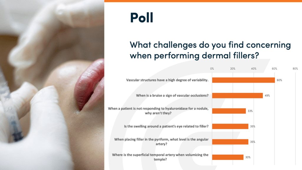
Poll Indicates Variability of Anatomy is a Common Challenge for Safe Injections
We conducted a webinar poll asking about the challenges attendees are faced with when injecting dermal fillers. Not surprisingly, despite knowledge of anatomy, variability of vascular structures appears to be the most common challenge.
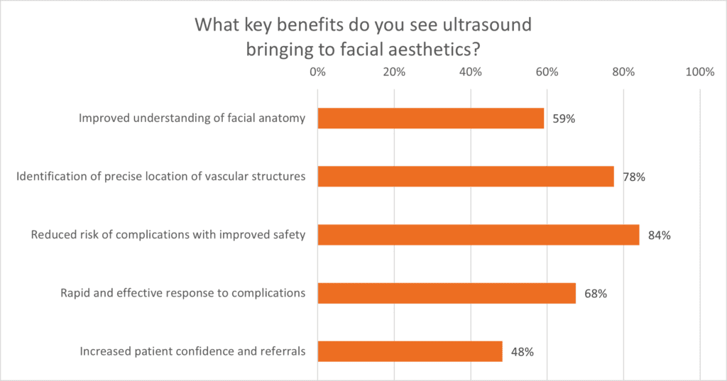
In this recent webinar, we also asked what physicians saw were the key benefits to bringing ultrasound to facial aesthetics. The most common result was reducing the risk of complications with improved safety, followed by the identification of vascular structures, and a rapid, effective response to complications.
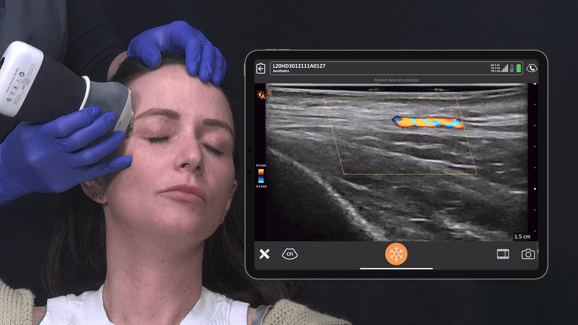
Vascular mapping with high-resolution ultrasound has been a game-changer for Dr. Weiner. With the Clarius L20 HD3 scanner, he can easily locate important tiny vascular structures in the face prior to injection and confirm filler placement away from these vessels following injection.
Deep Piriform Space & Angular Artery
The angular artery lies superficial to the space in the majority of people. It typically runs medial to the NLF at 3 mm in depth.
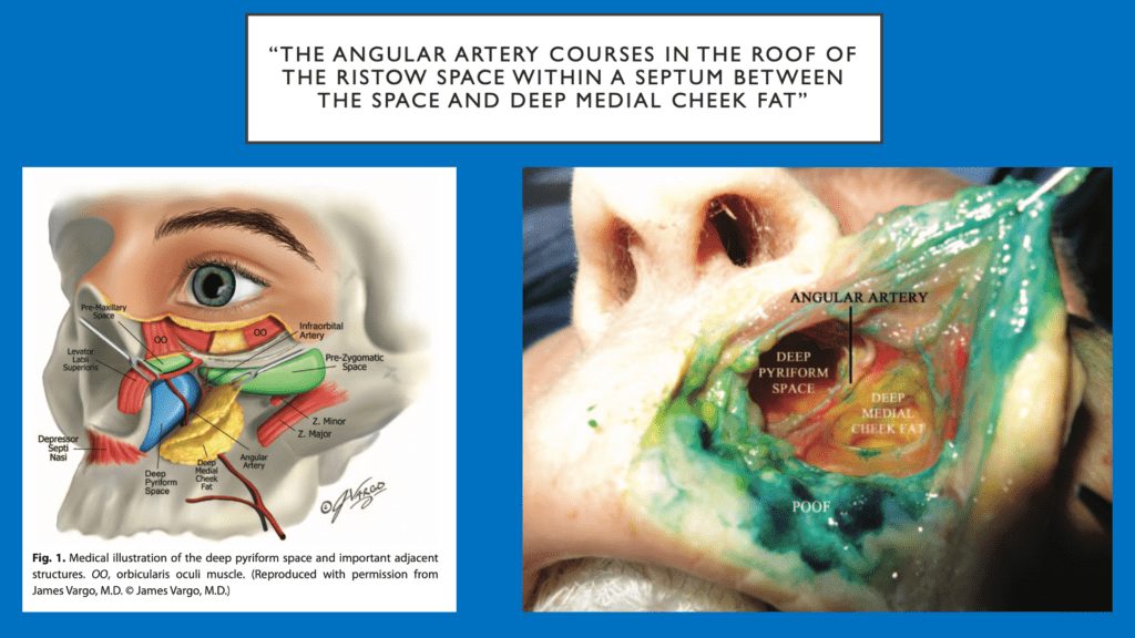
However, there are several anatomic variations including 1 – 3 branches. There can also be variations in the same patient from side to side.
30% of the time, there’s no artery. 1% to 2% of the time, it’s in the exact spot that you want to inject. So how do you know that? Well, you need to use your ultrasound to determine that,“ warns Dr. Weiner.
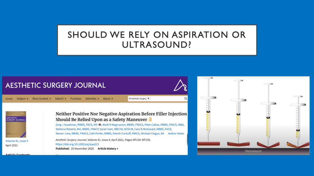
Should We Rely on Aspiration or Ultrasound?
As you can tell by the following illustration, on the right side, it’s possible that you can get a negative aspiration by pinching off the vessel with the needle and still injecting inside the vessel.
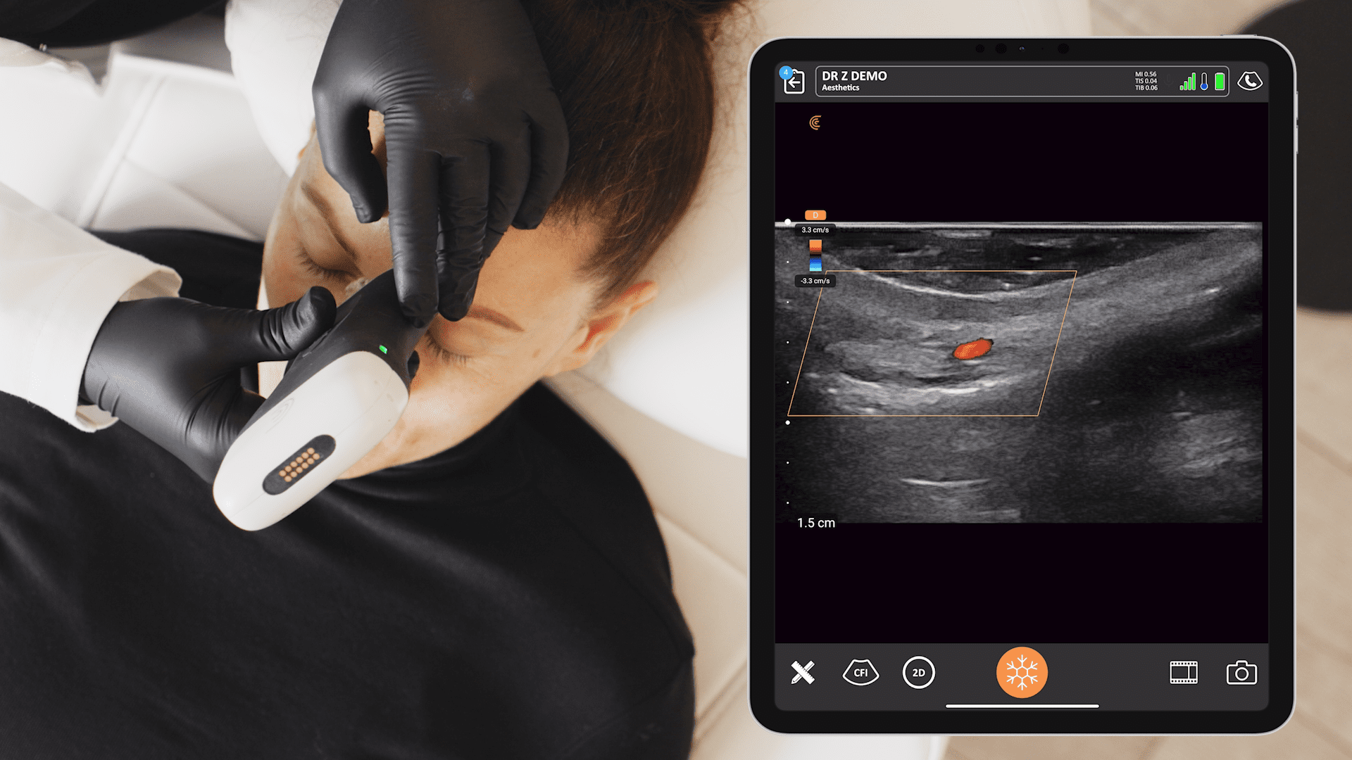
Video Demonstration: Piriform Injection
Piriform injection is done straight down and onto the periosteum.
In this video, Dr. Weiner uses ultrasound to locate the angular artery about halfway between the skin and the periosteum. The bolus of hypoechoic HA filler can be seen along the periosteum, and the artery is intact.
You can confirm to the patient that you saw everything, and it gives the patient confidence that the procedure was done safely,” says Dr. Weiner.
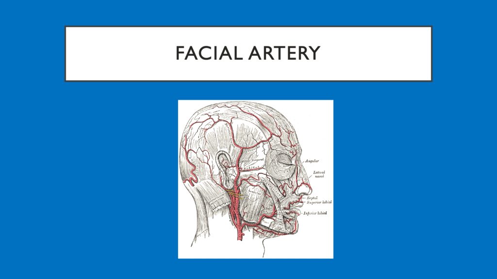
Jawline Injections & Facial Artery
The Facial artery is a branch of the external carotid artery. It is the main supplier of blood to the anterior face. The facial artery typically crosses the mandible in the antegonial notch. There can be variations in the location and branching of this vessel as well, so Dr. Weiner recommends confirming its position with ultrasound prior to jawline injections.
Planes of Injections include:
- Superficial (subcutaneous or supra-platysma) plane in superficial fat
- Safe – Facial artery is deep
- Better correction per CC
- Deep onto the periosteum
- Less noticeable filler
- Can’t place deeply in antigonial notch
- Combination
In the next video, Dr. Weiner marks the location of the artery and vein so he can safely inject posterior to them. Watch the video to see his technique.
5 Ways Ultrasound Can Help Your Practice
- Improving patient safety with vascular mapping
- Mitigate the risk of filler complications
- Solve complications with real-time imaging
- Increase patient confidence with high-definition imaging
- Increase billings and referrals
Ultrasound is the gold standard for evaluating patients, particularly with filler complications,“ he says. „But it’s better to be pre-emptive by evaluating vascular structures using vascular mapping in advance to avoid these structures when you’re doing the injection.”
Watch the full webinar, „Using Ultrasound for Safer Injections of the Lower Face: Deep Pyriform Space and Jawline„, to learn more about piriform and jawline injections.
About Dr. Weiner
Dr. Weiner practices at the Aesthetic Clinique in Florida, a company he began over 15 years ago.
He uses the Clarius L20 HD3 high-frequency scanner, which he considers ideal for facial aesthetic procedures with its ability to provide excellent detail of superficial structures.
Dr. Weiner offers a course called Sonosthetics with courses in Los Angeles, Vancouver, Chicago, New York, Salt Lake, Hickory, and Atlanta.
Improve Patient Safety for Facial Aesthetics with Ultrasound Guidance
Thanks to miniaturization and innovation with handheld ultrasound, high-definition imaging is now easy and affordable, offering savings up to 80% on the cost of a traditional ultrasound system.
To learn more about the high frequency Clarius L20 HD3 handheld ultrasound system that Dr. Weiner uses, and the Clarius 9.0 App with Advanced Plastic Surgery Package, contact us today or request an ultrasound demo.
We would love to show you the difference ultra-high-definition imaging makes for facial aesthetics. Contact us or visit our Aesthetics Specialty Page to learn more.


