

Making a confident diagnosis and treatment plan for MSK injuries is easier than ever with handheld ultrasound. But learning to scan remains a barrier for many clinicians.
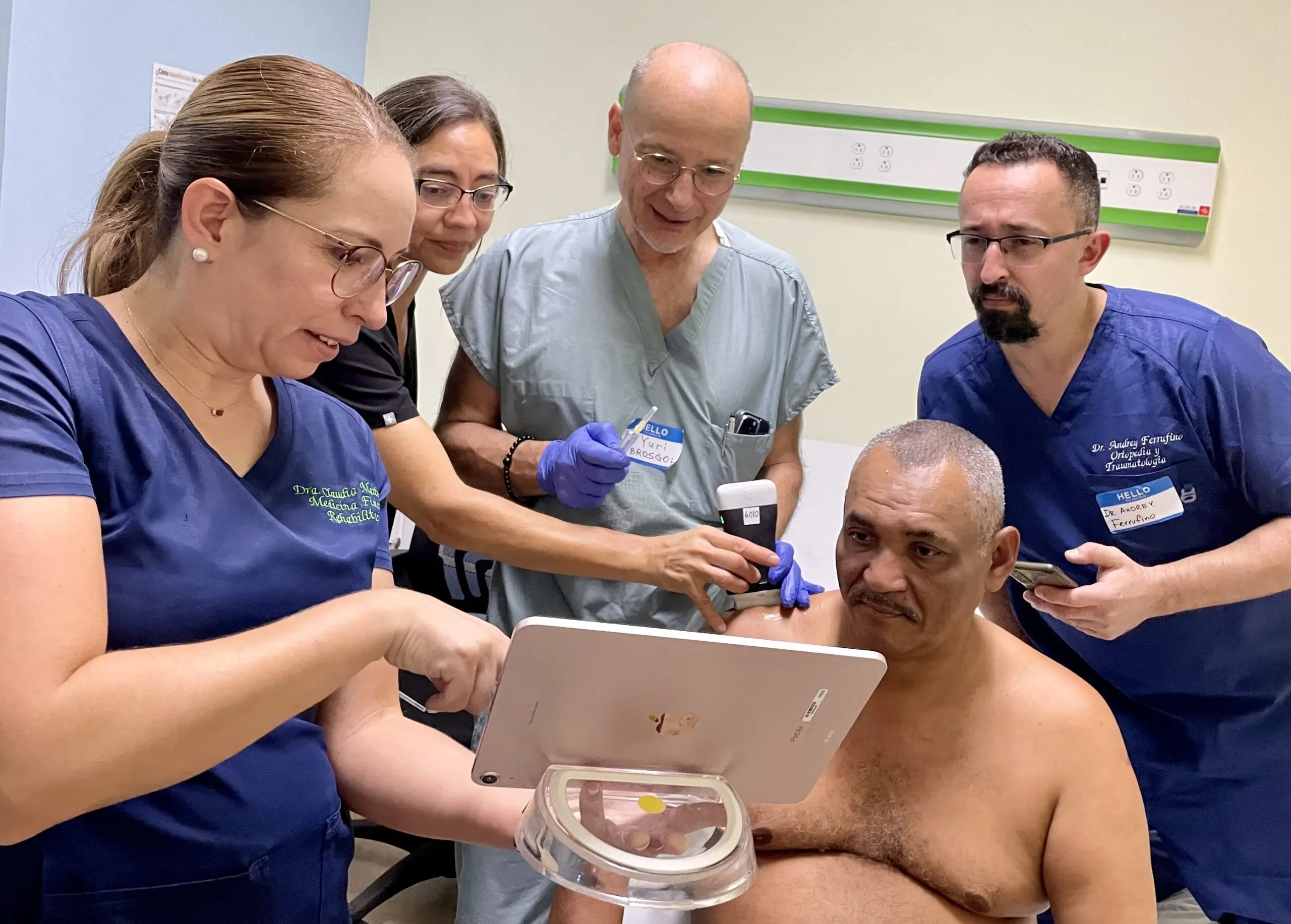
We recently invited renowned SonoSkills educator, Marc Schmitz, to co-present a webinar: — Pragmatic MSK Ultrasound: Scanning the Rotator Interval, Common Extensor Tendon and Patellar Tendon. Participants walked away with practical tips on how to scan joints, assess the rotator interval and look for pathology in the elbow and knees. Watch one-hour webinar or read this article for highlights.
Background on MSK Ultrasound
In our background research for this MSK webinar, we found that ultrasound is used in clinical practice to diagnose everything from fractures and dislocations to tendon and ligament injuries. It has become the primary diagnostic tool for many traumatic inflammatory and degenerative soft tissue conditions. Ultrasound helps to help make the diagnosis on the first visit and is subsequently used to monitor progress with ongoing quantitative measurements and reevaluation of the patients every time they come into the clinic. It’s easy to track progress and improvement or deterioration over time.

Key Challenges Facing MSK Clinicians
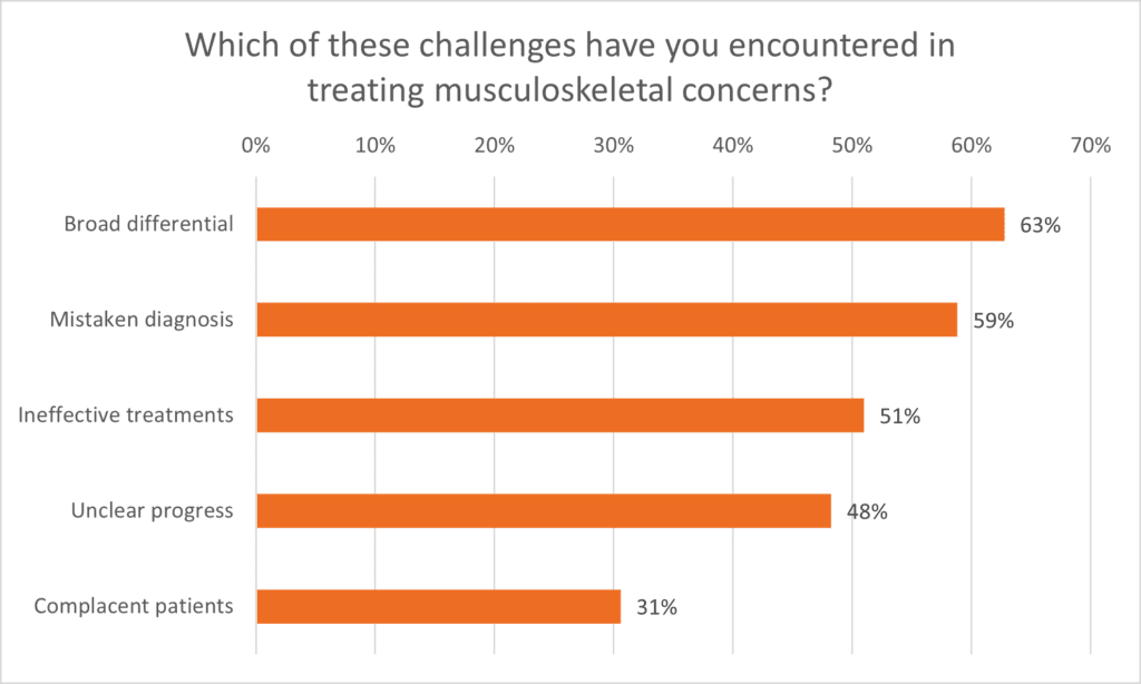
We kicked off the webinar with a poll of the attendees to identify the key challenges MSK clinicians face in clinical practice. Of the clinicians attending, 63% reported having challenges treating musculoskeletal concerns due to a broad differential diagnosis and 59% reported having made a mistaken diagnosis.
The webinar’s presenter, Marc Schmidt, learned early in his career that using ultrasound helped him to accurately diagnose MSK injuries. His interest in functional human anatomy grew while dissecting and studying cadavers. It was here that his interest in observing functional human anatomy in «real-time» peaked, which led to the start of embracing musculoskeletal ultrasound. He now teaches with a team of 22 expert trainers through Sonoskills, established in 2010. Functional anatomy is core to his ultrasound courses. Read on to learn what he had to say about the Common Extensor Tendon.
Anatomy of Common Extensor Tendon
The common extensor tendon is actually a combination of the extensor digitorum, the extensor digiti minimi, the extensor carpi ulnaris and also the extensor carpi radialis brevis. These are the common tendons originating from the lateral epicondyle and then at one point they transform and separate muscle bellies.
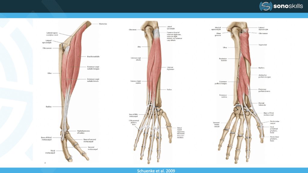
And you can see in this diagram that these have a proximal distal orientation going towards the wrist and the fingers and so they are truly extensors of this region.
One level more proximal is the extensor carpi radialis longus. This is really separate from the extensor carpi radialis brevis and the common extensor tendon but this can also mimic lateral elbow complaints and sometimes you can also find pathologies there.
We can also see one level deeper into the depth closer to radial supinator muscle and look at, this is a oblique course of fibers meaning it has a different function, it’s not an extensor but this is a supinate or pronator for the forearm.
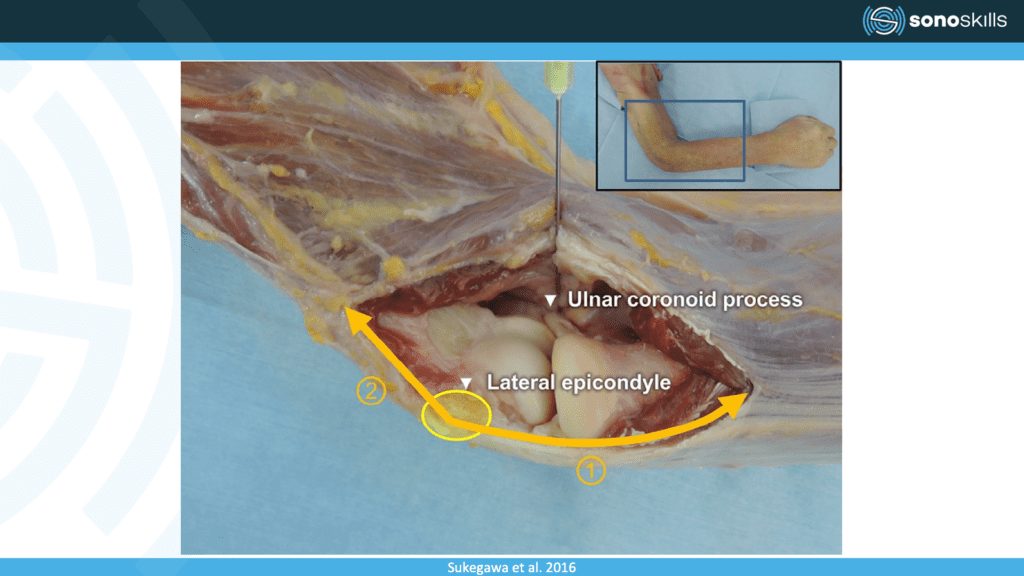
Really close to the bone and really close to joint is this a common extensor tendon, and because the lateral epicondyle is roughly there, here we can see the joint between the humerus and radius, and of course where we have a synovial joint, this is a synovial joint.
We always have a capsule and also reinforced with ligaments and in this case is reinforced with the collateral ligament and this collateral ligament is really close to the tendon and they have a similar appearance. So this is a pitfall that you might confuse ligament for tendon or vice versa.
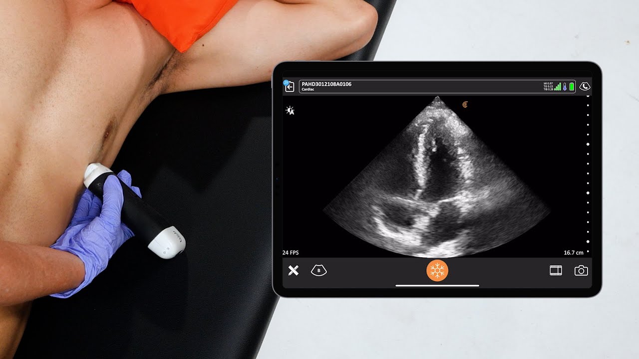
VIDEO: How to Scan the Common Extensor Tendon
Marc Schmidt demonstrates how to examine the common extensor tendon. Watch the 2-minute video.
Examples of Common Sonopathology
During the webinar, Marc Schmidt offers detailed commentary on ultrasound images of common pathologies of the extensor tendon, patellar tendon and rotator interval.
Watch the webinar to learn how to recognize common MSK pathology using ultrasound: Pragmatic MSK Ultrasound: Scanning the Rotator Interval, Common Extensor Tendon and Patellar Tendon.
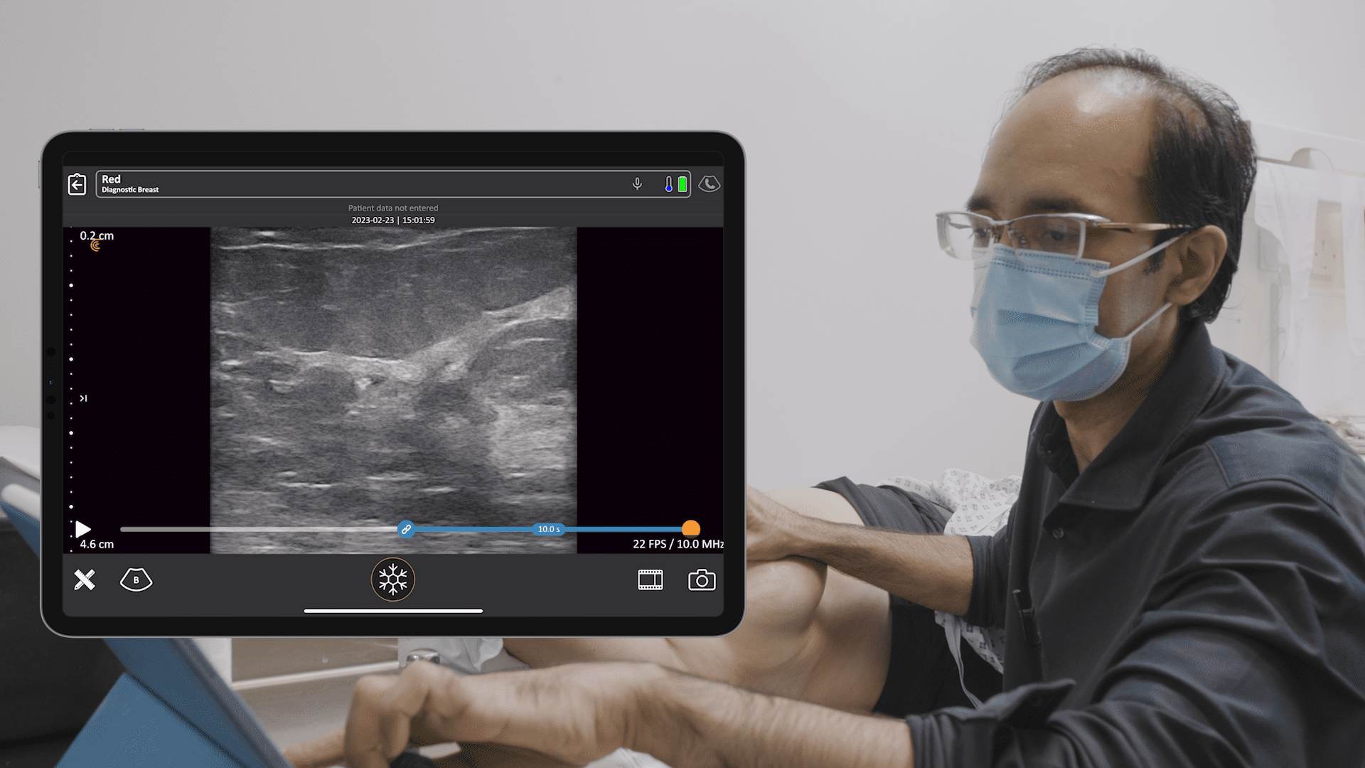
Handheld Ultrasound Designed for MSK Clinicians
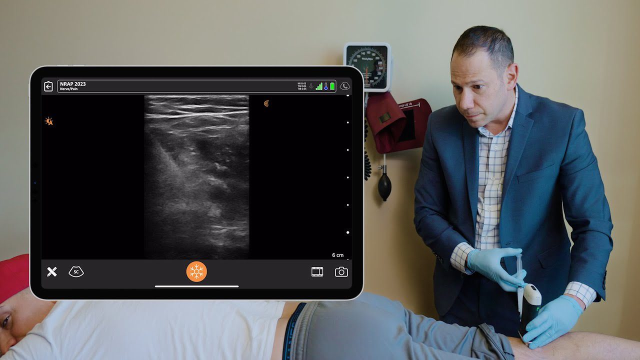
Getting detailed ultrasound images of musculoskeletal anatomy to make a confident diagnosis has never been easier. Ideal for MSK anatomy down to 7 cm, the Clarius L15 HD high-frequency ultrasound scanner is available with an optional Advanced MSK Package for diagnostic and interventional procedures.
Clarius offers four ultrasound scanners that are suitable for MSK applications. Learn more about which Clarius Handheld Ultrasound scanner is right for your practice. Or contact us today to request an ultrasound demo.


