By providing my email, I consent to receive Clarius webinar invitations, case studies, whitepapers, and more. I can unsubscribe anytime. Privacy Policy.
FREE WEBINAR
Small Animal Ultrasound: Mastering the Aspire UCS 8-Step Abdominal Ultrasound Survey Technique™
Watch On-Demand
Watch Now
Note: This webinar does not provide CE or CPD credits.
We’re excited to welcome seasoned sonographers turned passionate veterinary ultrasound educators Angie Lloyd-Jones and Julie Burnage to teach ultrasound image optimization, and Steps 1 – 3 of their trademarked veterinary abdominal ultrasound survey.
They’ll guide you through their proven wireless ultrasound techniques to simplify canine and feline abdominal imaging, making it more consistent, teachable, and diagnostically powerful. In this 1-hour webinar, Julie and Angie will provide detailed instruction and live scanning demonstrations. Attendees will learn how to:
- Optimize images by understanding basic ultrasound system settings (knobology)
- Apply steps 1 – 3* of the 8-step technique to both routine wellness exams and complex diagnostic cases.
- Optimize scanning efficiency while maintaining a thorough, standardized survey.
- Improve diagnostic accuracy with a repeatable, teachable approach to abdominal ultrasound.
- Leverage handheld ultrasound technology to streamline abdominal imaging in busy clinical settings.
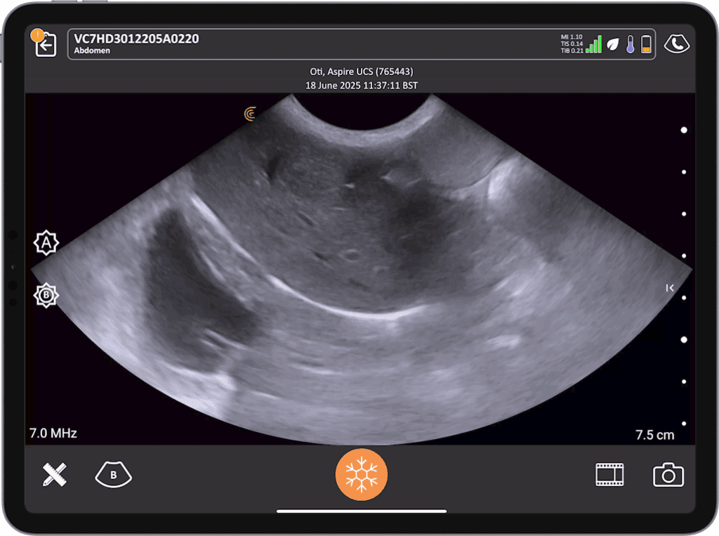
Abdominal ultrasound is one of the most valuable diagnostic tools in veterinary medicine, offering real-time insights into canine and feline patient health. Yet, many clinicians struggle with a systematic approach that ensures accuracy, efficiency, and repeatability in their scans.
The Aspire UCS – 8-Step Abdominal Ultrasound Survey Technique™ is a structured protocol designed to simplify abdominal imaging and improve diagnostic confidence. This proven method breaks down the complexity of abdominal ultrasound into eight practical, sequential steps, enabling veterinarians to consistently capture critical views, identify abnormalities, and reduce oversight.
- Step 1: Diaphragm, liver and gallbladder, cystic and common ducts, portal vein and lymph nodes
- Step 2: Stomach, pylorus, proximal duodenum, left pancreatic limb and lymph nodes
- Step 3: Spleen (head, body, tail), diaphragm, splenic vein and lymph nodes
Whether you’re new to veterinary ultrasound or looking to refine your skills, this session will provide actionable knowledge you can immediately implement in your practice. Join us to gain confidence in abdominal imaging and deliver more precise, timely care for your patients.
Clinical Lead, Aspire UCS
Angie Lloyd-Jones, DCR DMU
Angie Lloyd Jones began her career as a Diagnostic Radiographer in the late 1980s, starting her ultrasound journey in West Sussex in the early 1990s. Over the years, she worked across Wales and London, leaving behind skilled ultrasound practitioners in various specialties. Angie helped set up ultrasound services in numerous NHS hospitals, becoming a clinical tutor and lecturer for CASE-accredited programs. Later, Angie joined Julie as Head of Ultrasound for a UK-wide imaging company, further honing her business and clinical management skills. She eventually transitioned to small animal ultrasound, recognising the challenges primary care vets face in training. Angie is passionate about quality, standards, and competency-based learning. During the Covid-19 lockdown, she and Julie co-authored professional guidelines for small animal abdominal ultrasound, endorsed by ECVDI and IVUSS. She is an award-winning professional speaker, and the visionary behind the 'Lillie' Canine Ultrasound Phantom
Education Lead, Aspire UCS
Julie Burnage, DCR DMU FETC
Julie Burnage began her career as a Radiographer in North West England in the early 1980s, moving to North Wales in 1987. She started her ultrasound training in 1988 while also training to be a teacher to share her passion for ultrasound. In 1995, Julie founded an ultrasound company offering diagnostic scans in GP surgeries and clinics, advocating for ‘care closer to home’ despite skepticism from the imaging community. With support from forward-thinking radiologists, the business grew, eventually merging with another provider in 2013 and being acquired by venture capitalists in 2020.
Medical Sonographer
Shelley Guenther, CRGS, CRCS
Shelley Guenther worked as a Nuclear Medicine Technologist for 2 years before entering into the ultrasound program at the Royal Alexandra Hospital in Edmonton. After graduating with specialties in general ultrasound as well as echocardiography, she worked as a clinical expert in the commercial world of ultrasound for over 25 years. As Clinical Manager at Clarius, Shelley Guenther is dedicated to providing the highest quality educational content for clinicians looking to add wireless ultrasound to their practice, including practical webinars and Clarius Classroom video tutorials.