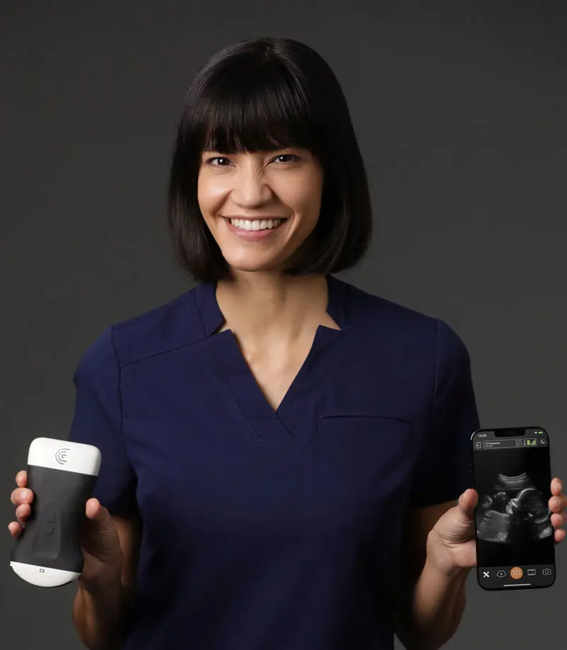
The introduction of the portable ultrasounds completely changed that. It means that now within an affordable price, medical aesthetic practitioners can purchase a portable device and now are in a fortunate position where there is more training available. And I think that it has elevated aesthetic practice a lot. There is an increase in use as a precautionary measure prior to injections. There is an increased use of ultrasound guided injections where necessary or where it is deemed helpful. There is an increased predictability in the way that we manage complications as an industry and there is also a greater understanding of dynamic anatomy as a result of being able to see the anatomy in real-time.”
Témoignages

Anish P. Keshwani
MD
I feel more confident with my ultrasound exams since I’ve started using OB AI. I love how the AI makes the app light up when things are perfectly lined up – I can see this really helping both seasoned clinicians and those who are starting their ultrasound journey.”

Oron Frenkel
MD
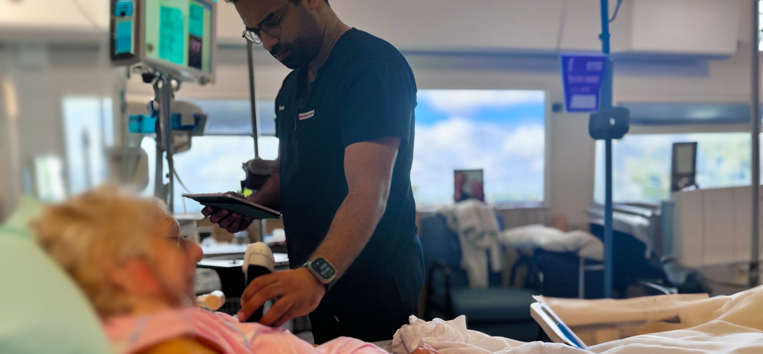
It really is my go-to friend for every patient. There is really no situation that I can imagine where the scanner wouldn’t be able to deliver what I need, either on the same patient, if I need to scan different parts of them during a single presentation, or going from bed to bed during a shift, I don’t ever have to switch out, and it really makes my workflow seamless.”

Zainab Al-Mukhtar
BDS, MJDF, RCSEng, MFDS
I now make a point of pre-scanning the temple, the nose, and the piriform fossa and indeed, the center of the chin if I’m using a bio stimulator that is not easy to dissolve. But always, the nose, the piriform fossa, and the temple. So, I pre-scan. In some situations, I use it for ultrasound-guided injections of filler and unique circumstances where I see that it’s necessary. So, for example, in the nose when I’m treating the lateral aspect of the nose, when I am treating the whole nasal labial fold subcutaneously and when I’m treating the temple in the subcutaneous or interfacial plane.”

David Rosenblum
This is something that has really changed my practice, increased my efficiency. It’s like having a third hand. I just use my voice and the commands happen, and it captures the image and it does everything my assistant would normally have helped me with during a procedure.”

John Arlette (T-Mode)
MD , FRCPC, FAAD, FACMS
The T-Mode AI color assist for facial ultrasound imaging in aesthetics is a significant advancement in training for those early in their scanning career. The demonstration of fascial planes, fat compartments and muscle rapidly leads to confident interpretation of the important structures below the skin.”
Hani Mikhail
MD, Critical Care physician
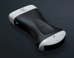
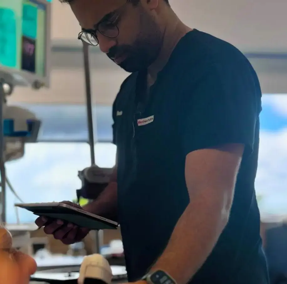
The Clarius PAL is the only scanner I know of currently that provides a feature set comparable to full-size ultrasound machines while remaining in such a portable form factor – all without compromise on image quality, ease-of-use or battery life.”

Christoph Kühnle
Diplomate ECVS
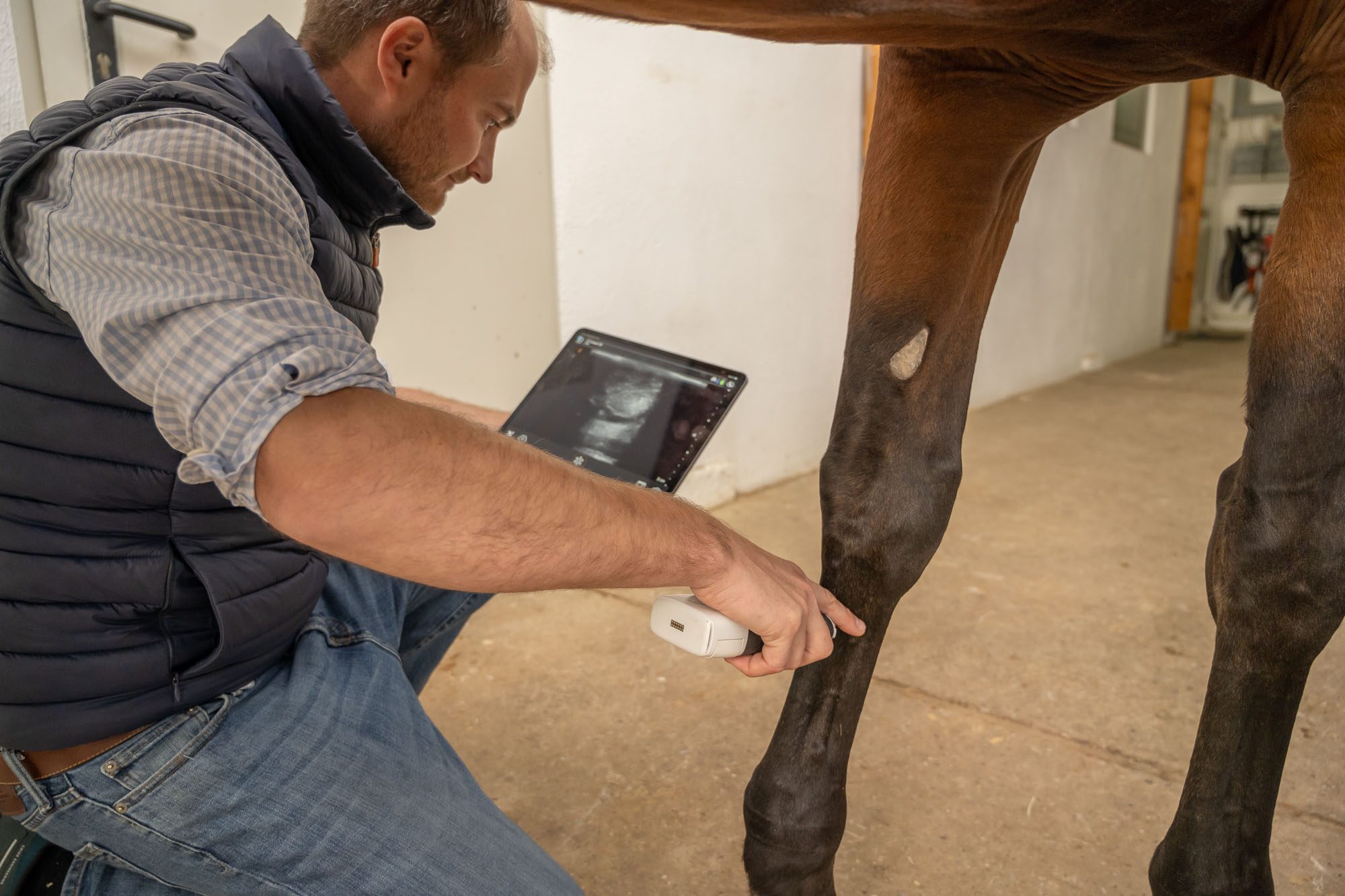
With the Clarius Scanner we have found a mobile solution for ultrasound imaging. Not only the fast operational readiness, but also the flexible use of the device is convincing. Larger diagnostic devices with cable are less practical , especially for sterile injections. Thanks to the Clarius scanner, we have the possibility to perform these treatments under the best conditions. Experience has shown that the acceptance of the horses is significantly higher than with conventional devices, which simplifies the treatment in many aspects and is less stressful for our patients. The dynamic image adjustment finds always the best setting, which makes a scan more efficient. »

Dr. Brian Johnson
Emergency Physician
It’s nice to get a nice snapshot of a critically ill patient right away so that helps me with the workflow. I can walk out of the room and say this guy is in pulmonary edema or this guy looks like he has a ruptured AAA. That can help guide my next steps of management for the patient. So, it’s hitting a lot of good spots for me.”
