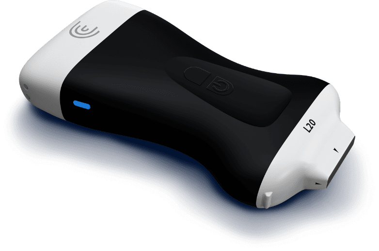
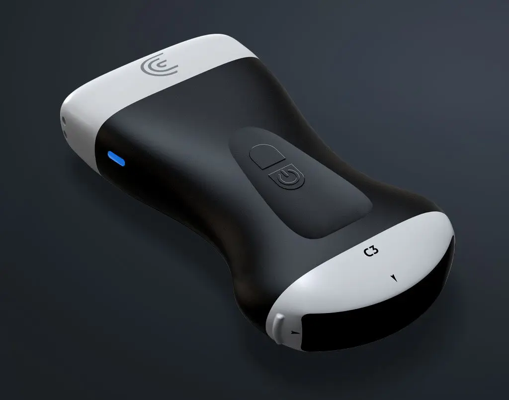
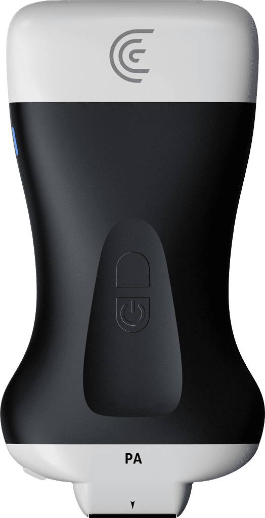
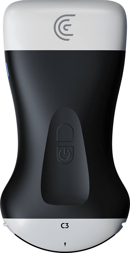
Lung ultrasound is being used as a fast and effective alternative to X-Ray and CT, particularly with COVID-19 patients. Clarius handheld ultrasound systems offer the added benefit of easier infection control measures. They can be completely encased and quickly disinfected after an exam as you move from patient to patient. With lung pre-sets on Clarius phased array, curvilinear and linear scanners, you can start scanning within seconds. And with Clarius Telemedicine, you can monitor, guide and review ultrasound exams from a distance.
COVID 19: Clarius Ultrasound for Lung Pathology
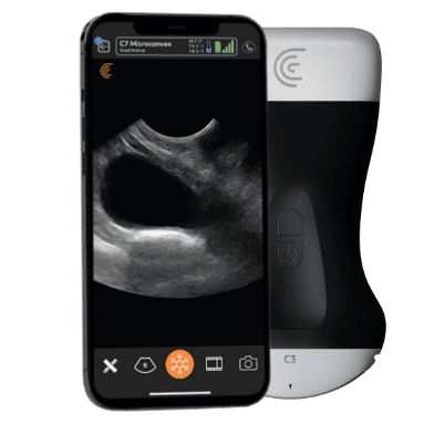
Ultrasound Lung Scanning Tutorial
Dr. Dan Kim uses a Clarius handheld scanner to show how you could examine the lungs of a patient with COVID-19. He discusses lung settings, a systematic approach to scanning the lungs and interpretation of lung images.
Lung Exams Using Clarius Scanners
Following are examples of lung pathology posted on our Clarius Cases community forum. For comparison, we’ve posted a clip from an exam of a healthy model.
Jochen Neumann
COPD
64y w, hx COPD; acute dyspnea for 2 d; initial bt 39°C, chest pain Sono: shred sign -> dx of non-translobar pneumonia Admission to hospital
Healthy Lung
Lung from a healthy 27-year-old male. Scanned using the Clarius HD C3 on the lung preset.
Jochen Neumann
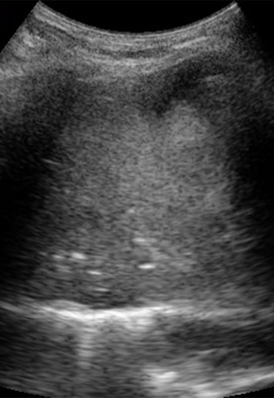
Pneumonia
74y w w/ dyspnea for 1 week -> first ever use of the C3 Scanner and the very first placement on left upper rib space anterior: Consolidation with positive air bronchogram – auscultation was semi-suspicious at best. Dx of pneumonia within 30 s, which may have been overlooked otherwise or at least delayed. I like !!
Dr Shane Arishenkoff
Right sided lobar pneumonia acts as a window to visualize the heart from a right coronal view
Patient presented with cough, sputum, fever and hypotension. Large right sided lobar pneumonia identified. This is a coronal image taken from the right posterior axillary line. Initially the liver, diaphragm (right of the image) and hepatized lung (left of the image) can be seen. As the transducer is adjusted the beam is directed above the diaphragm and only the hepatized lung is seen. What is interesting about this image is the fact that the hepatized lung acts as a window to visualize the heart all the way from the right lateral chest.
One click lung scanning with Clarius
Each of the following Clarius Scanners offers an automated preset for lung scanning, so you can start scanning quickly.
Convex Scanner
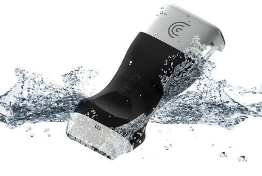
Cleaning and Disinfection is easy
Clarius is IPX7 rated – it’s waterproof and fully immersible for disinfection. Review the long list of cleaners and disinfectants that are qualified for use with the Clarius Scanner.
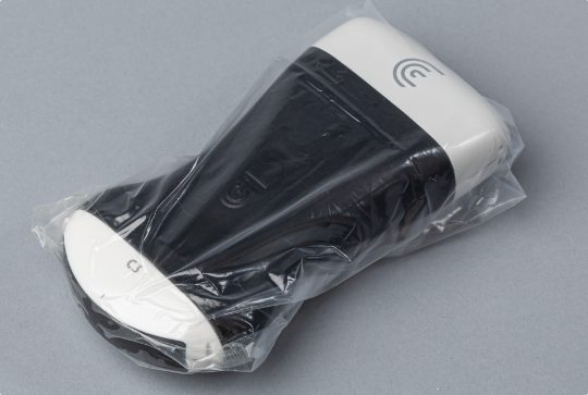
Encase the Whole Scanner
With no cords to worry about, place Clarius in a sterile bag to avoid contamination. If resources are scarce due to COVID-19, Clarius can also be encased in any airtight plastic bag such as Ziploc. Don’t forget to put gel in the bag before sealing.
Included with each scanner
Minimize Exposure with Telemedicine
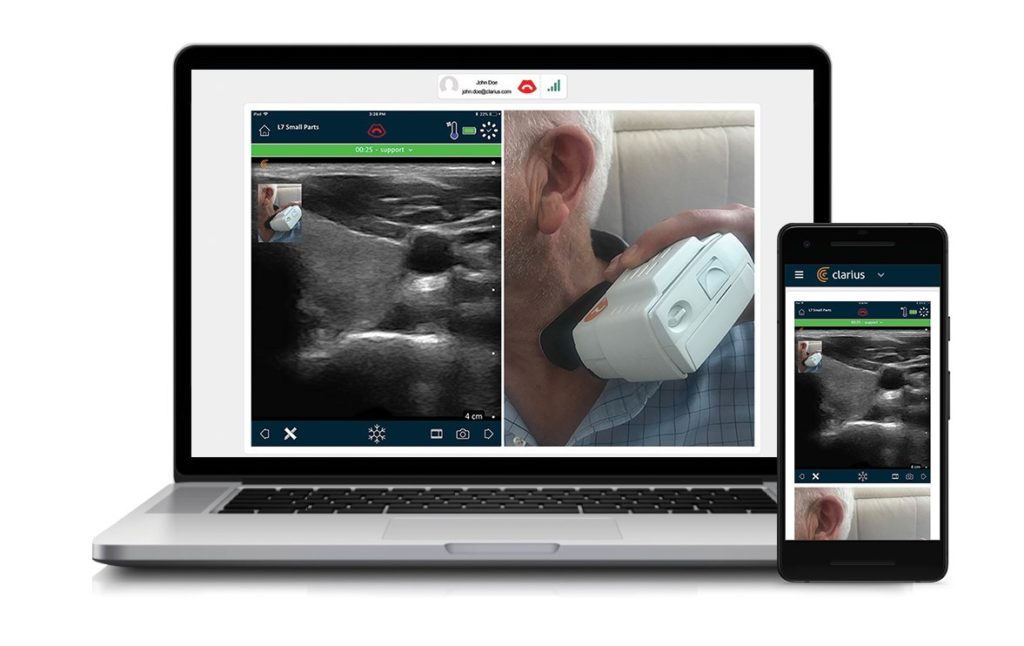
Struggling to limit exposure and be in more than one place at once? With Clarius Live Telemedicine, you can guide, monitor and review multiple ultrasound exams from wherever you are in real-time. Learn more.
Flexible storage solutions
Choose where to store and send exams from the Clarius App
Send exams to any DICOM-compliant server*, or store exams on your phone. Every Clarius scanner also comes with free cloud storage and exam management.
* DICOM is a premium feature.
Covid-19 Resources
External resources for information on the fight against Covid-19 in Point-of-Care.
Best Practices for COVID-19 Lung Scanning
An emergency physician’s quick guide on using ultrasound in the screening and monitoring of COVID-19.
Journal of Invasive Cardiology
Right Ventricular Dilation in Hospitalized COVID-19 Patients Can Be Indicator of High-Risk Cases
Bedside ultrasound of the heart shows right-ventricular-dilation may be a strong indicator of high risk COVID-19 cases. A team of doctors from the Icahn School of Medicine at Mount Sinai looked at the health records of 105 COVID-19 patients hospitalized at Mount Sinai Morningside in New York City between March 26th and April 22nd.
American Academy of Pediatrics
Lung Ultrasound in Children With COVID-19
… recent evolution in the ultrasound field allows the use of wireless devices, which, when available, are probably the most appropriate ultrasound equipment in patients with confirmed or suspected COVID-19. Both the wireless probe and the tablets are easily wrapped in disposable plastic covers, allowing simple sterilization procedures and reducing the risk of contamination…
Health Imaging
Lung ultrasound for COVID-19: Expert physicians propose new international standards
A wireless probe and tablet is the most appropriate type of ultrasound equipment to evaluate individuals with the coronavirus, and can be covered in single-use plastic for less contamination risk and easy sterilization. They’re also cheaper than traditional machines.
Diagnostic Imaging
COVID-19 and Lung Ultrasound: Experts Suggest New International Standards
When scanning patients, they advised, 14 areas – three posterior, two lateral, and two anterior – should be scanned for 10 seconds each. Scans should be intercostal and cover the widest surface area possible in one scan.
ACEPNow
COVID-19 for the Emergency Physicians: What You Need to Know
As public health and infectious disease specialists scramble to understand a novel viral disease with international implications, emergency and other frontline health care workers need accurate information to prepare their departments for the possibility of encountering patients infected with the virus.
The Lancet
COVID-19 outbreak: less stethoscope, more ultrasound
In our opinion, the use of ultrasound is now essential in the safe management of the COVID-19 outbreaks, since it can allow the concomitant execution of clinical examination and lung imaging at the bedside by the same doctor.
Talk to an Expert to Learn More
⚠️ Note: Clarius ultrasound is intended for use by medical professionals
