

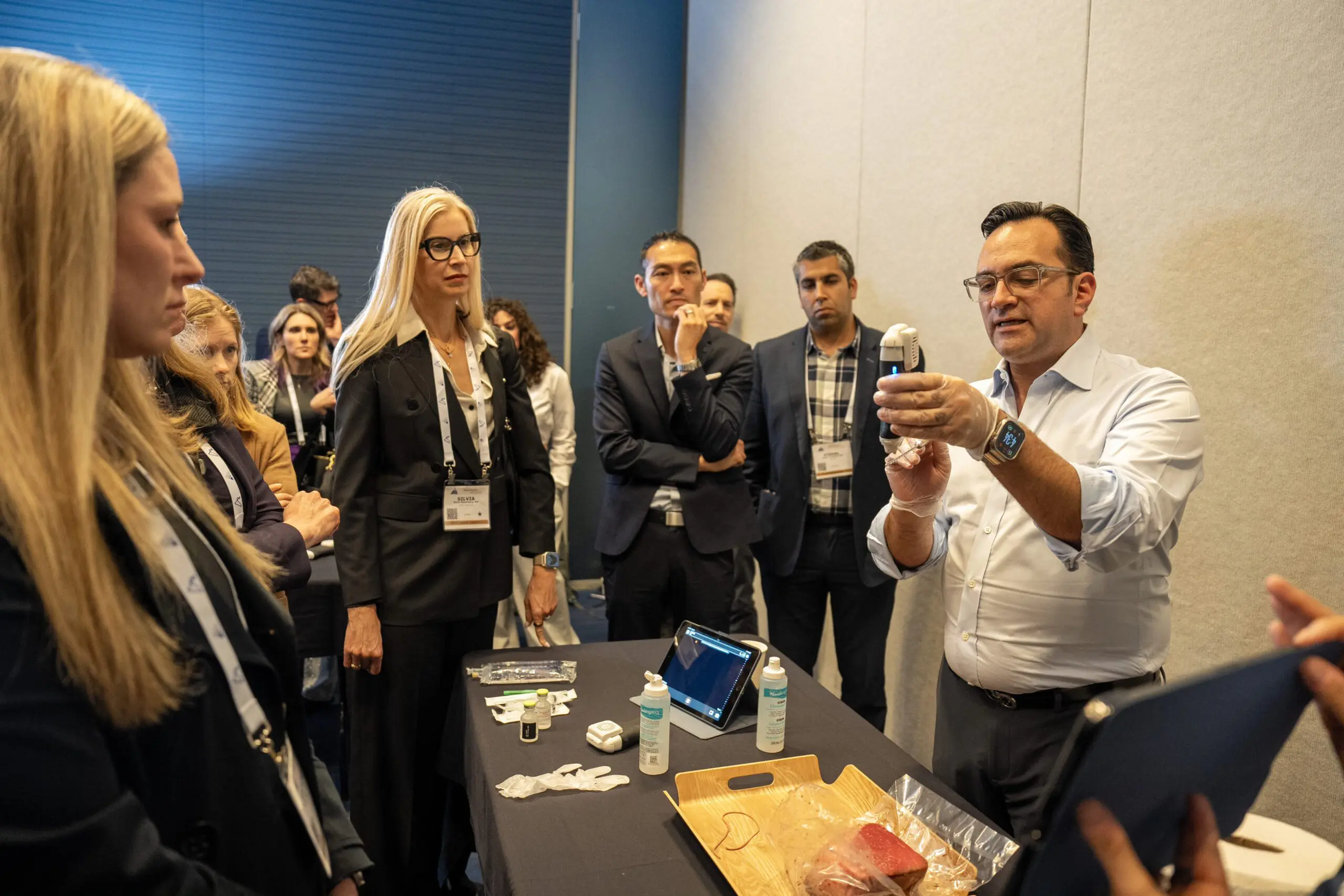
We were quick to organize a video interview with Steven F. Weiner, MD when we heard that he’s a big fan of the Clarius L20 high-frequency ultrasound scanner for cosmetic procedures. Dr. Weiner, board certified in Otolaryngology, Head and Neck Surgery, laid down his scalpel 15 years ago to establish the Aesthetic Clinique in Florida where he focuses on patient safety during treatments.
We asked Dr. Weiner how he’s using ultrasound in his practice. Watch our 3-minute video and read below to learn what he had to say.
How did you begin using ultrasound for your aesthetic practice?
“I started incorporating ultrasound approximately a year ago. I set out to learn from innovators in the field and spent three days with Dr. Leonie Schelke in Amsterdam who has been using ultrasound to treat dermal filler complications at her clinic for over 10 years. I realized that I wanted to do everything possible with ultrasound and fillers. To become proficient at using ultrasound, I also practiced on myself and my family.”
How long did it take you to become proficient at using ultrasound?
“I think it’s actually a very quick learning curve to learn how to use ultrasound for vascular mapping and to evaluate patients. But it takes a good three to six months of practice to get very good. I’m still learning and getting better and better the more I use it.”
When do you use ultrasound in your clinic?
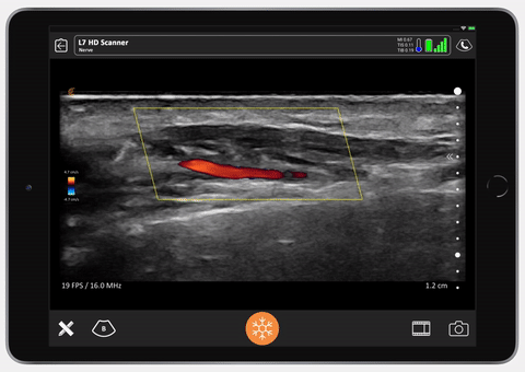
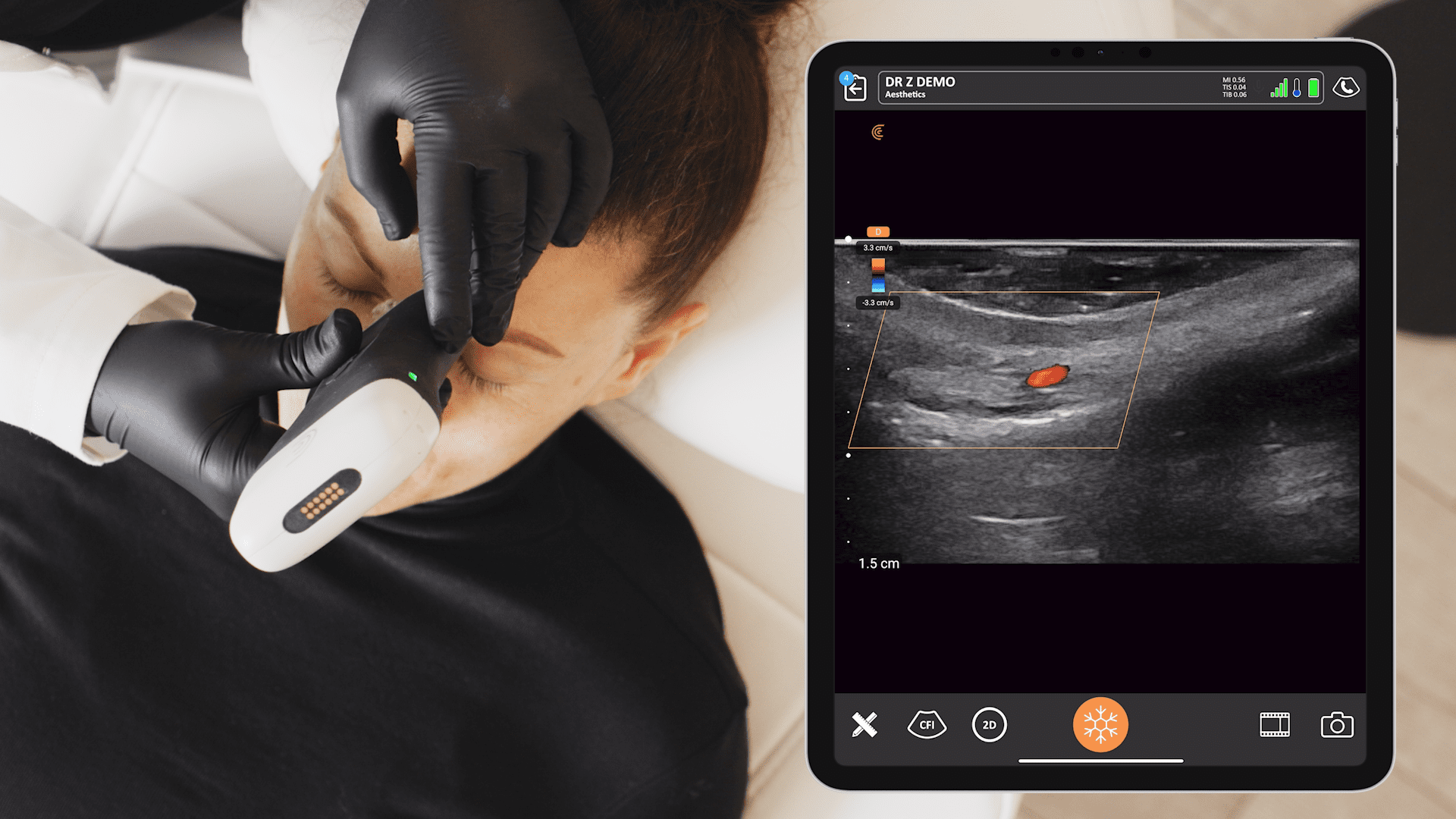
Ultrasound Evaluation of Vascular Structures
“Ultrasound is the gold standard for evaluating patients who have had filler complications. I also believe it’s better to use ultrasound to prevent complications. I like to evaluate vascular structures in advance. By using ultrasound for vascular mapping, I know where to avoid structures during injections. I know the depth of the vessels so I can avoid them by injecting either superficial or deep to these vessels.”
Evaluation of Non-Vascular Structures
“I actually use it daily to evaluate filler complications that aren’t necessarily vascular related, such as nodules, granulomas or just swelling related to fillers. Often, I see patients who come to me with swelling around the eye from filler that they had five to ten years ago. They don’t think it’s related to filler and believe they need surgery like a blepharoplasty. With ultrasound, I can see remaining fillers and I can dissolve it – the problem disappears without surgery!”
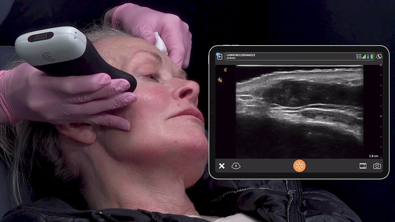
Measuring Dermal Thickness with Ultrasound for RF Micro-needling
“I also use ultrasound to get the maximum benefit from my RF micro-needling device as well. It’s critical to get the needles to the right depths. Dermal thickness varies from patient to patient from area to area. With ultrasound imaging I can ensure to use the right depth for each patient when performing micro needling.”
How does ultrasound improve patient outcomes?
“Ultrasound improves outcomes significantly in many ways. It allows us to avoid vascular structures in critical areas that are very superficial or very deep, which is where we normally place fillers. Physicians think that going down to bone is often safe, but there are studies that show that in 1% or more of patients, the angular artery runs along the periosteum. If you know where the artery is, you can either inject superficial or deep to it and avoid those vascular complications.
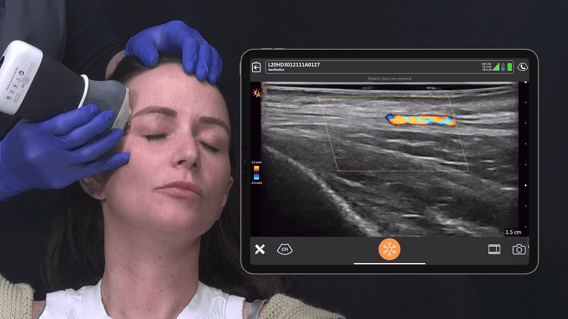
It’s also very critical to use ultrasound for injections around the nose, because it’s highly vascular in that area and injections in the wrong place can cause blindness. It’s safer to use ultrasound to see where the structures are so you can avoid them for safer procedures.
Overall, ultrasound enables you to inject with confidence because you know where to inject the filler. And in the case that there is a vascular complication, you’ll be able to identify it quickly and inject the pocket of filler using ultrasound guidance and eliminate the vascular occlusion very quickly and easily.”
How does Clarius compare to a traditional ultrasound system?
“Clarius is a very small compact, all-in-one ultrasound. I can throw it into my backpack. I can put it in my pocket. The ultrasound systems that I had previously were extremely bulky, required a cart, and they were much more expensive. Clarius was less than a third of the price, but it still delivered the same quality, high-frequency images that the very bulky $30,000 piece of equipment delivered.
The Clarius L20 high frequency scanner is what I would consider the go-to for people in the aesthetic field because it’s very portable, affordable and provides high quality images of vital vascular structures. It’s just the right frequency at 20 megahertz. It images to a maximum depth of about four centimeters, which is perfect for imaging the whole face.”
What do you consider some of the biggest advantages of using Clarius?
“Clarius is very user friendly and produces very high-quality imaging. One of the disadvantages of my $30,000 ultrasound was that the images were very low quality and more difficult to save and store. I get much better images with Clarius.
With Clarius there are no cords and wires laying around to walk or trip over. It’s easy to clean without the wires and it’s waterproof so we can clean it thoroughly between patients.
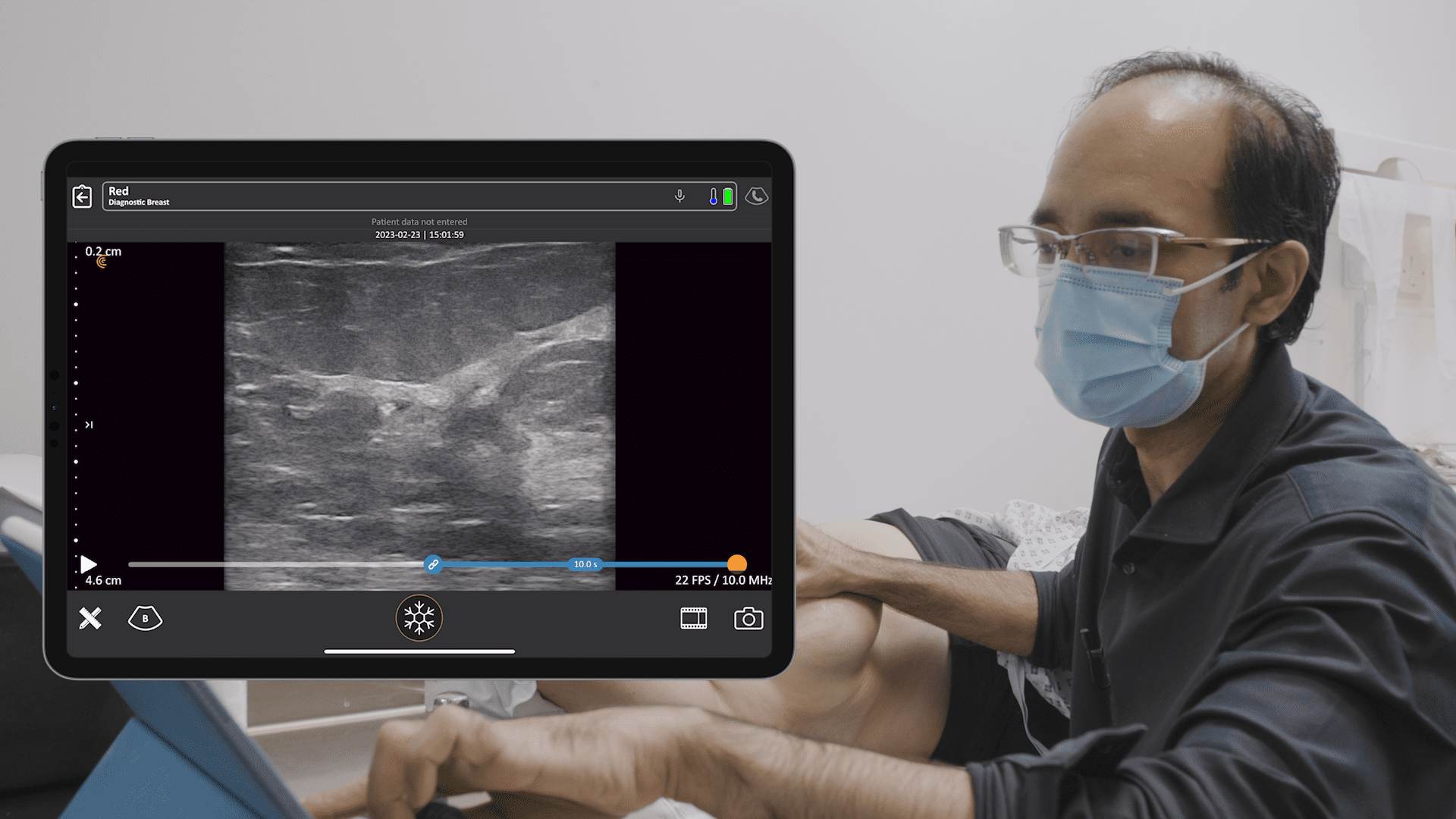
The other thing is that I’ve found very attractive is that Clarius is compatible with the iPad, which allows me to manipulate all the images very easily. I don’t have to deal with the constraints of DICOM.”
Do you use the Clarius Cloud image management system?
“Clarius Cloud is very easy to use. When I complete an examination, I hit a button and it transfers the images directly to the cloud. I can view the images from anywhere in the world — on my laptop or even iPhone. I can also give patients access to those images for consultations with other doctors. All the images are secure and password protected.”
With the miniaturization of high definition ultrasound and advances in artificial intelligence, it’s easy and affordable to add Clarius HD to your aesthetics practice. Contact us today or request an ultrasound demo to see the difference ultra-high definition imaging makes.
To learn more, watch our webinar on Ultrasound in Facial Aesthetics: Vascular Mapping, Evaluating Fillers and Complications with Dr. Steven F. Weiner.


