

Clarius Ultrasound Scanners are a mobile solution for medical schools, allowing students to have instant access to the technology in various settings. They play large role for California University of Science and Medicine’s year 1 and 2 curriculum through integrated active-learning sessions examining anatomy, physiology and pathology. The following article documents how the early exposure of ultrasound imaging techniques better prepare medical students for ultrasound encounters during their clinical years.
The original article can be downloaded here.
Faculty: Fauzia Nausheen, Rajunor Ettarh, Sherif Hassan, Robert Suskind, Alfred Tenore
Department of Medical Education, California University of Science and Medicine.
Introduction
At California University of Science and Medicine, School of Medicine, we have designed a clinical presentation-driven, system-based, fully integrated, active learning curriculum. The integrated active-learning sessions are conducted by teams of students that belong to specific learning units referred to as “colleges”. This curriculum is an amalgam of different pedagogical methods which also include modified versions of traditional team-based and problem-based learning. To complement the active, team- based learning pedagogy, ultrasound laboratory sessions are incorporated to support the learning of integrated anatomy, physiology and pathology by the interpretation of normal images during the system-based courses in year 1 and 2. The ultrasound anatomy laboratory sessions held each Friday will be aligned with the clinical presentations, clinical skills and case sessions of the week. Anatomy will also be taught on prosections, models, multimedia, online anatomy as well as radiology images. The objective of this study is to evaluate the student and instructor perception of teaching and learning of integrated basic sciences (Anatomy, Physiology and Pathology) with the use of wireless portable ultrasound scanners that will work on computer tablets provided to each team in the anatomy laboratory.

Method
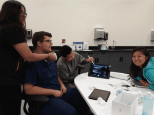
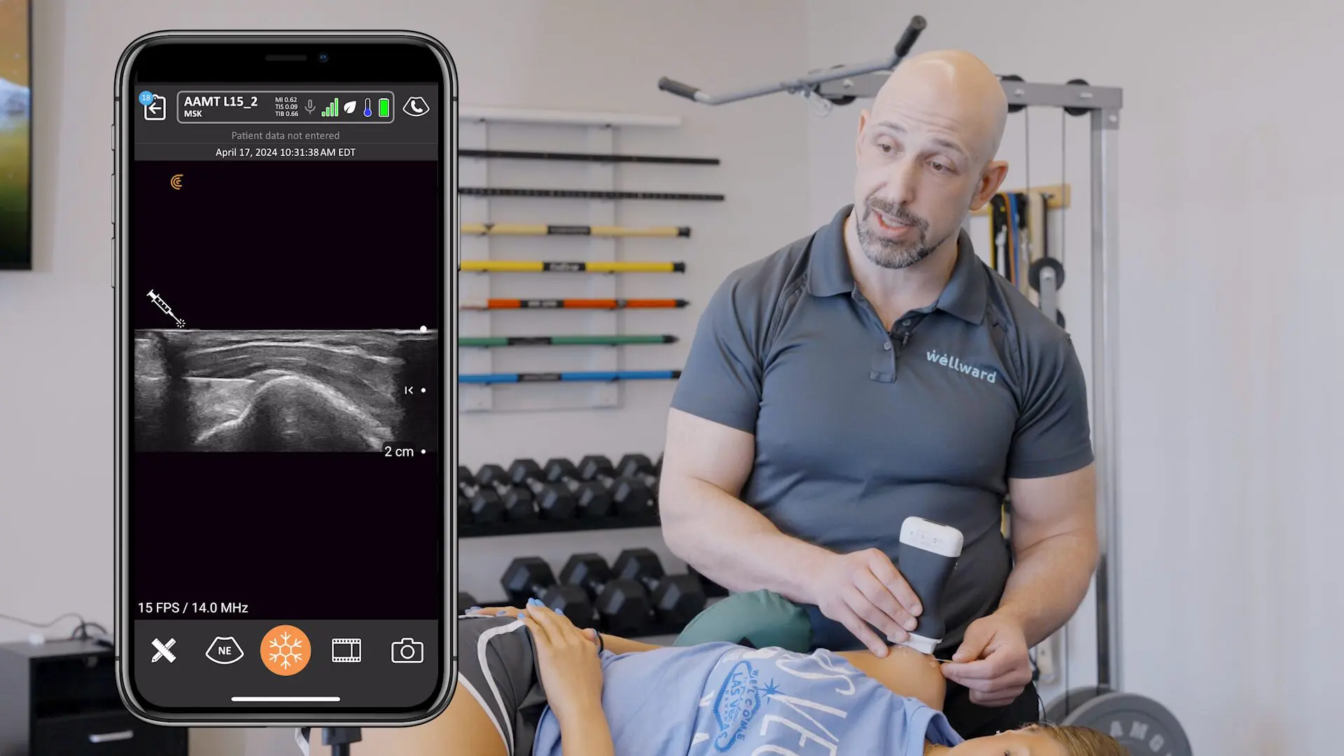
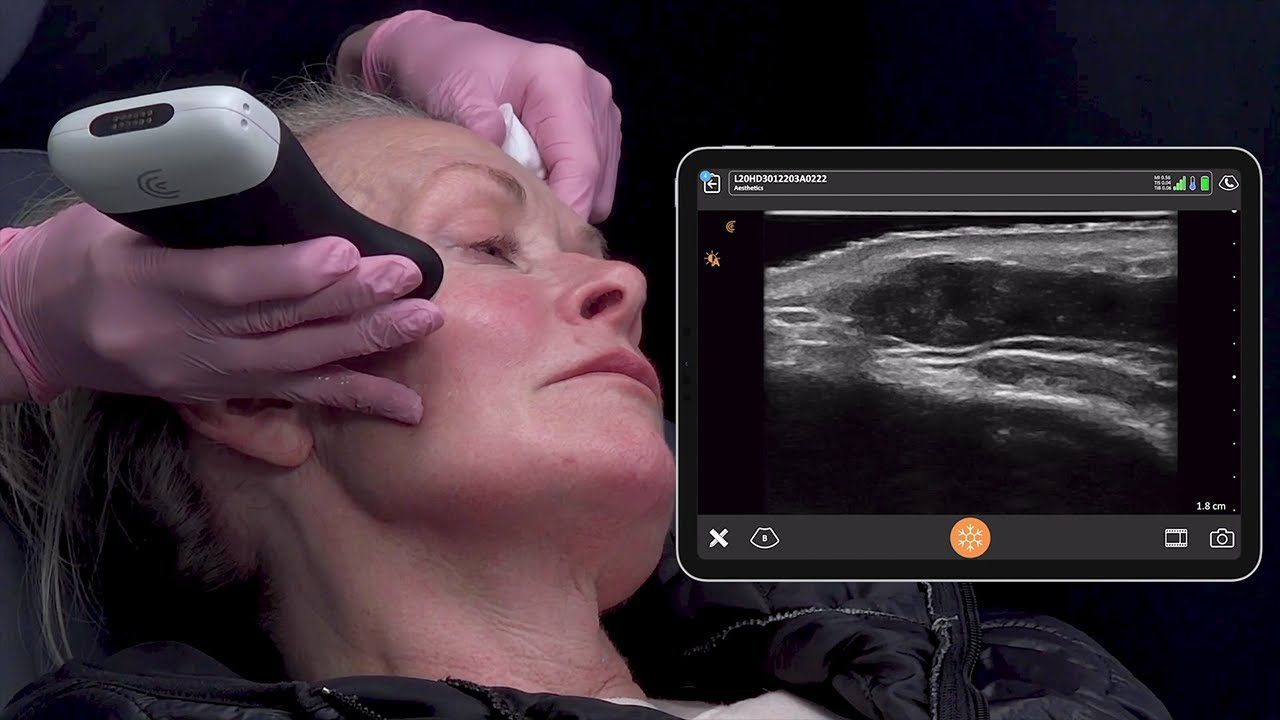
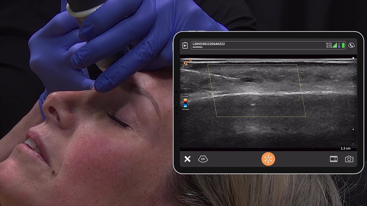
The inaugural class have 60 students, who is divided into 6 colleges. Each college is composed of 10 students divided into two teams of 5. The members of each team work together during lecture discussions in flipped classrooms, clinical case analysis and clinical skills activities which include role playing sessions and integrated laboratory sessions. Because of the increasing role of ultrasound in the clinical evaluation of patients, almost all system-based courses have developed and incorporated hands-on ultrasound experience. In each anatomy session, each team have 2 wireless portable ultrasound scanners to perform ultrasound on each other or simulated patients and identify the structures at different plane of sections for that specific lab as described in lab manual.
The scanners have linear, phased and convex array with the ability to scan superficial and deep structures in different systems of the body. They are required to identify the structures for the specific session and have their findings confirmed by the facilitators. To achieve the designated learning outcomes, the ultrasound lab activities have been divided in three sessions:
1) information gathering and independent self-directed learning: self-study from resources.
2) identification of structures on ultrasound and confirmation by instructors: The lab manual includes a guide to use ultrasound on each table.
3) interactivity and integration followed by presentation and discussion: the focus of this session is on problem solving exercises, critical-thinking and presentation by the teams.
The problems focus on related anatomical appearances on ultrasound. During the discussion the students are encouraged to justify their answers based on ultrasonographic principals and extra bonus points will be given by identifying the abnormality or related structures on ultrasound images
Results
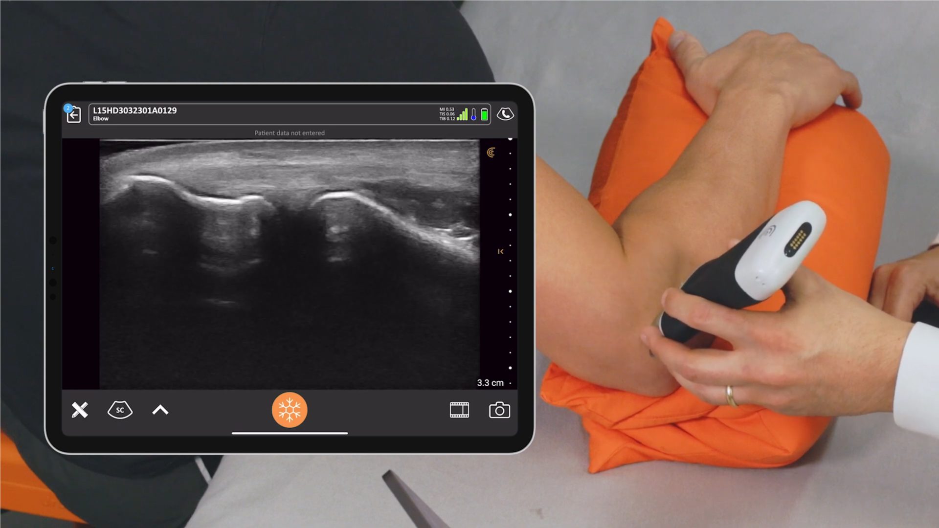
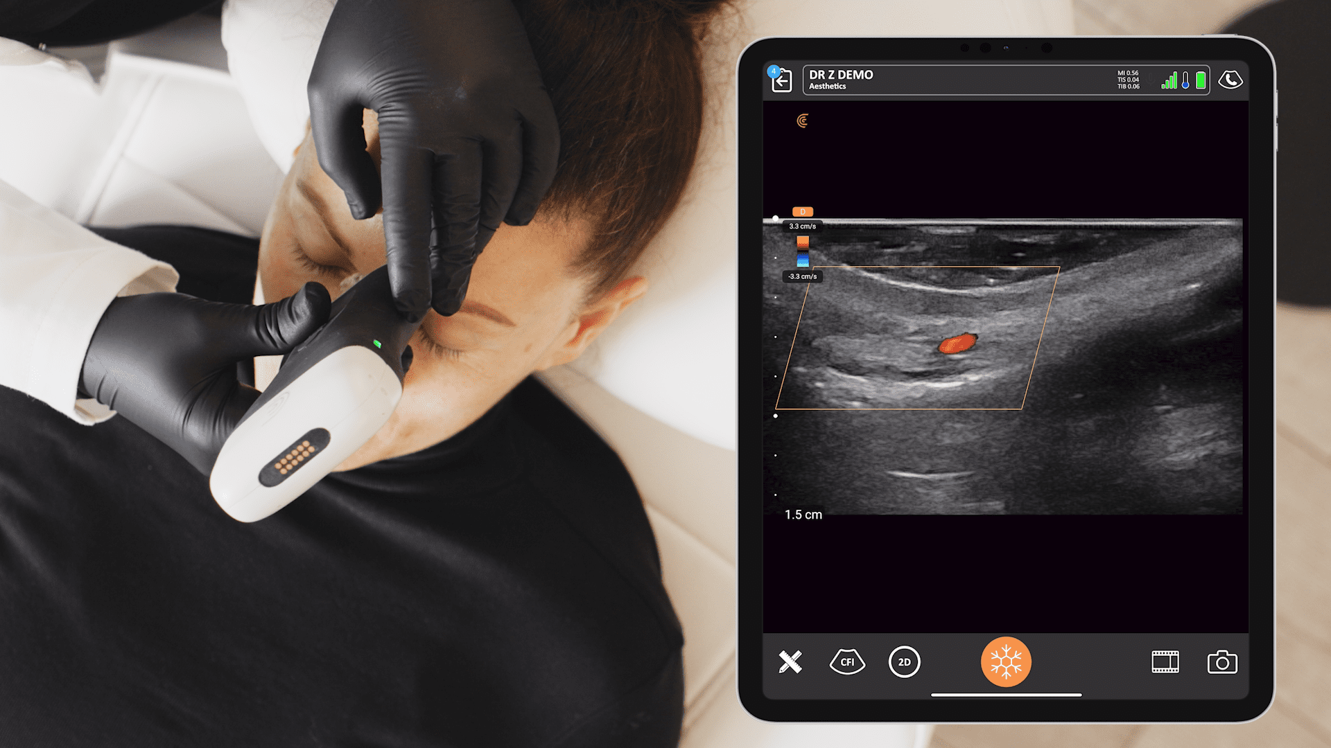
Years 1 and 2 have 11 system-based courses and each course has multiple ultrasound sessions depending on the weekly clinical presentation theme. Live ultrasound scanning is synchronized with the anatomy curriculum of each system by anatomic sections and location of different body parts that form the system. The following is an example of sessions and overall outcomes for ultrasound hands-on lab session in some of our systems-based courses; In “The Scientific Foundation of Medicine:” course , we have ultrasound sessions on introduction to medical imaging, doppler ultrasound, knobology, ultrasound physics and basics of ultrasound imaging. The learning outcome is : to differentiate the hyperechoic organs from the hypoechoic organs and explain the reason of their appearance based on ultrasound imaging principals. In “The Structural Integrity of the Human Body”; (i.e., Integumentary and musculo-skeletal systems), we will have session on ultrasound of shoulder, wrist, knee and ankle joints with identification of the bones and ligament and muscles moving the joints. The learning outcome is : to identify and differentiate the normal appearance of bones, muscles and tendons of shoulder, elbow, knee, hip and ankle joints. In “The Sustenance of the Body “(i.e., Gastrointestinal system and nutrition), we will have session of ultrasound on major abdominal vessels and organs including the Morison pouch, and introduction to FAST exam. The learning outcome is : to locate liver, gall bladder, spleen, pancreas, aorta and IVC on abdominal ultrasound and perform Doppler scan to show the direction of blood blow. In “The Depurative Functions of the Body” (i.e., renal system), we will have session on ultrasound of kidneys and urinary bladder. The learning outcome is : to locate kidneys, and urinary bladder on abdominal ultrasound. In “The Transport and Delivery of the Life’s Elements “(i.e., cardiorespiratory systems);we will have session on ultrasound appearance of normal lungs, ribs and pleural cavities and cardiac ultrasound four chamber view, cardiac cycle and valvular events. The learning outcome is : to locate four chambers of heart and identify the valves on subcostal and parasternal cardiac view. In “The Propagation of Life “(i.e., reproductive system), we have session of ultrasound of the pelvic organs and breasts. The learning outcome is : to locate uterus ovaries and prostates on pelvic ultrasound. In “The Foundation of Life’s Functions” (i.e., endocrine system), we have session of ultrasound of neck (thyroid glands). The learning outcome is : to locate thyroid gland on neck ultrasound. In “The Integration of Life’s Processes” (i.e., neurosciences), we have session of ultrasound of major vessels in the neck (carotids, jugular veins). The learning outcome is : to locate major neck vessels and perform doppler scan to show the blood flow. In “The Continuum of Life” (i.e., from the beginning to the endo of life), we have ultrasound session of introduction to POCUS (point of care ultrasound). The learning outcome is : to review the principals of point of care ultrasound. The laboratory exam will assess the student’s skills to perform ultrasound and locate the appropriate structure in the correct plane and identify the anatomical relations on simulated patients. Students and faculty will be given evaluation forms to get feedback about their perception of anatomy teaching and learning with wireless portable ultrasound scanners at the end of each system-based course and their suggestions to improve the sessions.
Conclusion
Wireless portable ultrasound scanners accessibility in all system-based courses in anatomy lab will be a bridging device between the integrated basic sciences and clinical practice. This early exposure of portable wireless ultrasound imaging technique in all organ systems will prepare our medical students for later ultrasound encounters of clinical years.


