

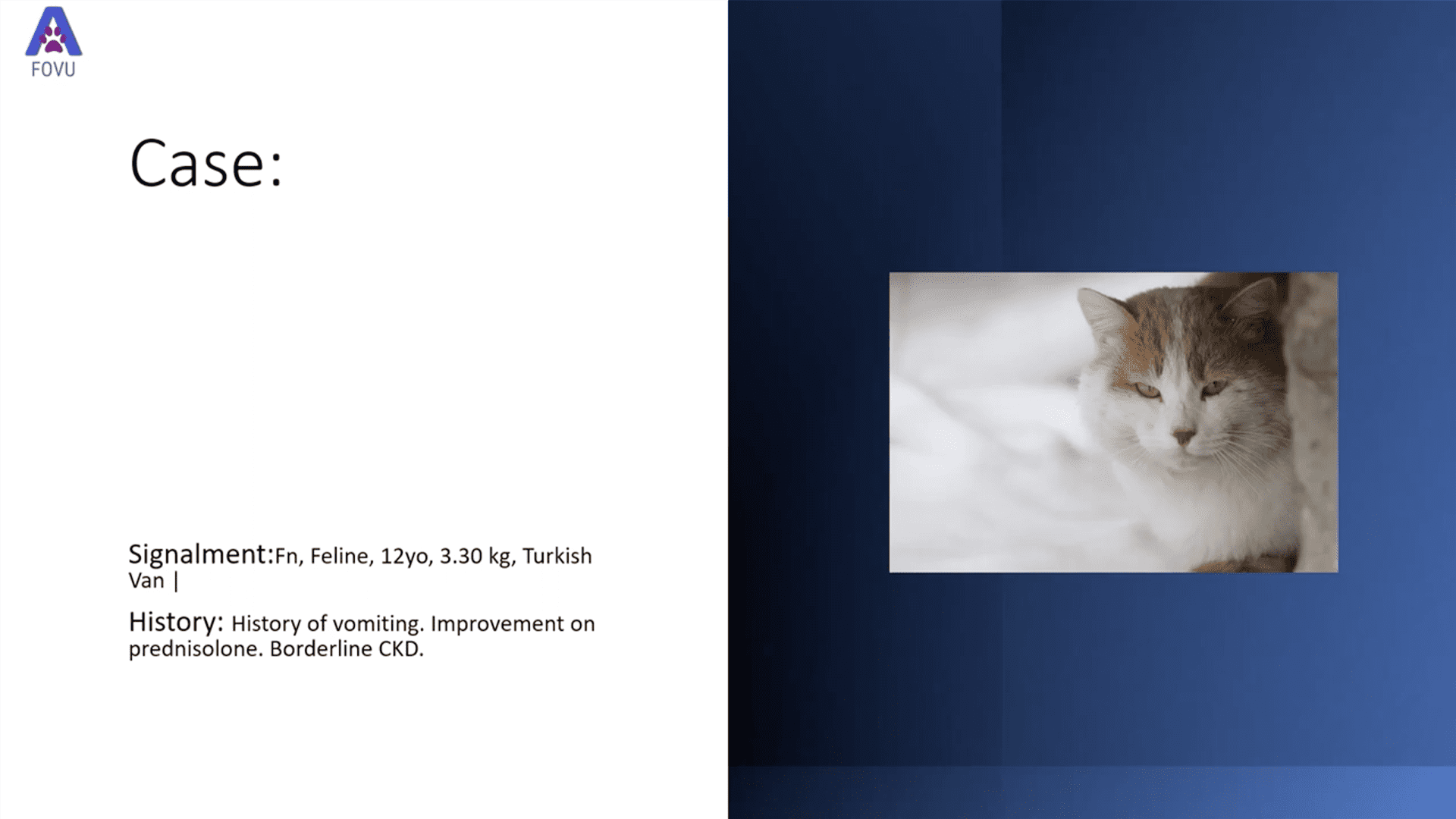
Dr. Camilla Edwards, DVM and founder of First Opinion Veterinary Ultrasound (FOVU) has shared a very interesting case of big bilateral nephrolithiasis and bladder thrombus in a dog. The ultrasound images are impressive, and combined with the dog’s medical history, make for a great detective story.
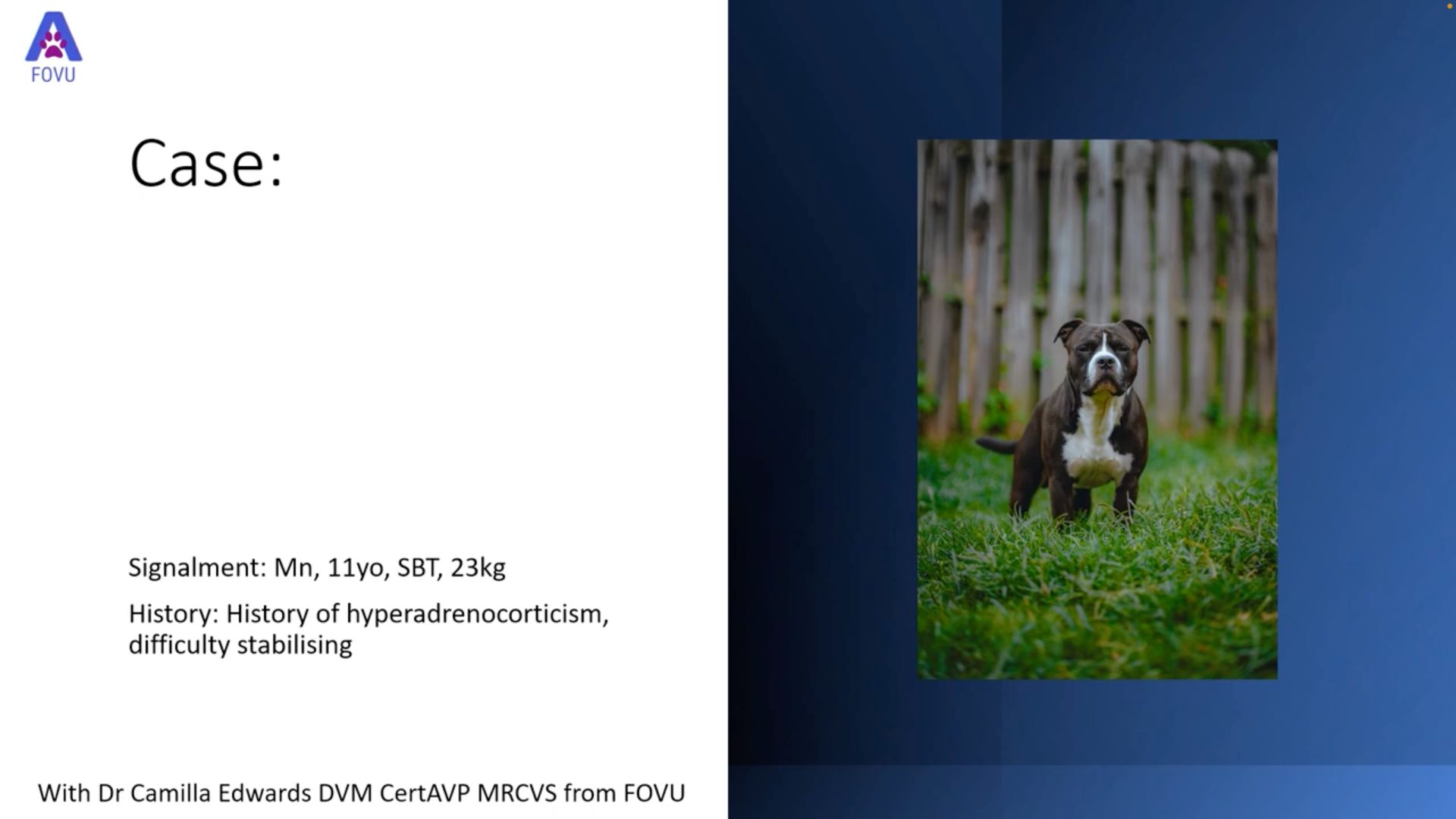
History: Dr. Edwards was asked to perform an abdominal ultrasound on a 6-year-old female neutered dog who was imported from Greece to the United Kingdom (UK) several years ago. She had a previous diagnosis of Leishmaniasis which is being treated with allopurinol and was recently diagnosed with Capillaria plica (bladder worm), which is uncommon in the UK.
Ultrasound Findings
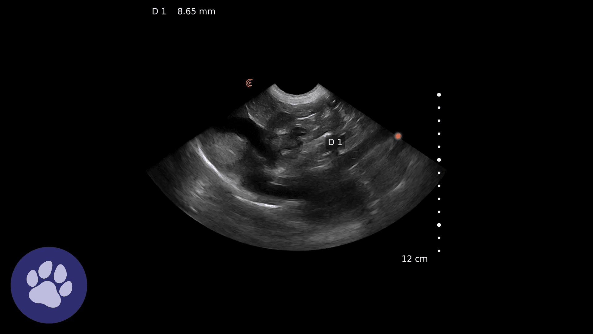
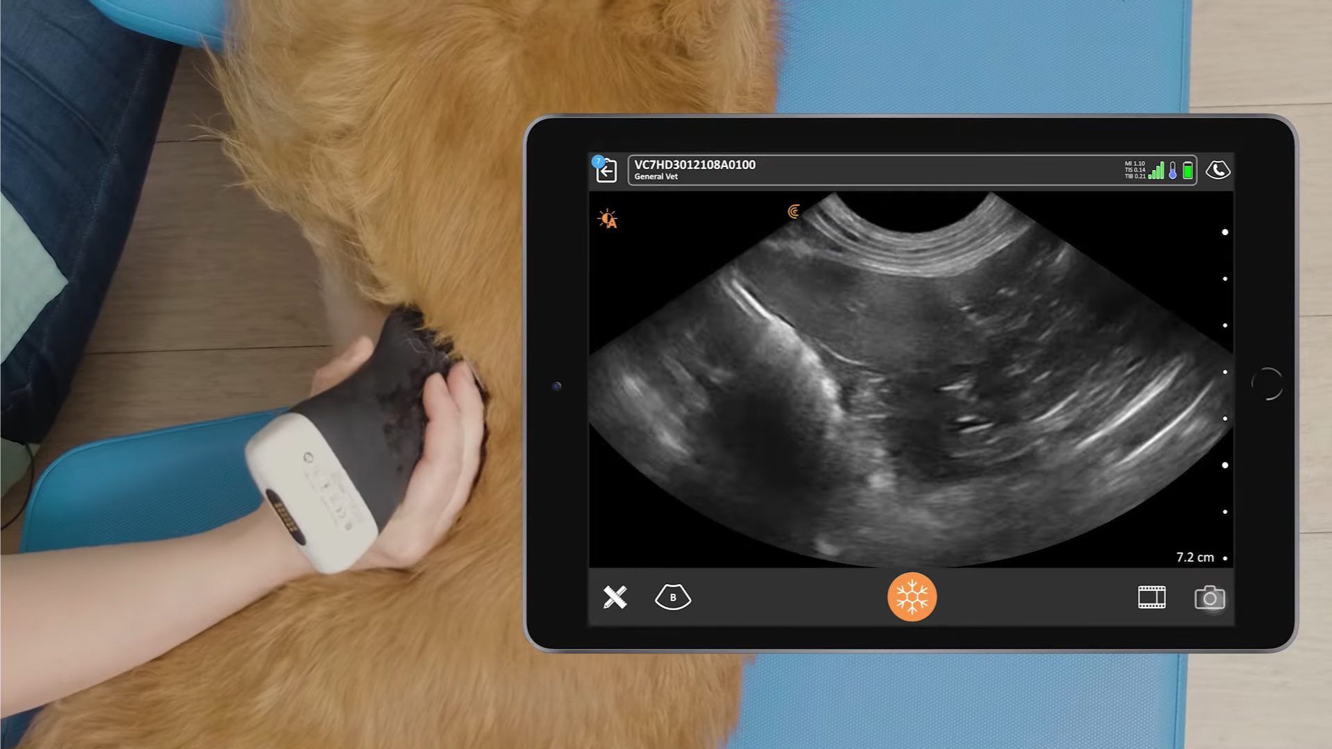
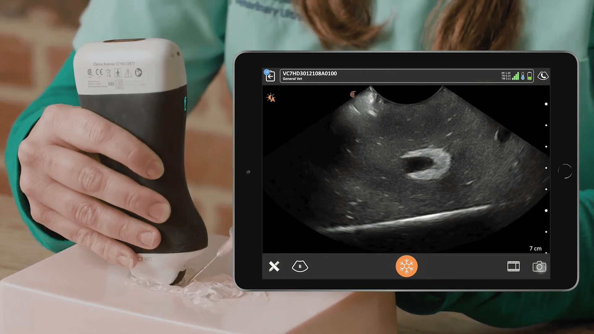
Ultrasound of the left kidney revealed a massive calculus with dense shadowing, occupying the entire renal pelvis. The renal cortex appears normal, and there doesn’t appear to be any evidence of ureteral dilatation suggesting obstruction. The right kidney had a similar appearance.
The urinary bladder wall appeared to have a normal thickness, but inside the bladder was an irregular mobile mass that fell to the gravity-dependent part of the bladder when the patient was moved. The mass did not show any vasculature with color Doppler.
Video: Ultrasound of the left kidney and urinary bladder
Outcome & Discussion
Canine Leishmaniasis is caused by Leishmania parasite and is transmitted by a sandfly bite. It is traditionally only found in the Mediterranean region, which is where our patient came from.
The treatment for the disease is allopurinol, which has known adverse urinary effects of xanthinuria with renal mineralization, as referenced in this paper from the Journal of Small Animal Practice titled Adverse urinary effects of allopurinol in dogs with leishmaniasis.
A blood clot in the bladder was assumed to be secondary to the nephroliths in the kidneys.
The dog was referred for surgery and because the nephroliths measured under six centimeters in length, the surgeons were willing to perform the surgery, which was successful.
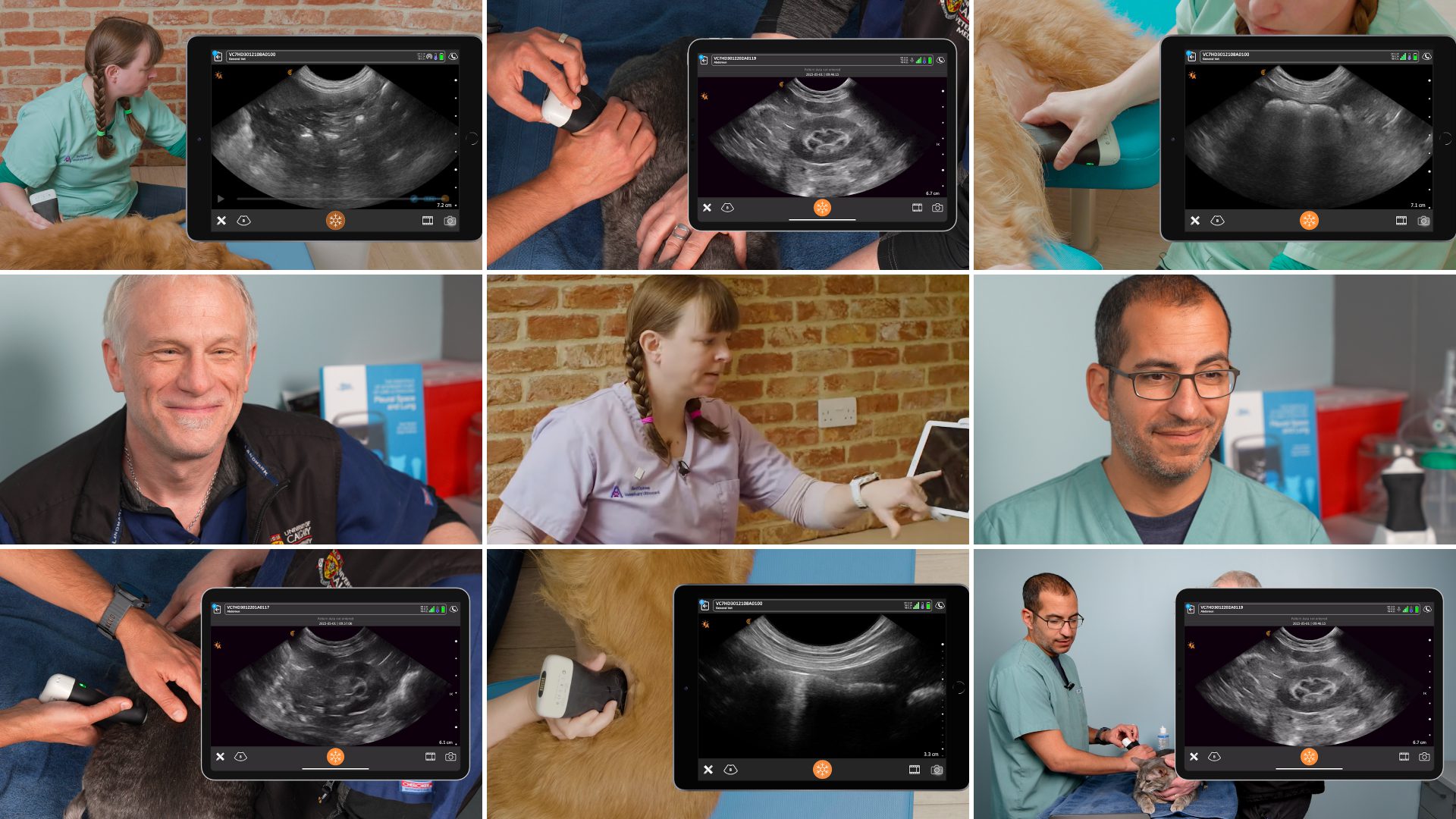
If you’d like to watch some quick instructional videos on how to scan the kidneys and bladder, and other small animal ultrasound demos, go to our Clarius classroom videos where we feature Dr. Edwards and her dog Pippi.
Video: How to Scan the Right Kidney
Video: How to scan the Left Kidney
Video: How to Scan the Bladder
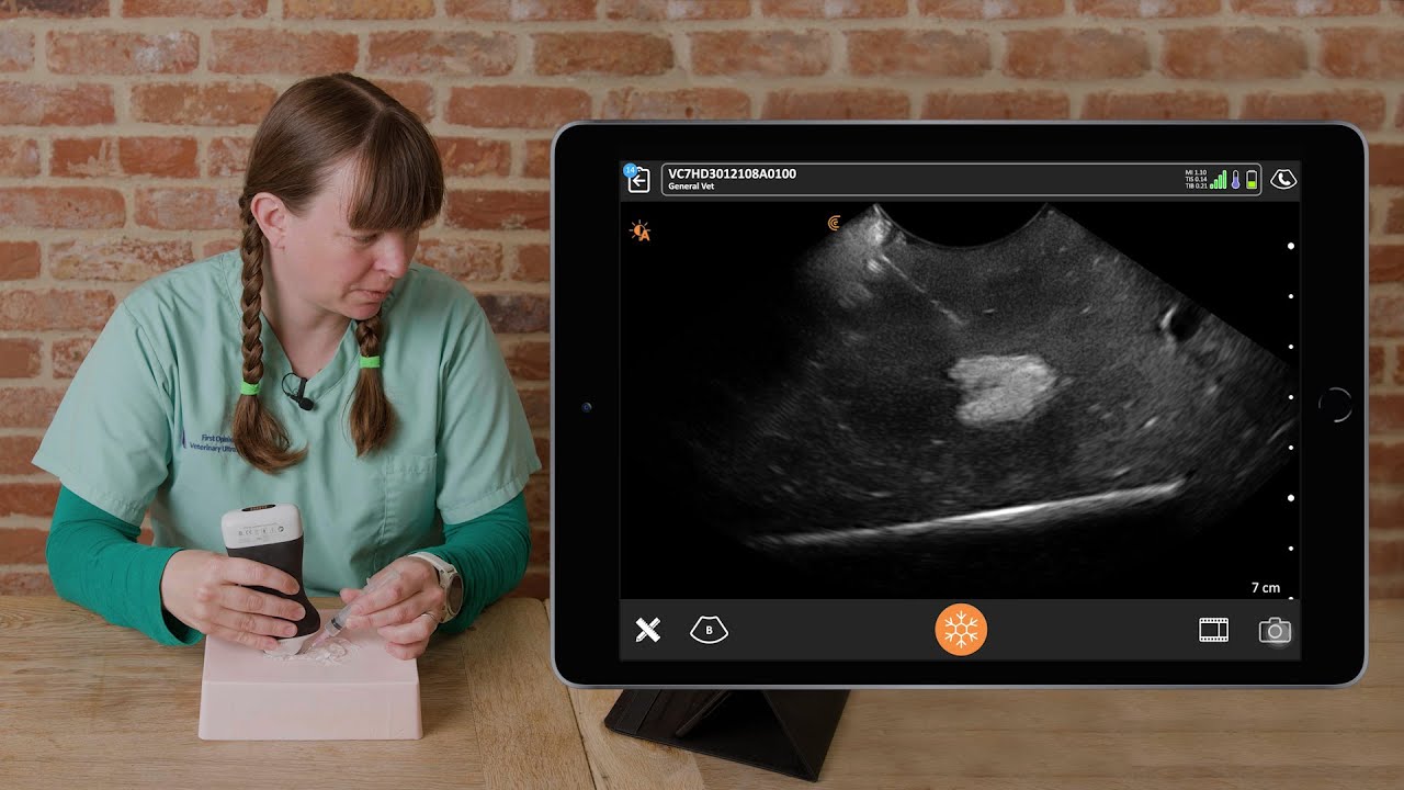
Dr. Edwards uses the Clarius C7 HD3 Vet, which is specifically designed for clinical imaging of small and medium-sized animals. Our Advanced Veterinary Package offers more flexibility for users who need additional presets and workflows for various animal exams.
To learn about how easy and affordable it is to add Clarius handheld ultrasound to your veterinary practice, visit our Clarius veterinary ultrasound page for product details. Or contact us today to discuss which scanner is right for your veterinarian practice.


