

Dr. Camilla Edwards, DVM, founder of First Opinion Veterinary Ultrasound (FOVU) has shared some great case studies with us in the past, and this new one does not disappoint!
History: This 19-year-old female neutered domestic short hair cat was seen by a vet because she stopped eating and had some weight loss. Dr. Edwards conducted an abdominal ultrasound with the Clarius C7 HD3 scanner.
Ultrasound Findings
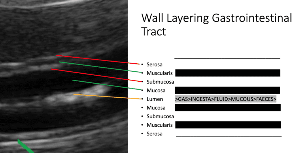
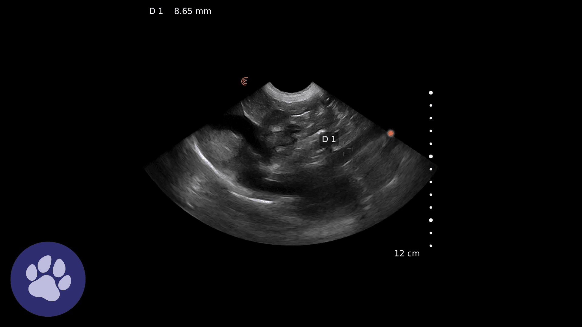
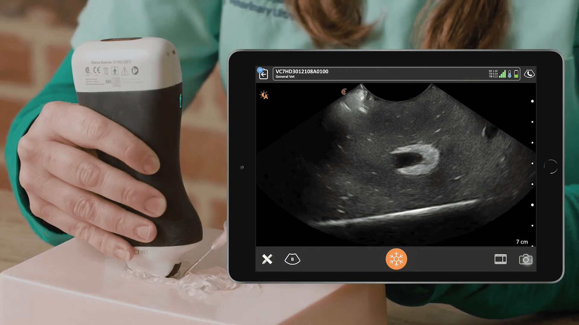
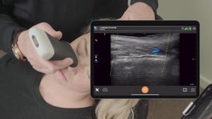
As the cat was a very skinny cat, so imaging was done at quite a shallow depth. One of the first things that is quite noticeable is the multiple loops of intestine, with clear visualization of the layers of the intestinal walls. From the lumen centrally, the mucosa, which is hypoechoic, then the submucosa, then the muscularis layer is also hypoechoic. This is surrounded by the serosa, which appears bright white on ultrasound.
This is an image from a webinar with Dr. Edwards where she describes wall layering in the GI tract.
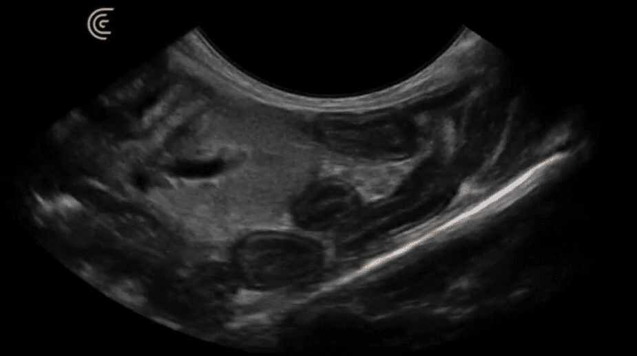
In this ultrasound case, the muscularis layer appeared mildly thickened, and
Dr. Edwards also noticed an enlarged jejunal lymph node. So, what are the next steps?
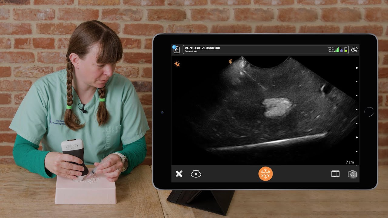
To get a better understanding and a diagnosis, she made the decision to perform ultrasound-guided fine needle aspiration (FNA) of the enlarged lymph node. During the ultrasound, Dr. Edwards demonstrates how she plans trajectory, determines needle length, and accurately guides the procedure.
Video: Abdominal ultrasound identifying pathology and an ultrasound-guided FNA
Outcome & Discussion
The findings of thickened muscularis layer throughout the jejunum and an enlarged hypoechoic jejunal lymph node put lymphoma high on the list of differential diagnosis. Another possibility was inflammatory bowel disease.
FNA of the lymph node required only mild sedation and was not terribly invasive, and in the case of this senior cat, was the kindest and most logical course of action. The aspirate revealed mild reactive lymphoid hyperplasia with no cells suggestive of neoplasia, which was good news for this feline. She would be treated for inflammatory bowel disease, with no worry of neoplasia.
You might be wondering why Dr. Edwards didn’t perform FNA of the intestinal wall, but in this case, it was deemed that the intestinal wall was not thick enough to guarantee a puncture into the lumen and potentially cause peritonitis.
If you’d like to learn more about scanning the jejunum watch this Clarius classroom video with instruction from Dr. Edwards.
Dr. Edwards uses the Clarius C7 HD3 Vet, which is specifically designed for clinical imaging of small and medium-sized animals. Our Advanced Veterinary Package offers more flexibility for users who need additional workflows for various animal exams.
To learn about how easy and affordable it is to add Clarius handheld ultrasound to your veterinary practice, visit our Clarius veterinary ultrasound page for product details. Or contact us today to discuss which scanner is right for your veterinarian practice.

