


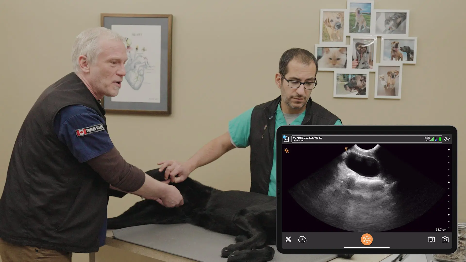
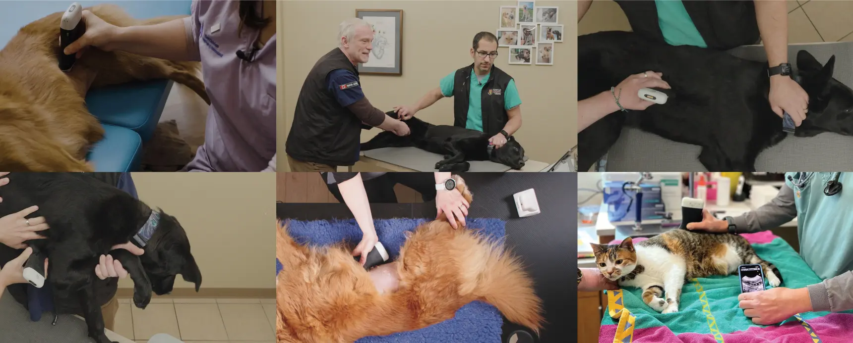
The journey to mastering veterinary ultrasound can feel daunting. That’s why veteran sonographers Angie Lloyd-Jones and Julie Burnage of Aspire UCS have developed a systematic survey technique for veterinarians seeking to transform ultrasound from an intimidating tool into a diagnostic powerhouse.
For Angie and Julie Burnage, a full abdominal survey is the gold standard that yields holistic, reliable results, granting the operator a “superpower” in the clinic. This article breaks down the foundational steps for new users, focusing on image optimization and the first three steps of their Aspire UCS 8-Step abdominal survey. Register for this webinar for steps 4-8 on January 28th, 2026.
The article is based on a popular Clarius webinar presented by Angie and Julie, which is now available to watch on demand: Small Animal Ultrasound: Mastering the Aspire UCS 8-Step Abdominal Ultrasound Survey Technique™.
Read on for key clinical takeaways and brief video tutorials.
1. The Power of a Protocol: Why Go Beyond POCUS?
While Point-of-Care Ultrasound (POCUS) like FAST/AFAST exams are invaluable for rapid binary (yes/no) answers in critically ill patients, a complete survey is necessary for thorough diagnosis.
Julie Burnage highlights the efficiency gained: “We go in a clockwise direction always. We start with our patient in the right lateral.” This methodical approach ensures no organs are missed and justifies sedation for a longer, more compliant, and accurate examination.
Key Clinical Takeaway: “There is absolutely no such thing as a quick scan because there is lots to get through. Our aim really is to help people to master manageable, learnable little scan components that can then be bolted together to build confidence…” —Angie Lloyd-Jones
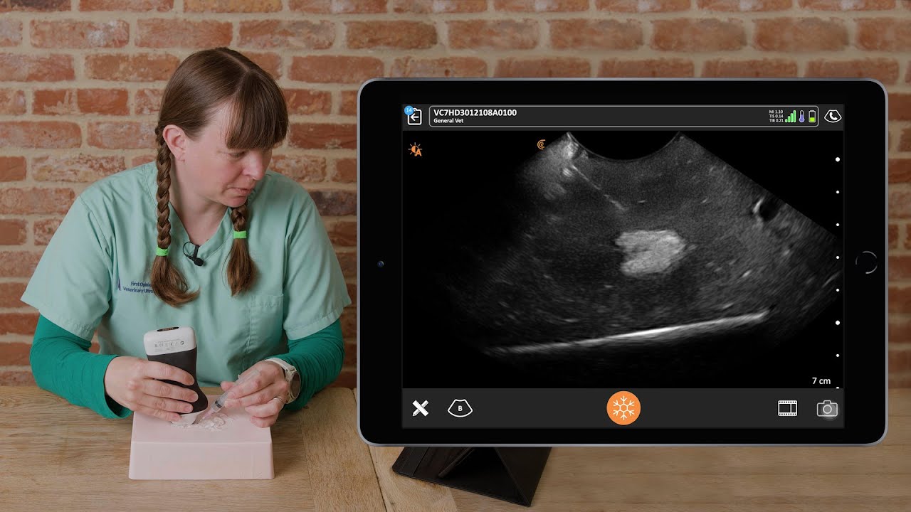
2. Mastering the Controls: Image Optimization (“Knobology”)
Ultrasound is operator-dependent; learning to “drive” your machine properly is non-negotiable for obtaining diagnostic-quality images.
Essential Controls for Beginners
| Control | Function & Goal | Expert Advice |
| Depth | Sets the display range. Must be adjusted for every organ. | “Put the region of interest in the middle of the screen so that because that’s where your eye is drawn automatically…” —Julie Burnage |
| Frequency | Determines image resolution vs. penetration. | “Higher frequency. Less depth of penetration but much better resolution.” —Julie Burnage |
| Overall Gain | Amplifies all returning signals equally (the image’s overall brightness). | Set the gain at about two thirds to three quarters of the range. |
| Focal Zone | The depth at which the ultrasound beam is narrowest, providing the best detail. | “The aim is to have the focus ideally at the region of interest.” —Angie Lloyd-Jones |
| Dynamic Range | Controls contrast by altering the range of grayscale displayed. | “If we have fewer grays, we have a more contrasty image… that can really make structures pop out and make them so visual.” —Angie Lloyd-Jones |
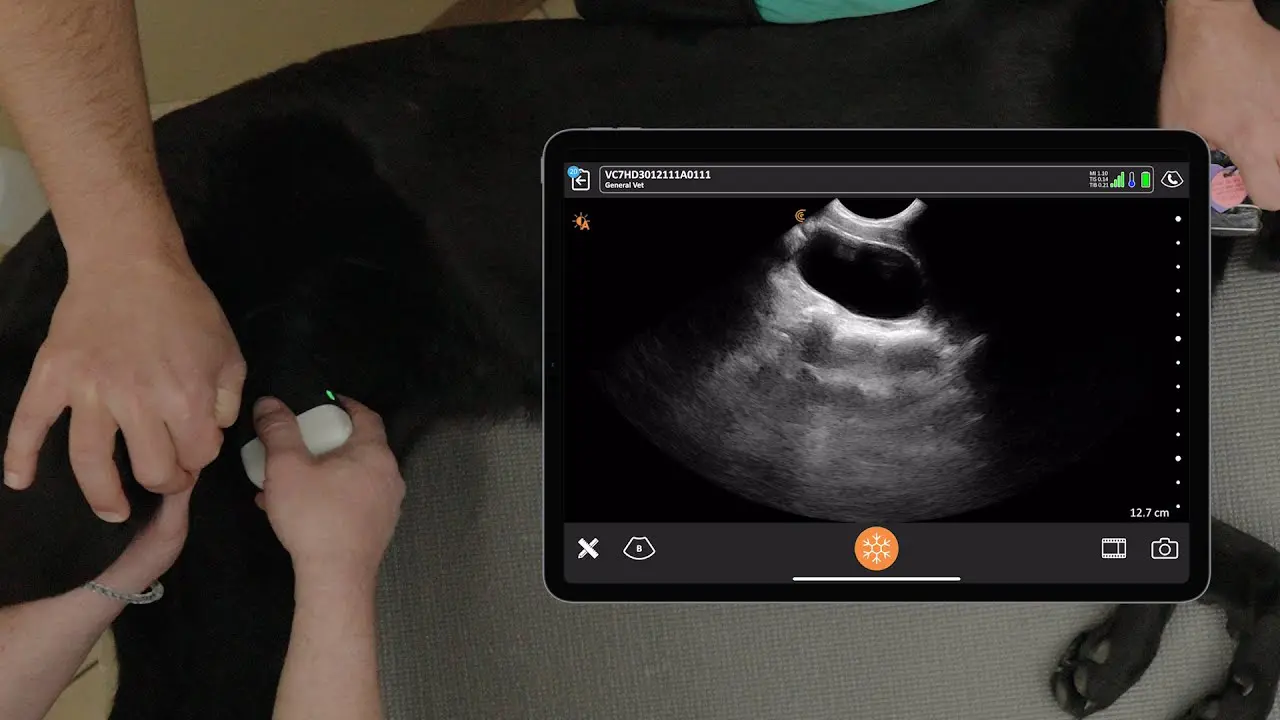
3. Survey Steps 1–3: The Cranial Abdomen
The systematic survey begins with the patient in right lateral recumbency.
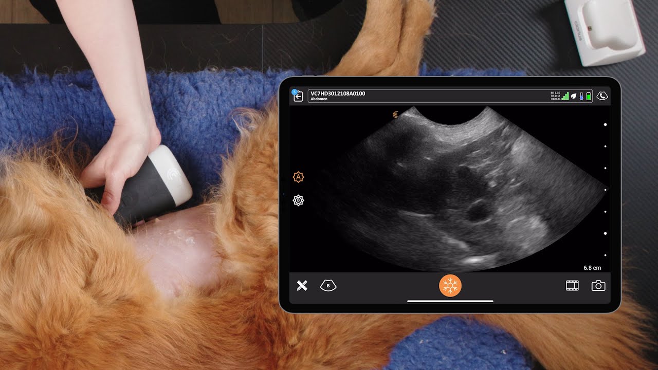
Step 1: Diaphragm, Liver, and Gallbladder
Scan the liver by moving the probe in long axis (sagittal) from the midline, fanning down toward the table, and then up toward the ceiling. Rotate to short axis (transverse) and sweep cranially to caudally, making sure to overlap your sweeps.
- Gallbladder Tip: The gallbladder is a mobile, gravity-dependent structure. “The gallbladder has flopped down towards the table to the gravity dependent.” —Julie Burnage
Video Tutorial: Scanning the diaphragm, liver, and gallbladder
Watch this 10-minute video to see Julie and Angie demonstrate their techniques for scanning the diaphragm, liver, and gallbladder in detail.
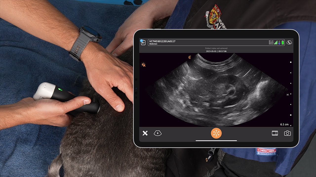
Step 2: Stomach, Pylorus, and Left Pancreas
The challenge here is navigating the gas-filled stomach and colon.
- Pancreas Location: Unlike most organs, “The pancreas and the stomach lie across the patient,” meaning a sagittal probe orientation produces a short-axis image of the pancreas.
- Anatomical Landmarks: Focus on locating these structures to bracket the pancreas:
- Caudal margin of the stomach.
- Gas in the transverse colon.
- The splenic portal vein.
- “In between those three landmarks… will be part of your left limb of pancreas.” —Angie Lloyd-Jones
- Prognostic Tip: “If you’re not seeing it greatly [pancreas], it’s probably because it’s normal.” —Julie Burnage
Video Tutorial: Scanning the Stomach, Pylorus, Duodenum, and Pancreatic Limb
In this next video, Julie and Angie from Aspire UCS demonstrate their ultrasound techniques to evaluate the stomach, duodenum, the pylorus, and the left limb of the pancreas.
Step 3: Spleen
The spleen is large in dogs, extending from the left to the right abdomen.
- Head of Spleen: This is the most challenging part to visualize due to the rib cage.
- “Don’t just scan to the costal margin angle underneath the costal margin.” —Angie Lloyd-Jones
- Vessels: Check the splenic vessels to rule out torsion.
- Margins: Look at the caudal margins of the liver and spleen; if they are rounded and blunted, it can indicate a disease process and enlargement.
Video Tutorial: Scanning the Diaphragm, Spleen, and Splenic Vein
Julie and Angie demonstrate techniques for scanning the left hemidiaphragm and the spleen, with tips on what to look for during the ultrasound evaluation in this 5-minute video.
Key Takeaways from the Veterinary Ultrasound Q&A Session
The webinar Q&A session with attendees addressed practical challenges, equipment features, and key anatomical landmarks for veterinarians learning ultrasound.
Anatomical and Scanning Advice
- Pancreas Identification: The ideal setting varies by equipment and patient size. For abdominal scanning, use the highest lower frequency you can to get better resolution. Look for the three key landmarks: the caudal margin of the stomach, the colon (indicated by gas), and the splenic portal vessel. The pancreas will be ventral to the portal vessel.
- Normal pancreas: If you’re struggling to see it clearly, it’s often because it’s normal and blends with the surrounding mesentery.
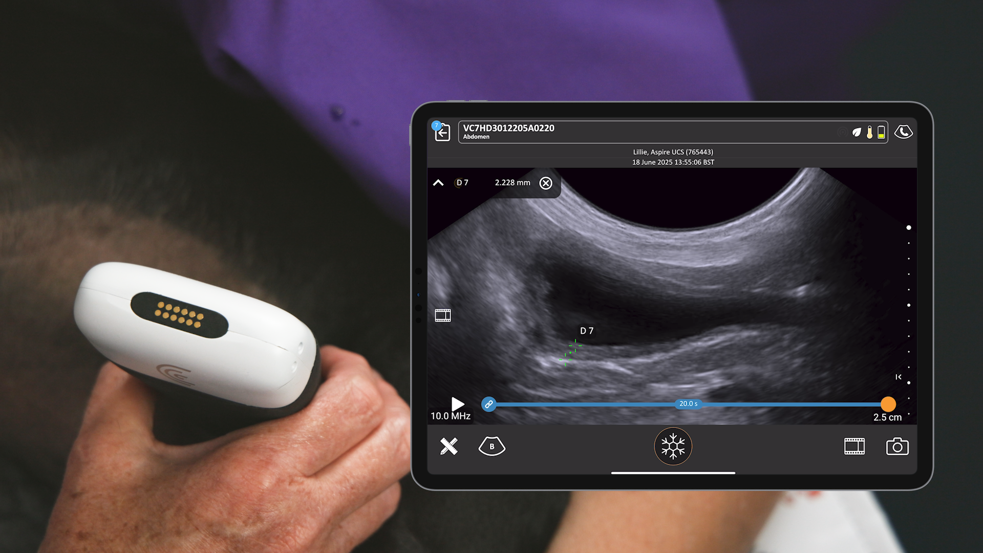
- Needle Aspiration (FNA): Geometry is crucial. With a micro-convex probe, insert the needle at a steep angle to maximize the chance of hitting scan lines. Changing the dynamic range will make the blacks (vessel lumen) and whites (needle shaft) stand out more.
- Patient Positioning: While the recommended approach is starting in right lateral recumbency, good sonographers must be adaptable. Be mindful that turning the patient changes the relative positions of mobile organs.
- Ferrets: Ferrets require a very high frequency probe because they’re so thin. Due to their flatter anatomy, starting in dorsal recumbent position may be suitable.
- Pylorus/Right Pancreas: To find these, scan underneath the patient’s right side in long axis, find the right kidney, and follow the descending duodenum up towards the stomach.
Procedural and Training Questions
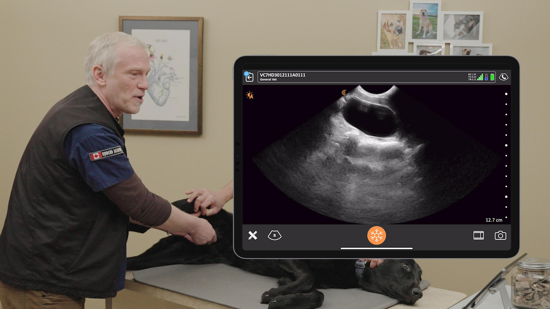
- Sedation: Sedation is generally recommended for a full abdominal survey. It ensures a compliant patient for the 40-minute duration, allows the operator to apply necessary pressure without causing pain, and promotes efficiency if FNA is needed.
- Eight-Step Approach vs. Binary Questions: The detail in the eight-step approach can seem daunting. The steps are intended as manageable, learnable components. Veterinarians should practice steps individually (e.g., Step 2 or 5) on routine cases, building towards the full survey.
- FAST/BFAST Protocols: For sick, unstable patients, established protocols are recommended, such as those by Dr. Shalhoub and Boysen. These are often found in educational resources like the Clarius Classroom and on YouTube.
Equipment and Features
- Scanning Tables: The presenters do not use a cradle; they scan patients lying flat on a wet bed on the table to allow the operator to scan up and underneath. Sandbags or wedges are used to support the patient and prevent falls.
- Auto Scan/Auto Preset AI: This feature that was demonstrated during the webinar is a toggle button on the Clarius system. When the auto scan is orange, it is on and automatically optimizes settings (gain, depth, etc.) as the anatomy is recognized (e.g., Cardiac, Lung, Bladder). When the button is white, the user can manually optimize the image using individual controls.
Video: Auto Preset AI for Veterinary Ultrasound
Watch this 2-minute video to see Shelley Guenther demonstrate how Auto Preset AI for VET recognizes the anatomy being scanned and selects the appropriate preset for hands-free image optimization.
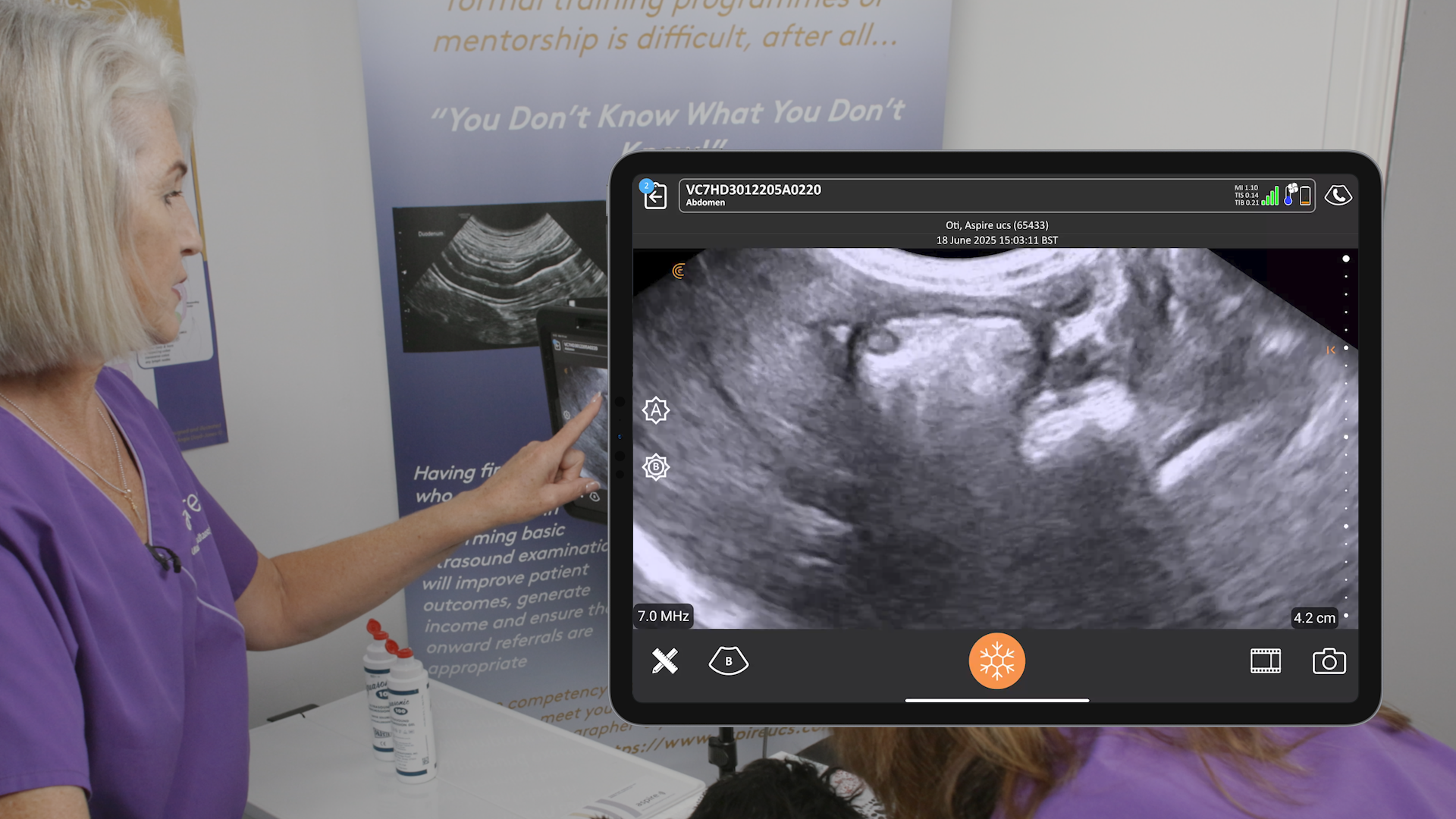
Final Clinical Takeaway: “Always annotate representative images of what you’re looking at because it’s evidence of what you saw and when you saw it.” —Angie Lloyd-Jones
Register for Aspire Webinar Part 2
Angie and Julie are back soon to co-present a webinar with Clarius. Register now to reserve your spot! Building on Part 1, this session will dive into Steps 4 – 8 of their proven approach to canine and feline abdominal imaging. Angie and Julie will share practical instruction and scanning demonstrations designed to help you advance your skills with confidence. Learn more.
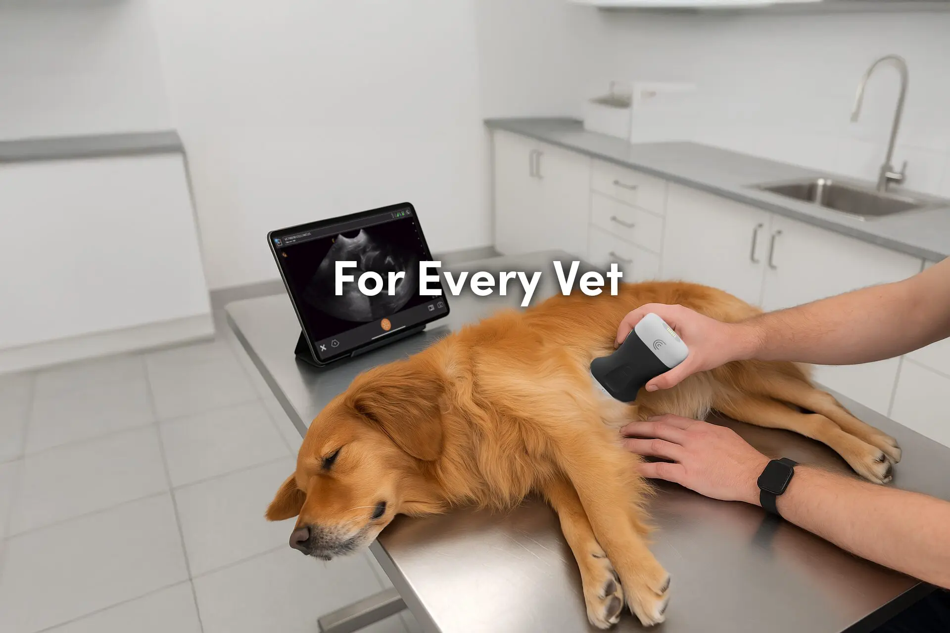
Master Veterinary Scanning with Wireless Ultrasound Specialized for Companion Animals
Clarius VET HD3 scanner is designed to deliver high-definition, wireless ultrasound imaging with the performance of a traditional system at a fraction of the cost. The wireless design and zero footprint make them highly portable, enabling clinicians to perform exams anywhere in the clinic or in the field without wires getting in the way or startling animals. The Clarius VET HD3 comes with an intuitive, AI-powered app for iOS or Android devices that streamlines workflows and automatically optimizes imaging. The Clarius ecosystem includes access to Clarius Cloud for unlimited exam storage and management, Clarius Classroom videos, and a one-click telemedicine feature with Clarius Live, making it a comprehensive and user-friendly solution for veterinary professionals.
To learn more about how you can add wireless ultrasound to your practice, visit our Veterinary Specialty Page. There you’ll have access to additional webinars and classroom videos. For more information you can request a personalized virtual ultrasound demo.
