











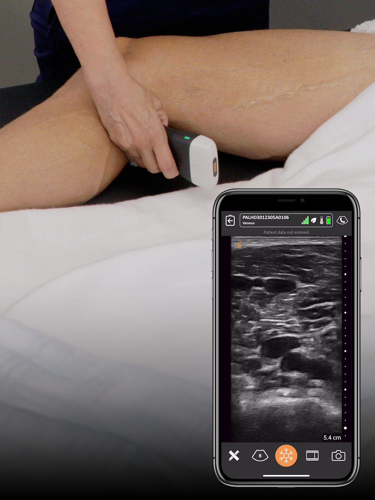

A powerful, portable ultrasound research toolkit built for universities, labs, and R&D teams.




A powerful, portable ultrasound research toolkit built for universities, labs, and R&D teams.
CORE ADVANTAGES


Develop custom ultrasound sequences with ability to adjust per-scanline transmit and receive parameters
Start with essential imaging or unlock full system control with raw data, APIs, and Texo II
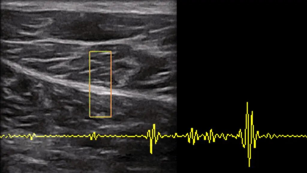
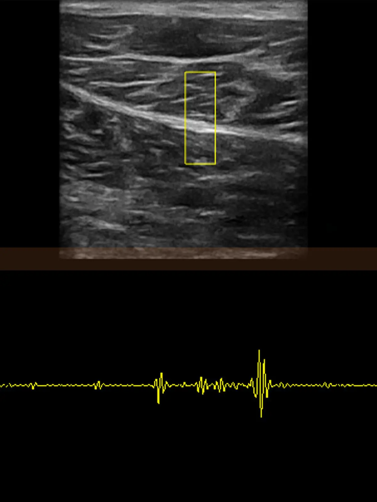
By using offline or real-time RF, run complex signal processing algorithms to help understand underlying properties of anatomy that is often hidden by the greyscale nature of ultrasound
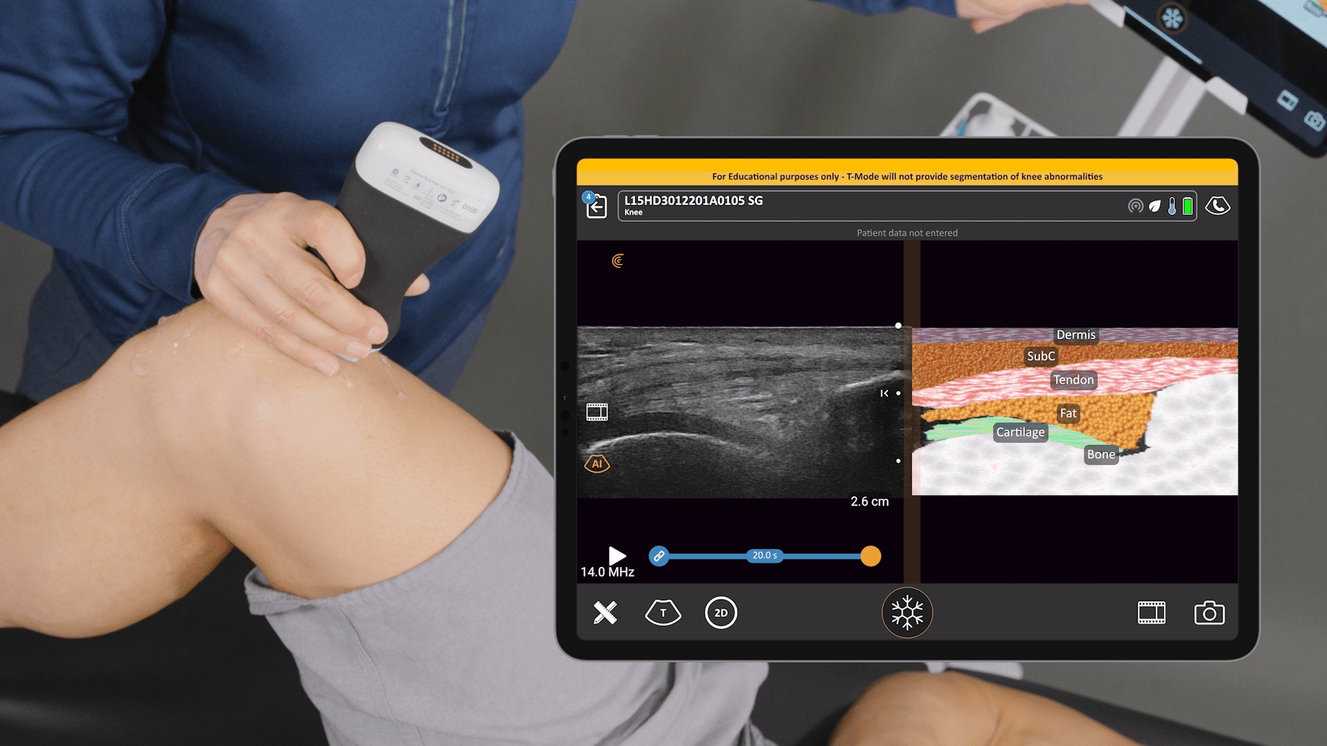
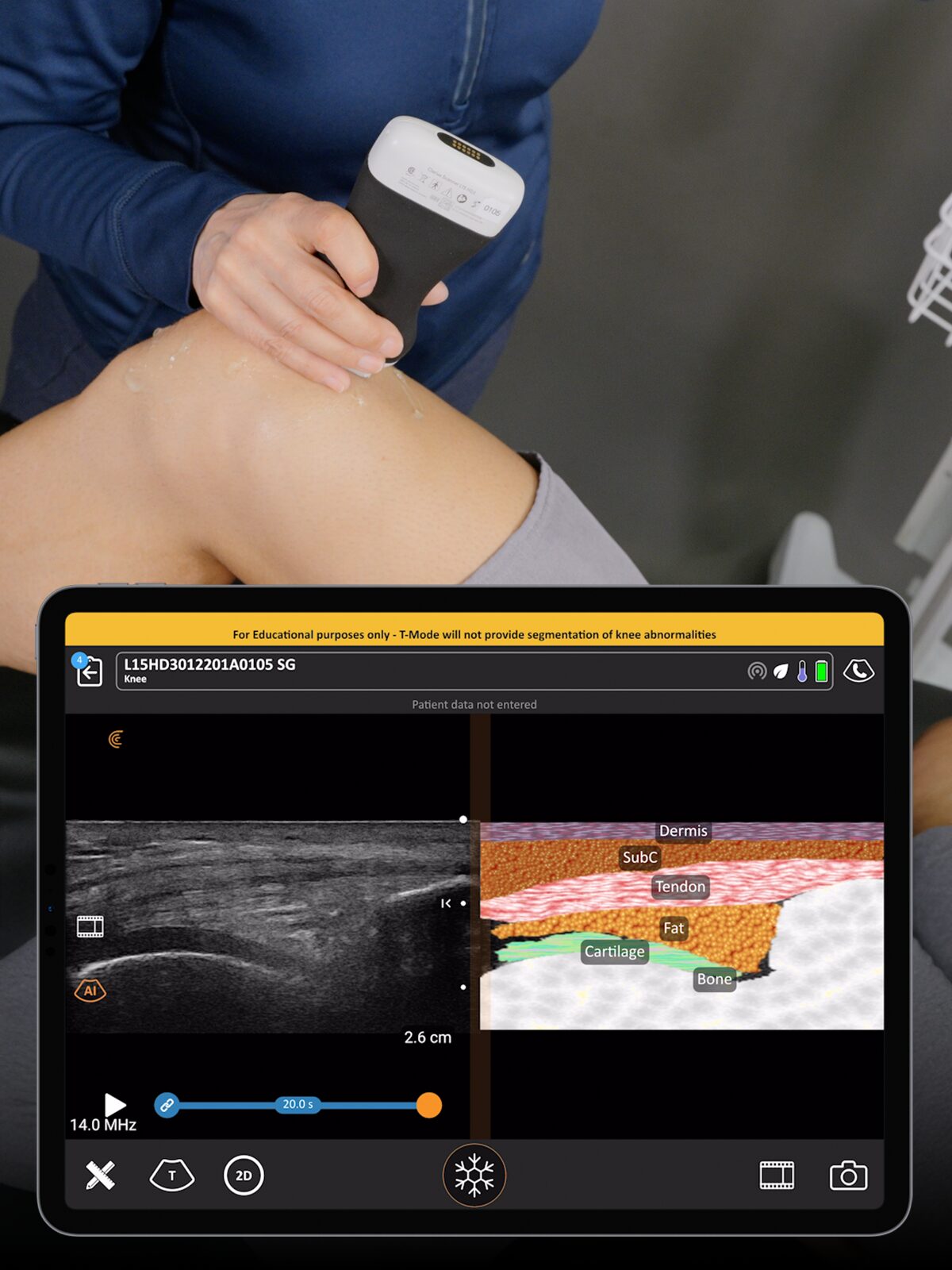
Train machine learning and deep learning algorithms to classify, segment, and interpret ultrasound data for predictive and diagnostic insights. Use the platform to curate comprehensive training datasets and evaluate the performance of AI algorithms for organ‑specific imaging, anomaly detection, and predictive analytics.
Investigate tissue properties using RF and Doppler signals to study biomechanics, pathology, and clinical outcomes. This research informs the development of quantitative tissue biomarkers and helps refine diagnostic protocols.
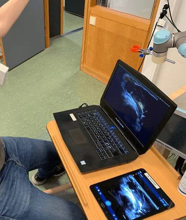
Integrate the 9‑DOF IMU for motion‑aware scanning, navigation, and robotic integration in research and teaching. Integrate ultrasound with robotics and surgical navigation systems to enable real‑time image‑guided interventions and dynamic tissue mapping.
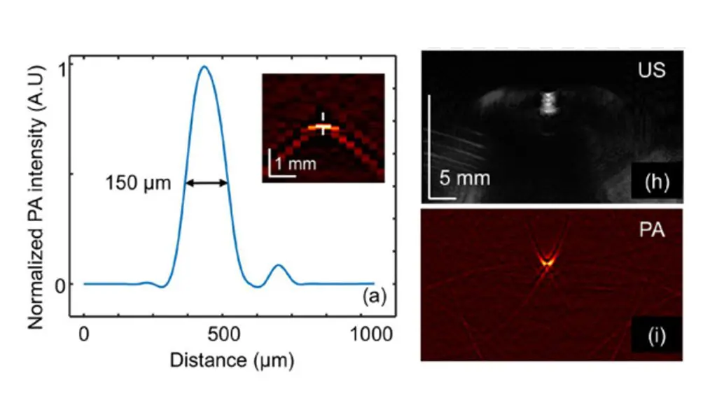
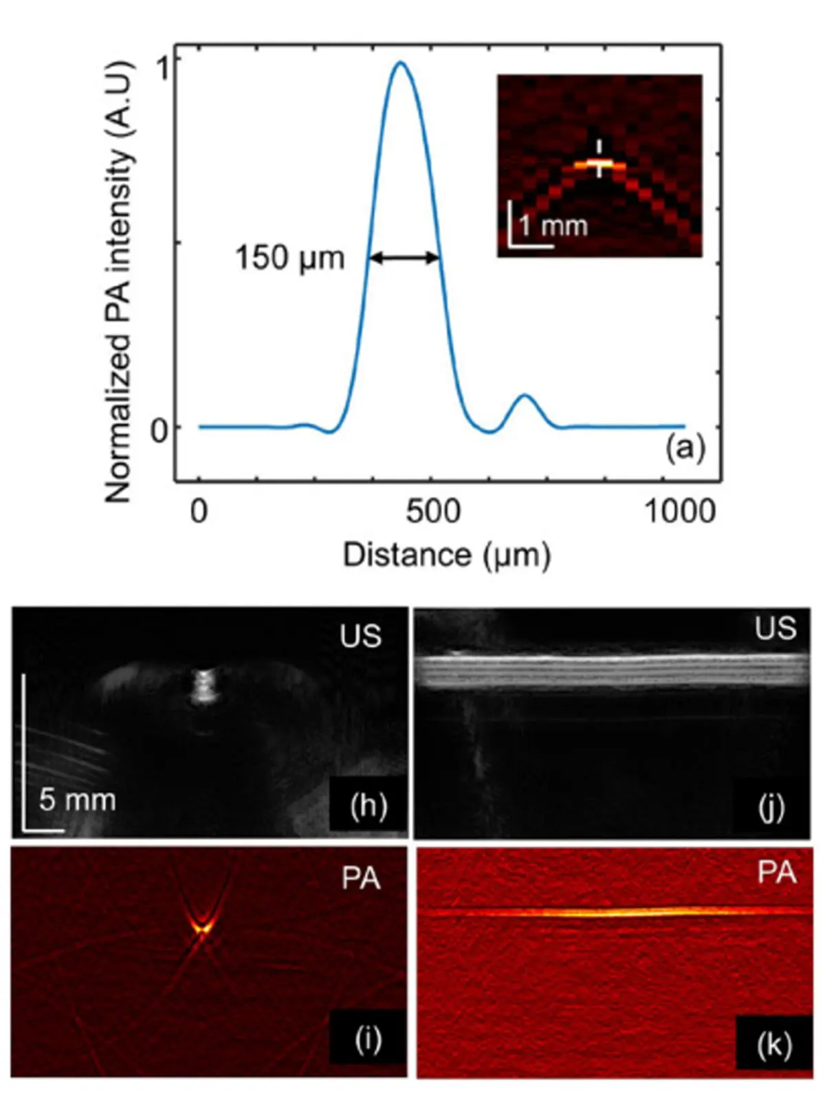
Capture and analyze photoacoustic signals to explore hybrid imaging modalities and energy absorption in tissues. Leverage hybrid imaging to map vascular networks, detect metabolic changes, and investigate optical absorption properties for early tumour detection.
Experiment with custom imaging parameters to tailor ultrasound performance for specialized applications across disciplines. Adjust scanning parameters such as frequency, power, and frame rate to fine‑tune the system for varied tissues, experimental conditions, and imaging modalities.
To support rapid development and integration, Clarius provides:
Thomas Lango
SINTEF Digital – Health Research
The wireless Clarius ultrasound imaging device offers many interesting features, as seen from a research perspective. Portable and wireless ultrasound with easy to use interface is perfect in quick demonstrations and development in a multidisciplinary team. We believe that the Clarius device will bring a lot more flexibility in our research and innovation projects due to its ease of use and high image quality.

Michael Friebe
Otto-von-Guericke-University, Magdeburg, Germany
Clarius is the ideal base system for our research engineers, as it is very easy to use and provides excellent image quality. Due to its small size and weight, it can be connected to our robots and holding arms, and subsequently be used as a hybrid imaging system in combination with our low-cost X-ray, Endoscopy and CT units.
Edmond Lou
University of Alberta
The portability, ease of use, and the high-quality imaging is what I love about the Clarius Scanner. As an engineer, I like their research package. With the availability of the raw data, a streaming API and 3D positional information, I am able to build custom software to reconstruct 3D images. The technical support and development team is excellent in response to user questions and demand. I recommend the unit to both clinical staff colleagues and researchers.


All data streams—RF, channel RF, IQ, greyscale, Doppler—include metadata in YAML format for easy parsing.
Yes. Clarius provides SDKs and sample code for both MATLAB and Python environments.
Absolutely. The Cast API supports real‑time data streaming and low‑level control to integrate with custom machine learning workflows.
Clarius software runs on Windows, Linux, macOS, Android, and iOS.
Yes, the Cast API and SDKs are designed to handle multiple probes or devices for synchronized data capture.