

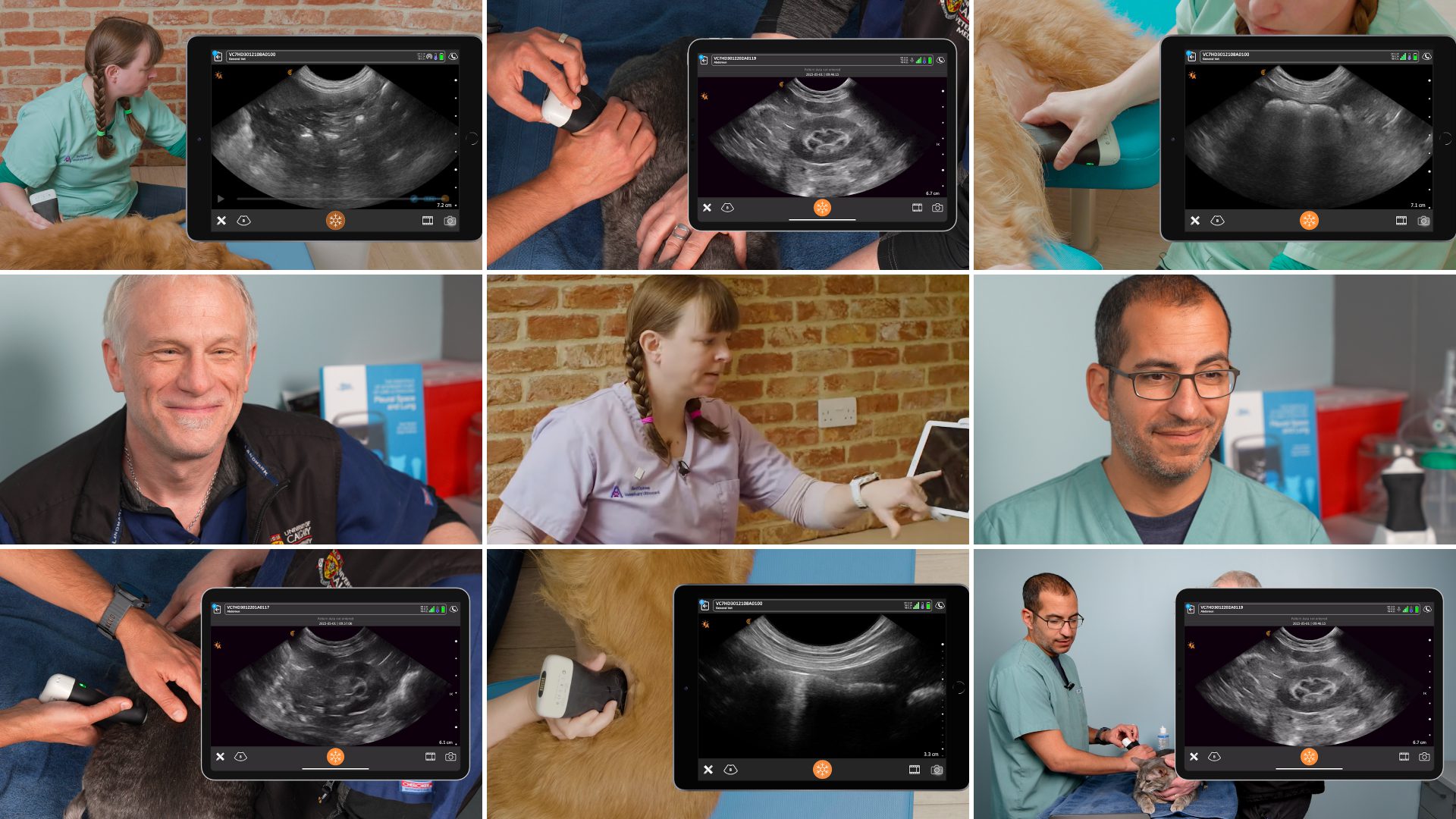
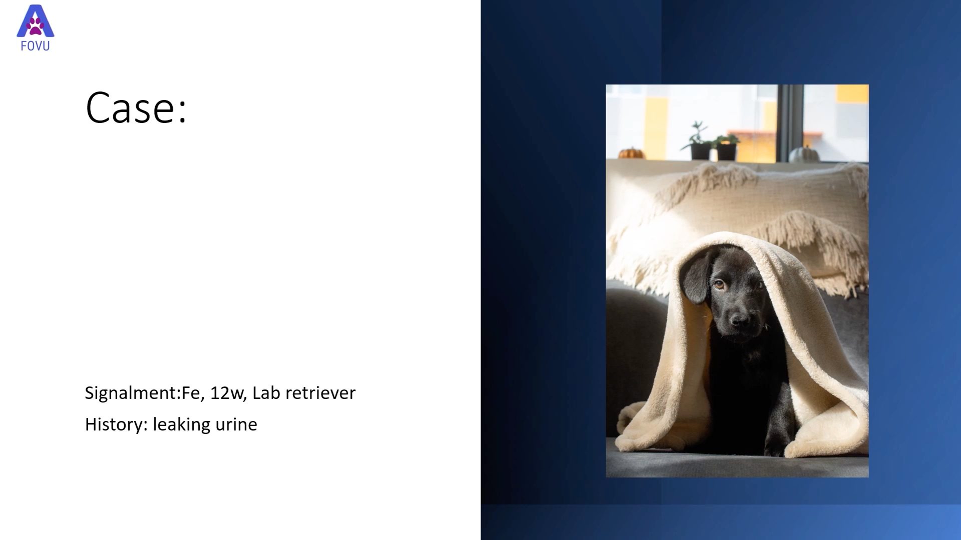
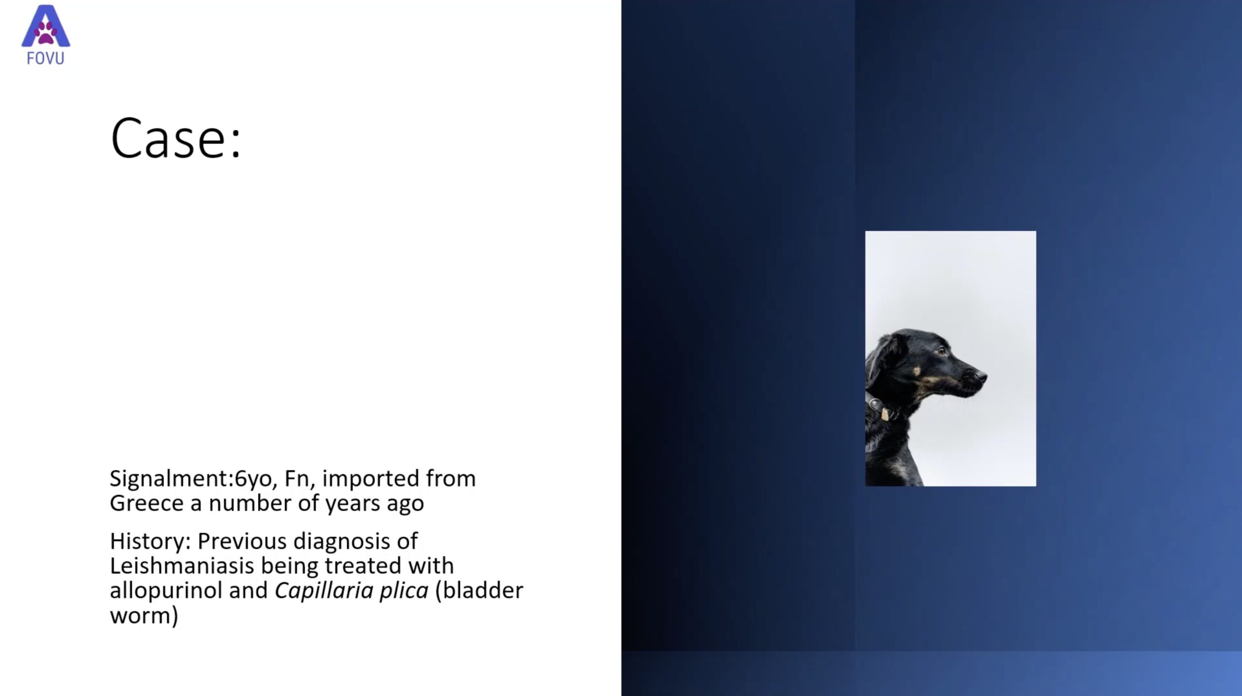
Gastrointestinal diseases, which are commonly seen in small animal veterinary practice, are easy to detect and diagnose with the right techniques using point of care ultrasound (POCUS). We recently invited Dr. Camilla Edwards, DVM, CertAVP, MRCVS, a popular ultrasound educator, and peripatetic veterinary ultrasonographer, to present a webinar featuring four real cases of GI tract abnormalities in cats and dogs. Based in Cambridge, UK, Dr. Camilla Edwards, travels with her dog Pippi to clinics within a 50-mile radius. A recording of this practical one-hour webinar is available to watch at your convenience featuring Dr. Camilla’s ultrasound scanning technique and her detailed overview of these cases:
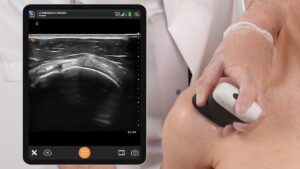
Case 1: A domestic short-haired cat presenting with a history of weight loss. Watch Dr. Camilla demonstrate how POCUS allowed her to identify and measure a section of thickened jejunal wall.
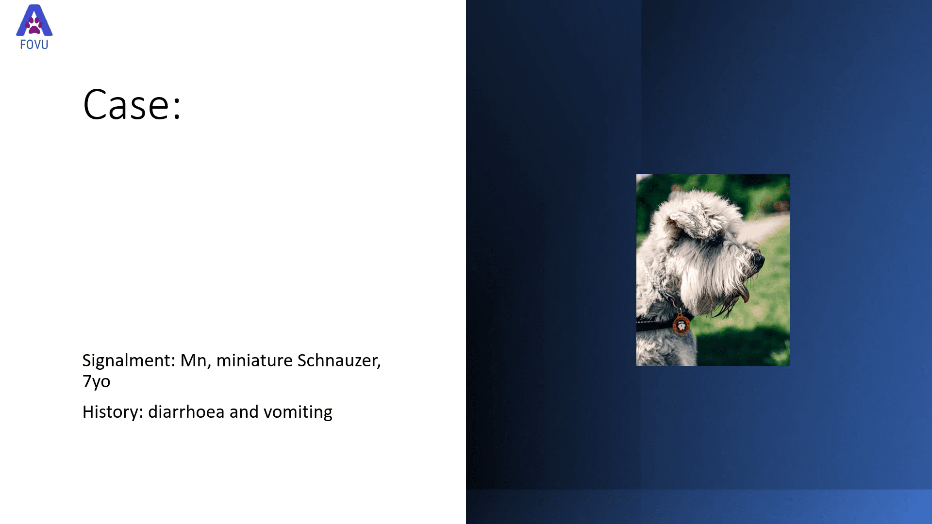
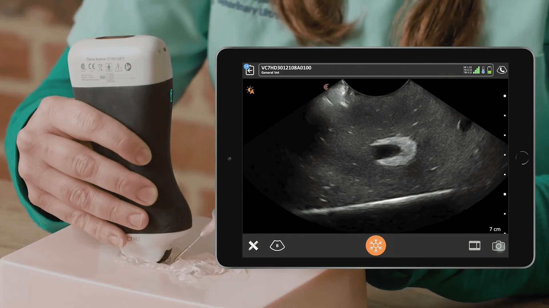
Case 2: An Australian Shepherd with chronic diarrhea. Dr. Edwards shares ultrasound imaging that showed pronounced thickening of the ascending colon wall, also noting the loss of wall layering and hyperechoic reactive fat in the region.
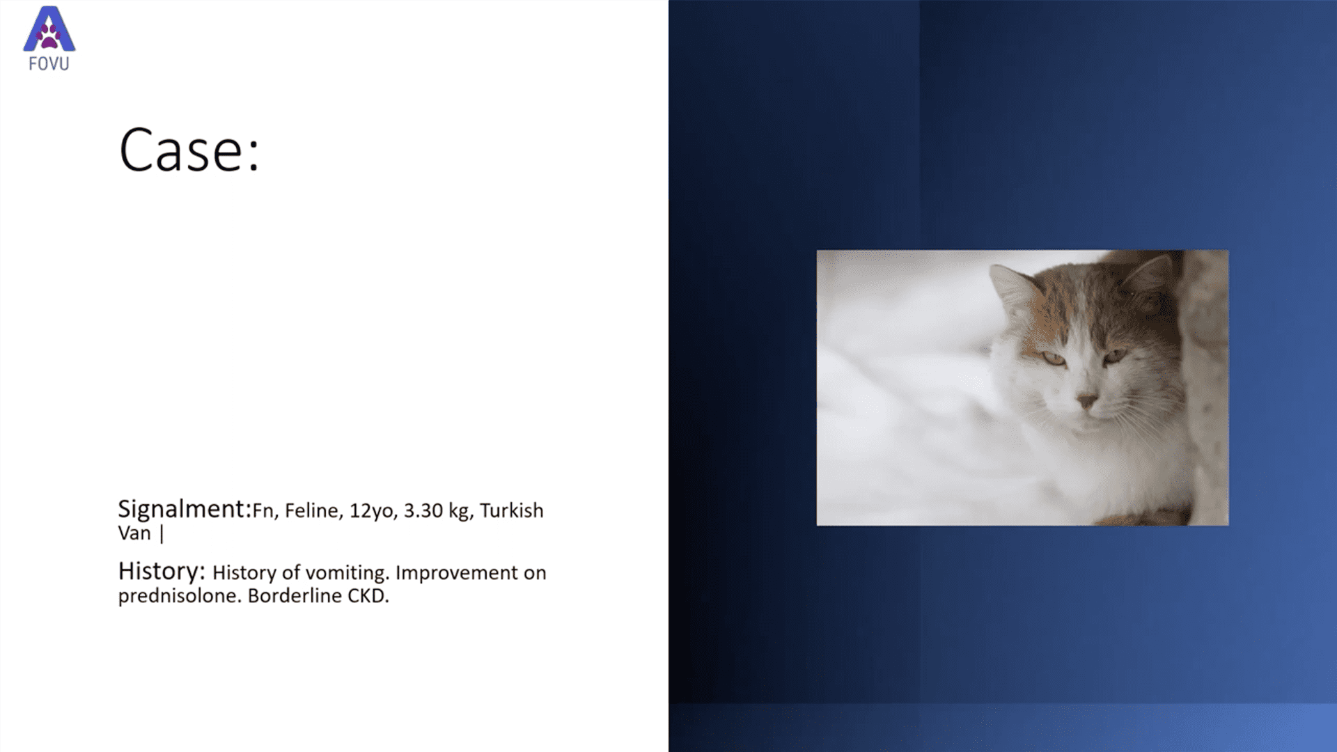
Case 3: A Turkish Van cat with a history of vomiting. Watch a POCUS exam that revealed a mass in the stomach wall and a large lymph node caudal to the mass.
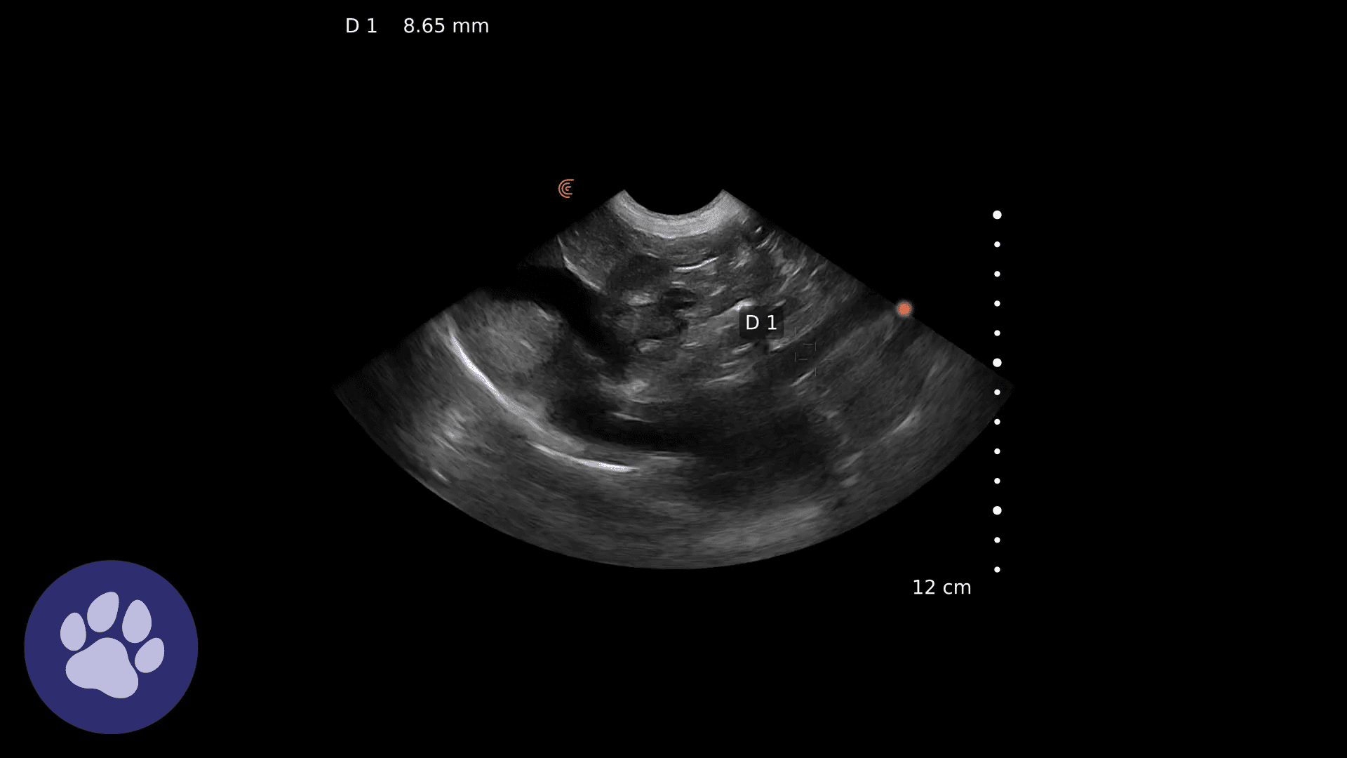
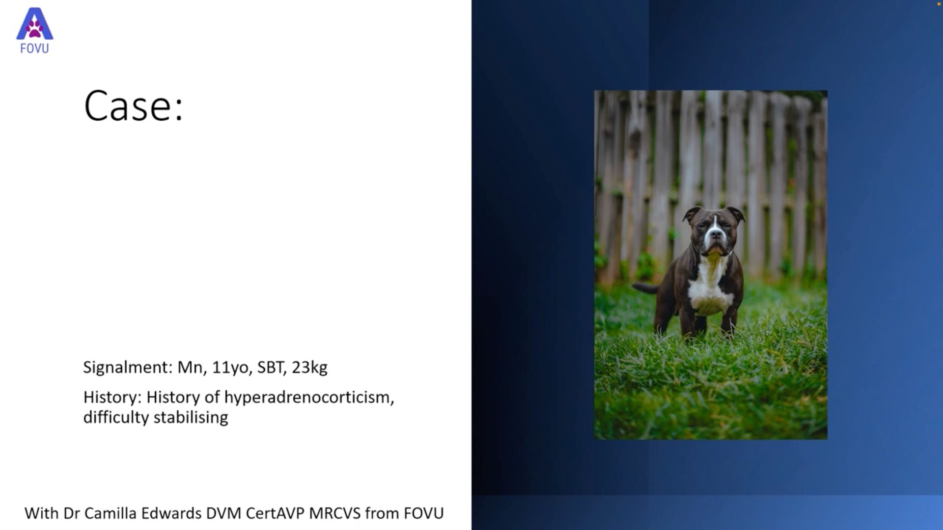
Case 4: A remarkable intestinal ultrasound in a Staffordshire Bull Terrier suffering dramatic weight loss and diarrhea. Dr. Edwards shows a large mass in the intestinal wall, which was confirmed as intestinal lymphoma after an ultrasound-guided fine needle aspiration (FNA).
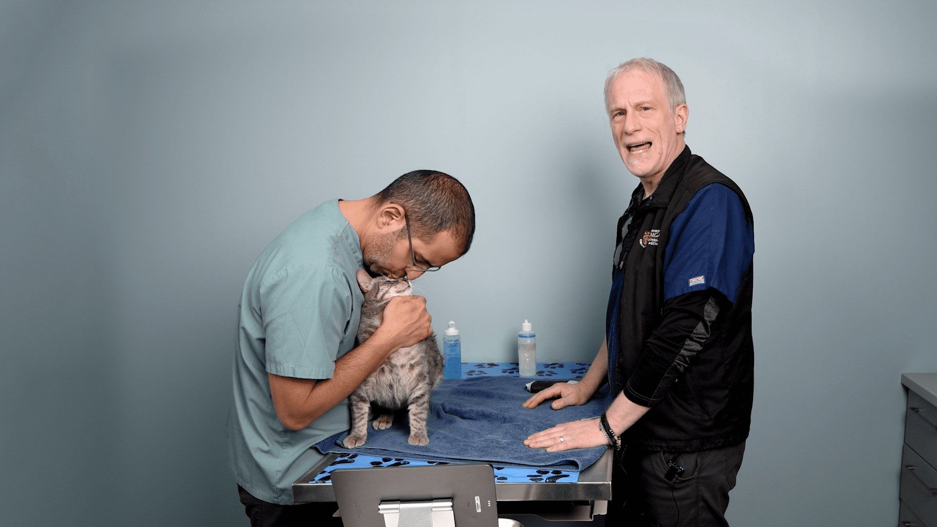
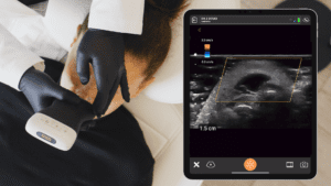
Video: Ultrasound-Guided Fine Needle Aspirate
If you’ve only got a few minutes to spare, check out Dr. Camilla’s scanning technique in this 90-second video. She teaches how and where to identify both needle and target when performing an ultrasound-guided FNA using a phantom.
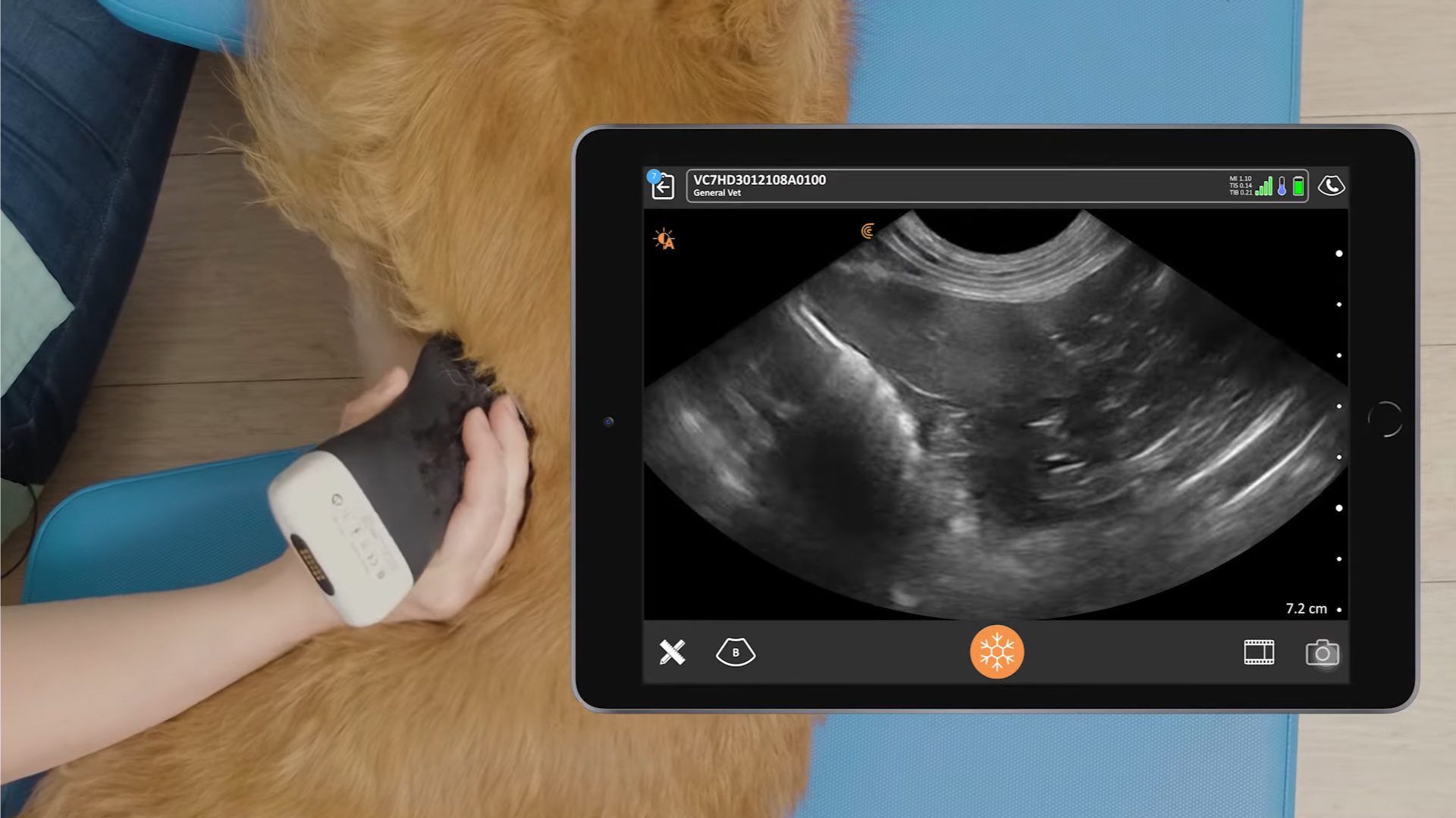
Make a Rapid Diagnosis Cage Side with Clarius Ultrasound
Optimize comfort with on-the-spot imaging and no wires to startle your furry patients. Ultra-portable and affordable, Clarius delivers the quality imaging and performance of traditional systems without complex knobs and buttons. The AI-powered app is as easy to use as your smartphone. Improve animal care with clear visualization of anatomy and pathology for a more accurate diagnosis on your patient’s first visit. We invite you to book a virtual demonstration to discuss which Clarius scanner is ideal for your veterinary practice. Or visit our Veterinary Specialty page to learn why Clarius ultrasound is a popular choice for veterinarians shopping for an ultrasound system.

