

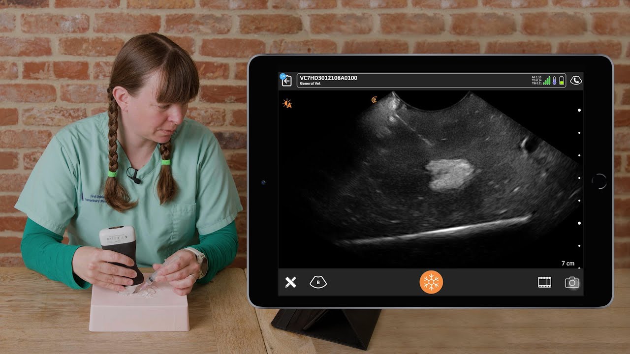
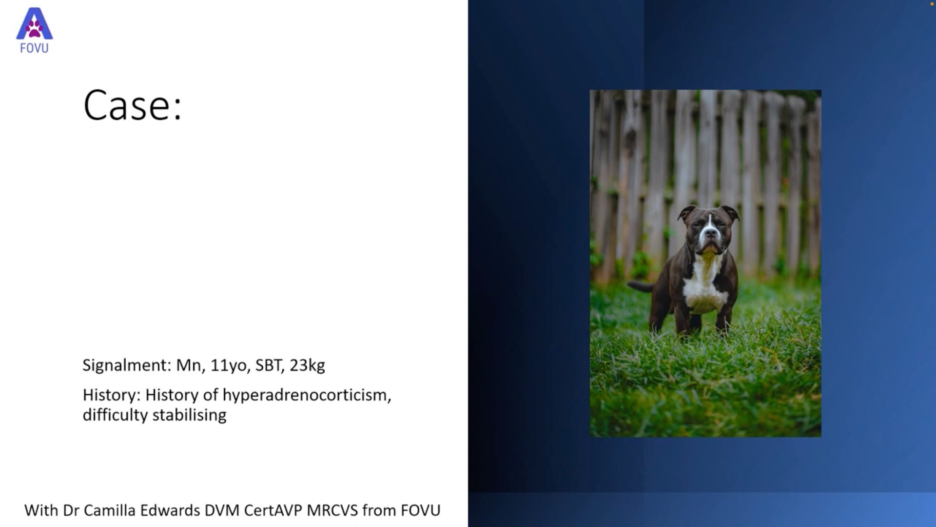
Dr. Camilla Edwards, DVM and founder of First Opinion Veterinary Ultrasound (FOVU) has been sharing some interesting case studies. This one is a really nice example of a dilated left renal pelvis and ureter.
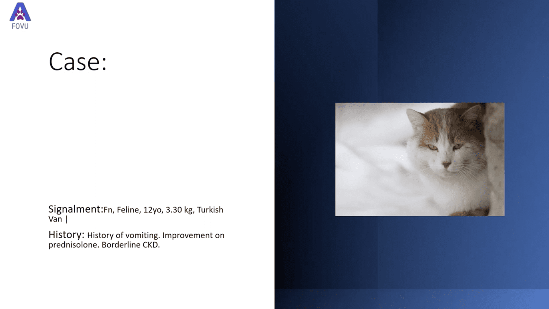
History: Twelve-week-old female Labrador Retriever, leaking urine.
Ultrasound Findings
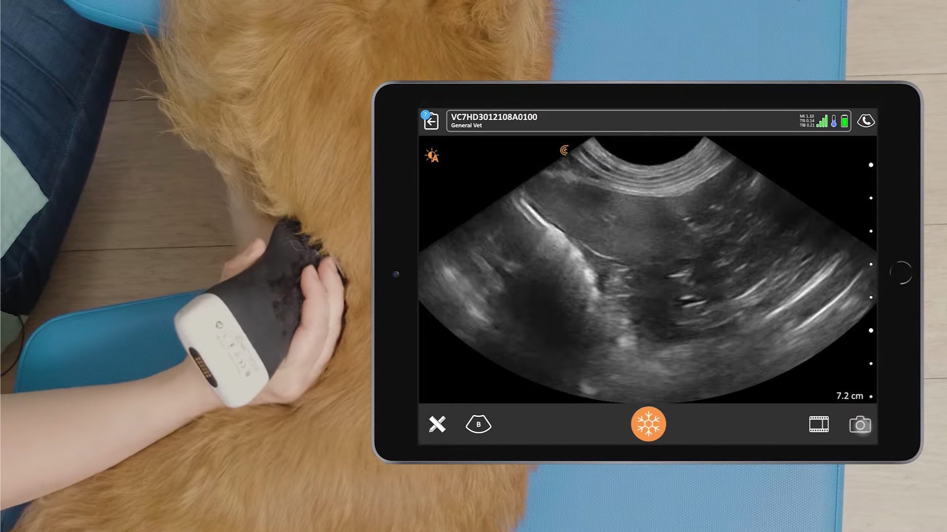
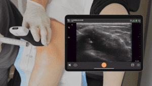
Dr. Edwards performed an ultrasound of the kidneys and bladder to attempt to determine a cause for the incontinence in this puppy.
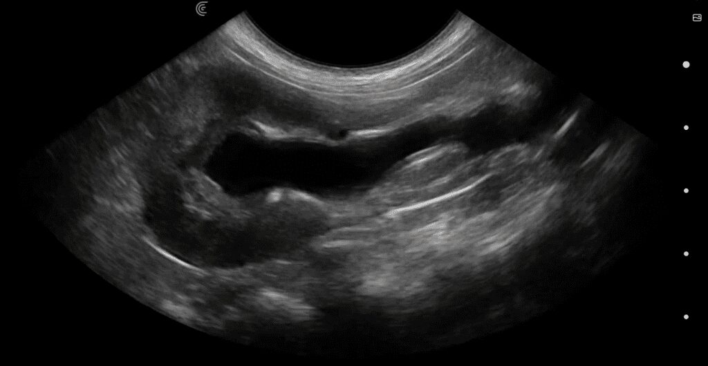
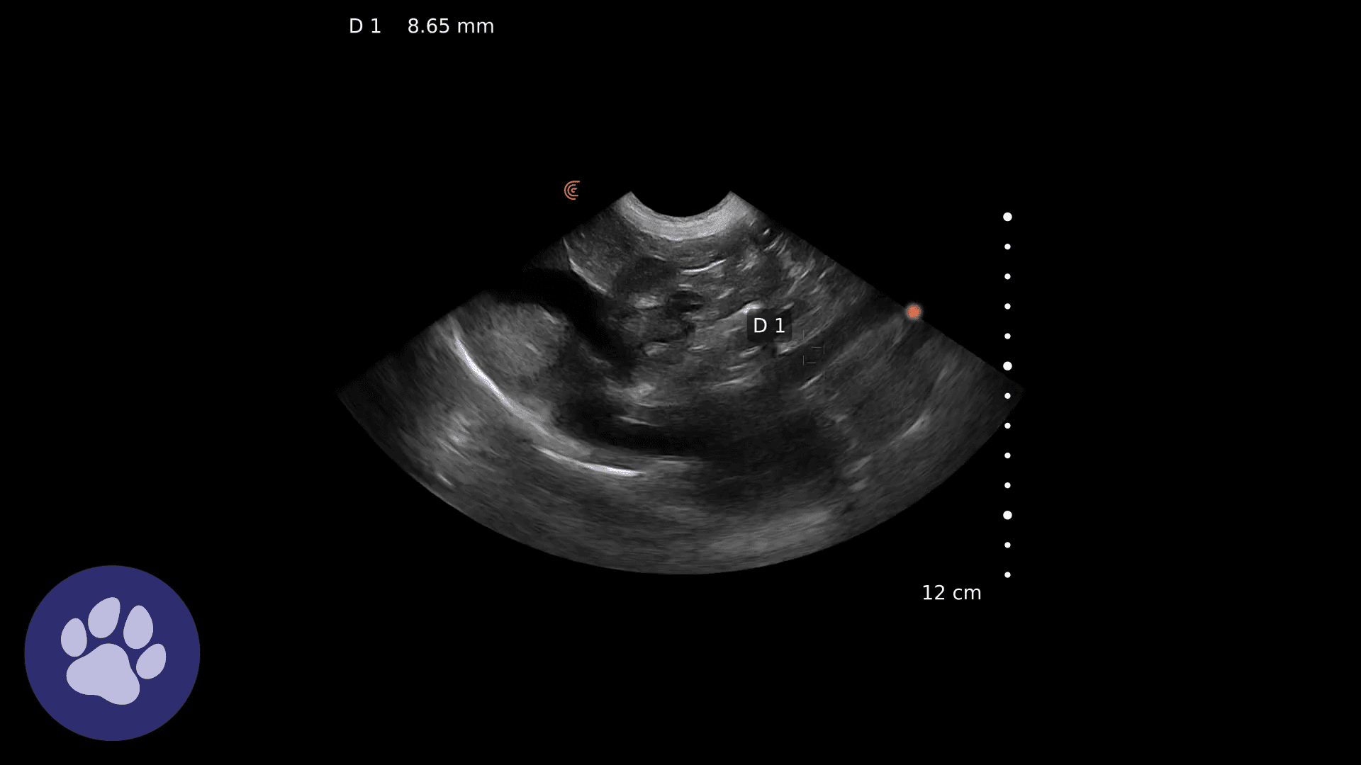
The left kidney demonstrated marked dilatation of the renal pelvis and ureter. This is a transverse plane to show the continuity of the collecting system and the severely dilated ureter. Although it’s not shown in the video, the ureter could be followed caudally and was shown to enter the urethra instead of the trigone of the bladder, indicating an ectopic ureter.
The right kidney and bladder appeared normal.
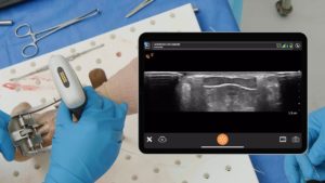
There’s an extra little pearl in this video. A short time ago Dr. Edwards shared her expertise on the topic of imaging abdominal lymph nodes in dogs. During Dr. Edwards’ webinar, “Practical Small Animal Ultrasound: POCUS Techniques for Imaging Abdominal Lymph Nodes”, she talked about how, in younger healthy dogs, the lymph nodes often appear enlarged.
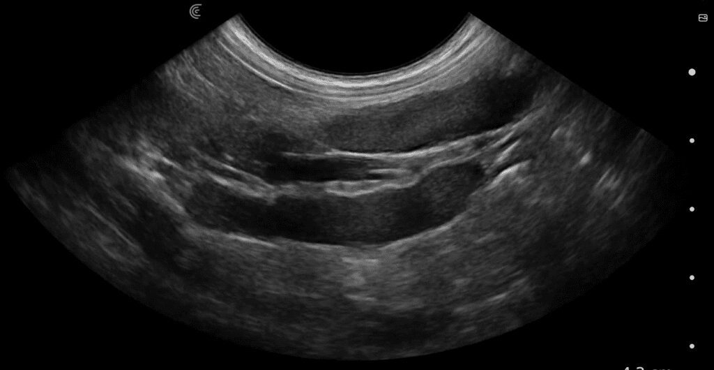
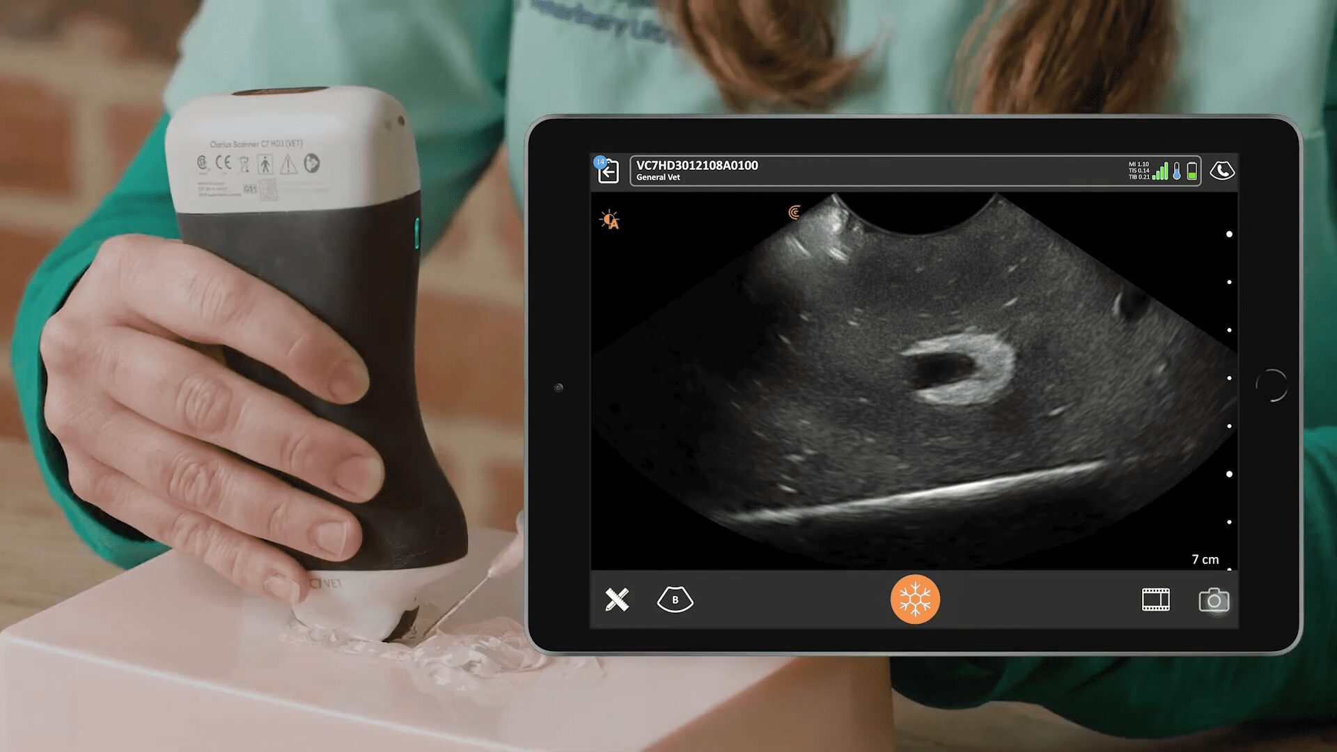
During this ultrasound exam, she was able to demonstrate very large, but normal, jejunal lymph nodes in this lab pup.
Discussion
Ultrasound is an extremely helpful tool for examining the kidneys and bladder in small animals. Fluid appears black or anechoic on ultrasound, so any degree of hydronephrosis will show a black area distending the renal pelvis. This area may or may not extend into the tubular-shaped ureter, depending on the level of obstruction. If the urinary bladder is full, it will appear black as well.
For a review of how to scan the normal kidneys and bladder, visit our Clarius Classroom, where Dr. Edwards shares her tips and tricks.
Dr. Edwards uses the Clarius C7 HD3 Vet, which is specifically designed for clinical imaging of small and medium-sized animals. Our Advanced Veterinary Package offers more flexibility for users who need additional workflows for various animal exams.
To learn about how easy and affordable it is to add Clarius handheld ultrasound to your veterinary practice, visit our Clarius veterinary ultrasound page for product details. Or contact us today to discuss which scanner is right for your veterinarian practice.

