

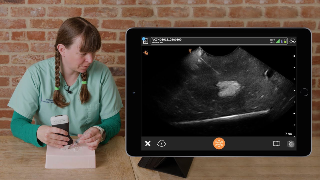
Dr. Camilla Edwards, DVM and founder of First Opinion Veterinary Ultrasound (FOVU) recently shared an interesting case study where she used ultrasound to identify an enlarged adrenal gland in a dog.
History: This male neutered 11-year-old Staffordshire Bull Terrier weighing 23 kilos presented with a history of diagnosed hyperadrenocorticism, or Cushing’s disease. His vets were having difficulty stabilizing him, so they consulted with Dr. Edwards who performed an abdominal ultrasound, with a focus on the adrenal glands.
Ultrasound Findings
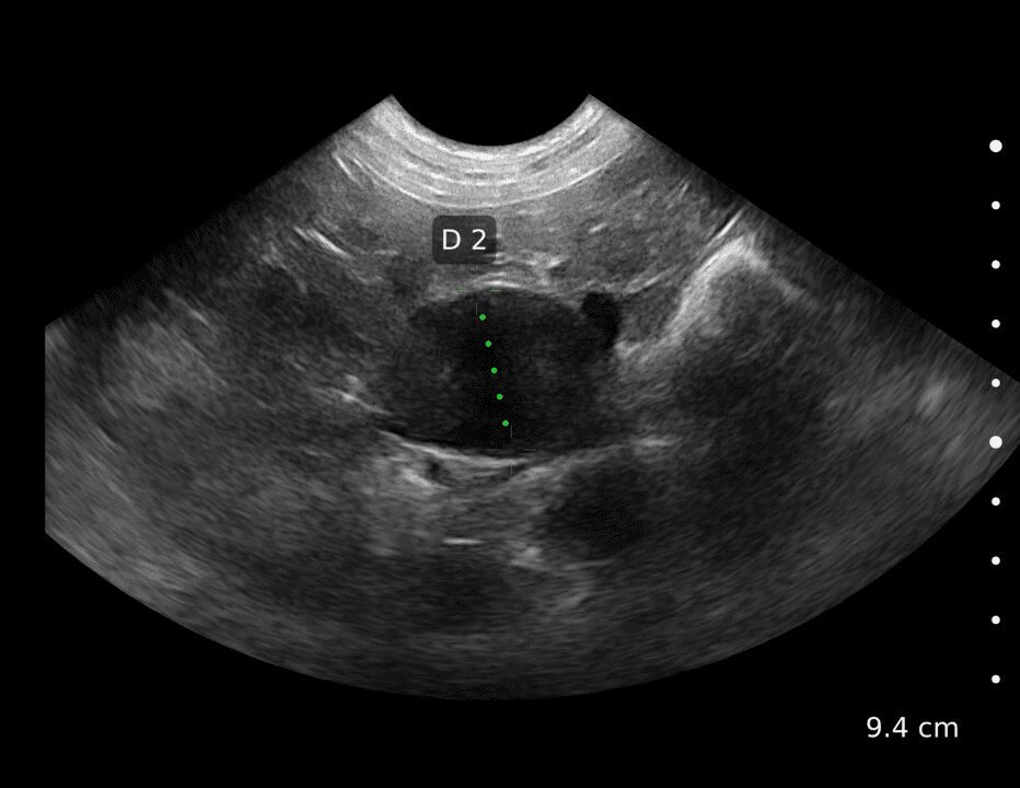
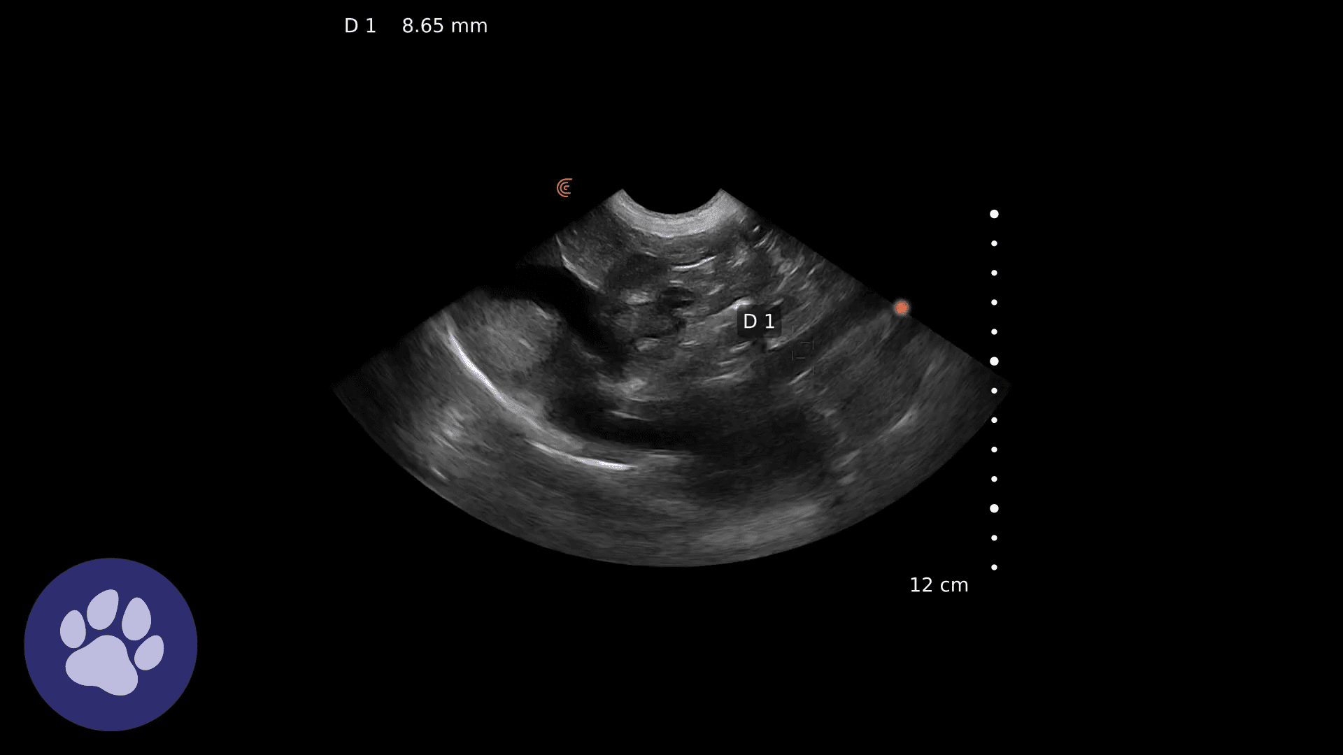
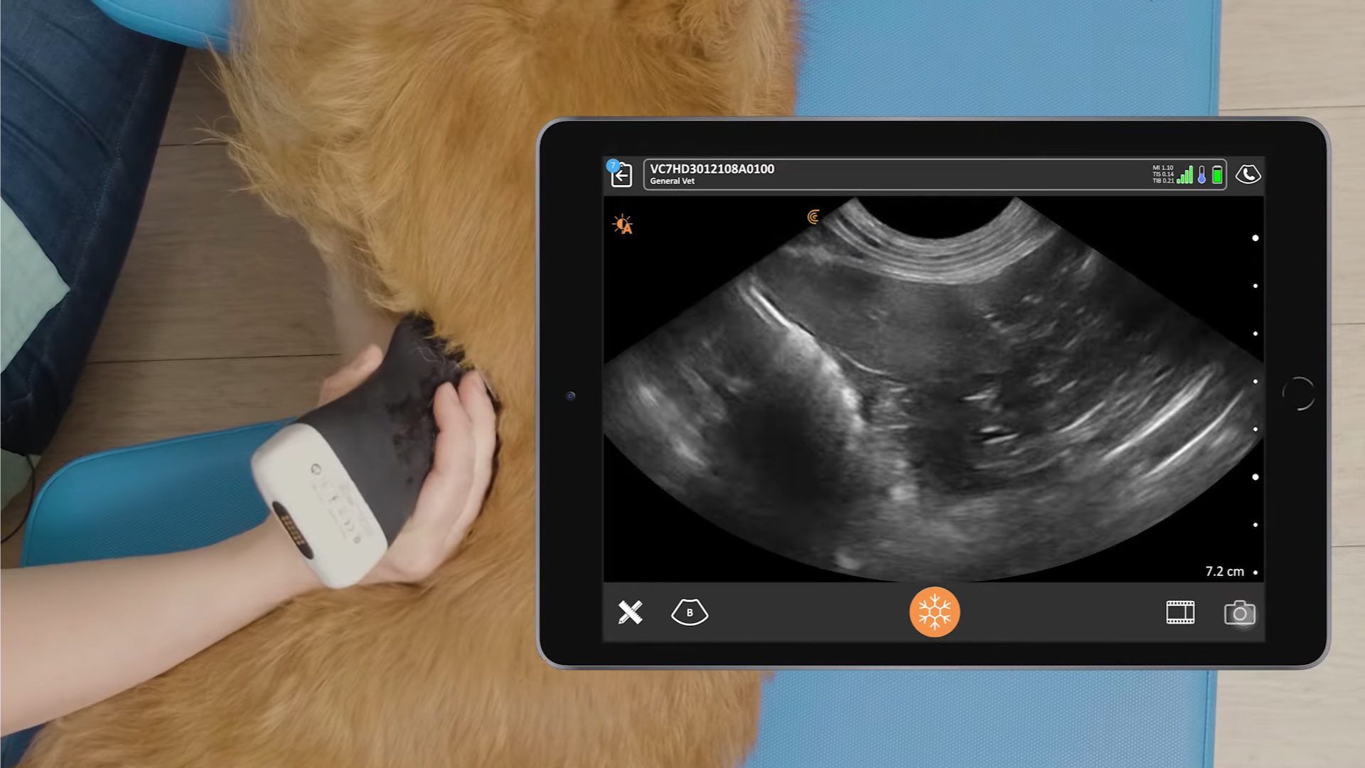
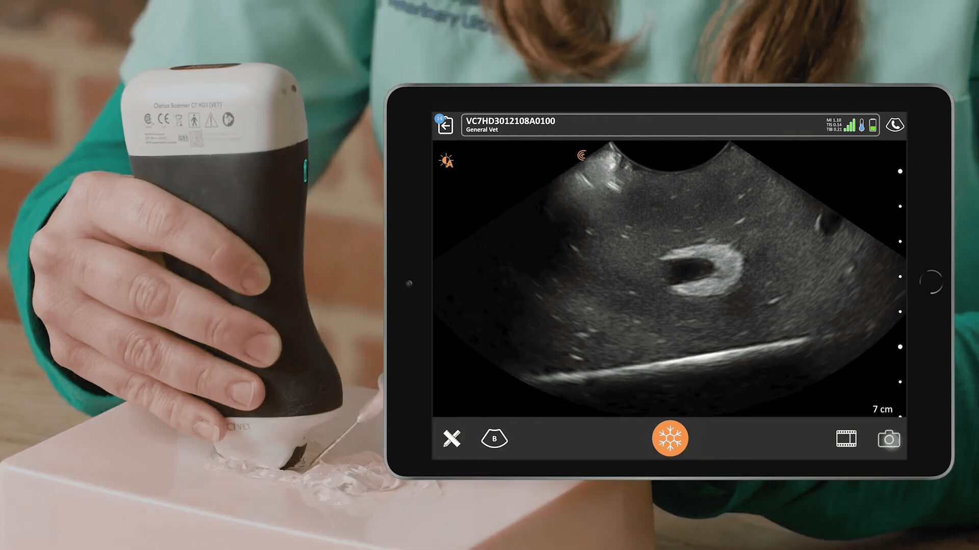
To locate the left adrenal gland, Dr. Edwards first identifies the aorta, the caudal vena cava, and the left renal artery. The adrenal gland sits just cranial to the left renal artery. In this case, there is a large hypoechoic structure in the region of the left adrenal gland. The cranial pole appears normal, but the caudal pole is massively enlarged. Dr. Edwards uses color Doppler to identify the phrenicoabdominal vein, which is a helpful landmark and crosses between the two poles of the gland, as well as to look for turbulent flow in surrounding vessels which may indicate local invasion. There is none.
The normal diameter of the caudal pole in a 23-kilo dog should be around 0.5 cm. In this case, the measurement was 2.75 cm, which indicates massive enlargement.
The right adrenal gland was scanned for comparison. The right adrenal can be visualized near the caudal vena cava and has a normal ultrasound appearance. Occasionally when one gland is over-functioning, the other can actually atrophy and appear smaller than normal.
Video: Dynamic ultrasound identifying a large left adrenal mass
Outcome and Discussion
As Cushing’s disease may result from an adrenal tumor, it’s important to image the adrenal glands with ultrasound. In this case, Dr. Edwards’ was able to image a large adrenal mass on the left side. There was no local invasion or metastases visualized, so it was determined to be a primary functional tumor causing adrenal hyperadrenocorticism. Fortunately, surgical resection of the left adrenal gland is likely to lead to a positive outcome for this dog.
Don’t miss Dr. Edwards’ upcoming webinar, Practical Small Animal Ultrasound: Guiding Diagnosis and Management of Palpable Abdominal Masses, where she’ll explain how to rapidly identify and characterize palpable abdominal masses in veterinary patients using wireless ultrasound. The session will cover systematic ultrasound examinations, distinguishing between different types of masses, investigating spread, performing ultrasound-guided sampling, and monitoring changes over time for better treatment decision-making.
Find Answers Under the Fur with Clarius Ultrasound
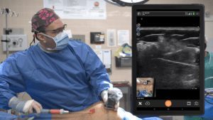
Dr. Edwards uses the Clarius C7 HD3 Vet, which is specifically designed for clinical imaging of small and medium-sized animals. Our Advanced Veterinary Package offers more flexibility for users who need additional workflows for various animal exams.
To learn about how easy and affordable it is to add Clarius handheld ultrasound to your veterinary practice, visit our Clarius veterinary ultrasound page for product details. Or contact us today to discuss which scanner is right for your veterinarian practice.

