[VIDEO] World-Renowned Plastic Surgeon Shares His Ultrasound Secrets
When Dr. Marc Salzman recorded ultrasound-guidance training videos for Clarius, he didn’t realize that his expert techniques would help surgeons worldwide gain confidence in new procedures.
I’ve had people from all over the world come up to me at meetings and say, “Thank you very much; I learned how to do a PEC block from your video”, says Dr. Salzman. “I guess it’s possible because we show and explain every step in the teaching videos and if you have the machine, it’s not hard to try. It gives you confidence when you watch another surgeon doing it.”
A double-board certified plastic surgeon with more than 30 years of experience, Dr. Salzman dedicates a great deal of his time outside his plastic surgery practice teaching his innovative techniques to residents, physicians, nurses, and paramedical cosmetologists. We are excited he will be sharing his expertise with us during a new webinar on October 23: Ultrasound-Guided Fat Grafting: Safe Techniques for BBL and Breast Augmentation. Register now to learn his proven ultrasound-guided techniques for improving the safety of Brazilian Butt Lift (BBL) procedures and autologous fat transfer in breast augmentations.
While we were at Dr. Salman’s practice to prepare for the upcoming webinar, we had the opportunity to capture some of his thoughts about how Clarius has made a difference in his practice over the years. We were delighted to hear he recommends Clarius ultrasound to his colleagues.
I always recommend Clarius Ultrasound to people who ask me. I’ve tried all the other ones and it just shines above the others. I like the wireless component to it. I like the choices of the three different models. I like the form factor. I like the fact that it is updated constantly. I haven’t, knock wood, seen one break yet or not be usable. So, I have nothing but positives to say.”
Watch this 4-minute video to hear how Dr. Salzman uses Clarius ultrasound to enhance patient safety.
Wireless, Pocket-Sized, AI-Powered Ultrasound for Plastic Surgery
Clarius delivers the high-definition imaging and performance of traditional ultrasound systems in a highly affordable ultra-mobile scanner. Specialized workflows and dedicated plastic surgery presets deliver fast and clear imaging on your iOS or Android device with an easy-to-use app that’s powered by artificial intelligence. Learn more about why Clarius ultrasound is the leading choice for plastic surgeons. Or talk to an expert about which Clarius scanner is right for your practice.
[WEBINAR] 1st Trimester Ultrasound: Essential Guide for Midwives
The rising popularity of point-of-care ultrasound (POCUS) for obstetrics and midwifery has the potential to improve fetal and maternal outcomes across the globe. It provides the ability to make quicker and more accurate assessments. And in the first trimester, ultrasound assessments by midwives can have a particularly significant impact on the health and well-being of both the mother and the developing fetus.
To address the growing need for ultrasound education, we recently invited midwifery expert and ultrasound educator Carolyn Gegor to present two webinars covering ultrasound essentials for midwives. The first one-hour webinar: Early Pregnancy Care: 1st Trimester Ultrasound Essentials for Midwives, is available now to watch on demand.
Scroll down for some key clinical takeaways and two demonstration videos.
POCUS in Early Pregnancy: Benefits and Cautions
Advantages:
Accessibility: POCUS devices are portable and easy to use, enabling bedside assessments.
Timely Information: Get immediate answers to clinical questions, expediting patient management.
Adequate Training: Hands-on training and understanding ultrasound principles are crucial.
Clear Guidelines: Adhere to established guidelines for POCUS in midwifery practice.
Equipment Maintenance: Ensure proper equipment maintenance for optimal image quality.
Timely Referrals: Recognize limitations and refer patients for specialized care when needed.
1st Trimester Ultrasound: Clinical Applications
Confirming Pregnancy Location:
Intrauterine Pregnancy (IUP): Visualize the gestational sac within the uterus, minimizing the risk of ectopic pregnancy.
Ectopic Pregnancy: Suspect ectopic if no IUP is seen with a positive pregnancy test; requires immediate referral.
Assessing Fetal Well-being:
Cardiac Activity: Document fetal heartbeat using M-Mode for accurate assessment.
Gestational Age: – Early Pregnancy: Use mean sac diameter (MSD) if no embryo is visible. – Later 1st Trimester: Crown-rump length (CRL) provides the most accurate dating. – After 12 Weeks: Biparietal diameter (BPD), abdominal circumference (AC), and femur length (FL) are used.
Identifying Complications:
Early Pregnancy Loss: Empty sac, absent cardiac activity, or lack of embryo development indicate loss.
Subchorionic Hemorrhage: Bleeding between the chorion and placenta; often resolves on its own.
Multiple Gestations: Twins require referral for a comprehensive ultrasound to assess for potential complications.
VIDEO: 1ST Trimester Ultrasound Scan
Watch Shelley Guenther, CRGS, CRCS perform a 1st trimester pregnancy ultrasound using the Clarius C3 HD3 wireless handheld ultrasound scanner in under 3 minutes.
Documentation: A Critical Aspect
Thorough Reporting: Include indication, patient history, exam findings, and follow-up recommendations.
Image Storage: Save images to correlate with the report.
Billing: Proper documentation is essential for billing purposes.
Recording patient data and notes is simple using the Clarius App. You can also record a full-length scan using the new Clarius Procedure Recording Mode. Watch this 1-minute video to learn more.
A 1st-trimester ultrasound scan is a powerful tool for midwives, enabling early detection of complications and accurate pregnancy dating. Integrating POCUS into your practice responsibly can enhance the quality of care you provide to both mother and baby. With some training, adherence to guidelines, and timely referrals, you’ll maximize the benefits of POCUS in early pregnancy care.
Get An Instant Window into the Health of Mothers and Their Fetuses with Clarius Ultrasound
Ideal for first visits, quick check-ups, and when you need to investigate pelvic pain, pre-term labor symptoms, and fetal viability, the Clarius C3 HD3 convex and EC7 HD3 endocavity scanners help deliver the image quality you need for a confident assessment.
Physicians on the Road for AAOM’s Mission Brigade to Cancun Use Clarius Scanners for Training and Patient Care
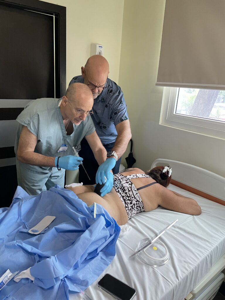
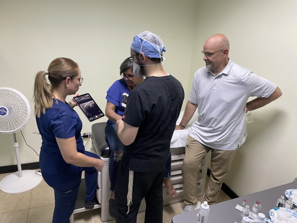
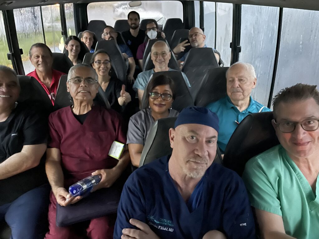
We were delighted to support the AAOM’s (The American Academy/Association of Orthopedic Medicine) 2024 international mission to Cancun with the loan of Clarius handheld ultrasound scanners. 12 instructors and 33 students recently returned after an intense week of learning, teaching, and healing patients suffering from musculoskeletal injuries and diseases.
The integration of Clarius ultrasound machines into the brigade’s mission was a pivotal element in achieving success,” reports Amy M Bucaida, Executive Director of AAOM. “These devices not only enhanced the educational experience but also played a critical role in patient care. The 12 hours of pre-training ensured that everyone was well-prepared to use the equipment effectively. Over the four days of patient care, the Clarius machines provided crucial guidance for evaluating, diagnosing, and administering treatments, making the process more precise and efficient. This hands-on experience with advanced technology undoubtedly contributed to the overall effectiveness of the brigade’s efforts and the quality of care provided.”
The team treated more than 300 patients and participated in educational workshops on preventive care and techniques they would use at home in their practices. The workshop’s curriculum focused on integrating ultrasound for patient evaluation and diagnosis, offering physicians valuable hands-on experience in pain management treatments, guided by experienced AAOM instructors. The provision of free medical care to patients was a significant aspect of the course, addressing chronic pain conditions that might otherwise remain untreated.
The course had a profound impact both on the participants and the patients they served,” Amy adds. “The blend of hands-on skills development in interventional regenerative orthopedic medicine, also known as IROM, with the personal satisfaction of making a difference in the patients’ lives creates a powerful experience. The moments of connection with patients and their gratitude are memorable and often stay with workshop participants beyond the technical skills acquired.”
If you’re interested in joining AAOM on a future brigade, let them know by completing this form.
Short-term Equipment Loans for Clarius Global Health Program
If you’re interested in borrowing Clarius scanners for a free short-term for a global mission, contact us to learn more.
Clarius offers three wireless handheld ultrasound scanners for pain management. Book a virtual demo with a Clarius expert to learn which Clarius wireless scanner is right for your practice.
Clarius Marks Ten Year Anniversary
When Clarius founder, Laurent Pelissier met with another ultrasound industry veteran Dave Willis 10 years ago, he had the glimmer of an idea for an ultrasound system that could be carried in a clinician’s pocket. By the end of their dinner together, they had come up with a rough sketch for a wireless ultrasound system on the back of a napkin. And a few weeks later, they had sourced some funding and assembled a team of five trusted engineers to prove that the concept of handheld ultrasound systems using smart phones for control and display could work.
It’s gratifying to look back at our many accomplishments since our humble beginnings in a small office working on an exciting project,” says Laurent Pelissier who continues to focus on ultrasound innovation as the Chief Strategy Officer at Clarius. “It’s a privilege to work with a great team of almost 150 people sharing the same vision to make ultrasound use ubiquitous.”
10 Years of Global Growth
Now, Clarius is the only handheld ultrasound company that offers 11 specialized AI-powered wireless ultrasound scanners for human and veterinary applications. Its products are sold in 96 countries and trusted by clinicians across 64 specialties. More than 28,000 scanners have been manufactured and used for almost 7 million scanning sessions during the last ten years.
In 2016, Clarius was the first to introduce a high-definition wireless ultrasound system that operated with both Apple and Android smart devices.
Since 2016, Clarius has introduced three generations of handheld wireless ultrasound scanners and several unique options for various clinical specialties. Its research platform enables affordable clinical research and its open software platform empowers AI-enabled ultrasound imaging innovation with numerous technology partners.
Clarius C3 Clarius C7 Clarius L7 Clarius EC7 Clarius C7 Vet Clarius L7 Vet
Clarius C3 HD Clarius C7 HD Clarius PA HD Clarius L7 HD Clarius L15 HD Clarius EC7 HD Clarius C7 Vet HD Clarius L7 Vet HD Clarius L20 HD (2019)
Clarius C3 HD3 Clarius C7 HD3 Clarius PA HD3 Clarius L7 HD3 Clarius L15 HD3 Clarius L20 HD3 Clarius EC7 HD3 Clarius C7 Vet HD3 Clarius L7 Vet HD3 Clarius C3 Vet HD3 Clarius PAL HD3 (2024)
Number of Scanners
6
8 (9 in 2019)
10 (11 in 2024)
Ultrasound Anywhere
As we mark our tenth year in business, Clarius is stronger than ever and we are excited about the positive impact our AI-powered ultrasound innovation will have on patient care in the years to come,” says Ohad Arazi, President and CEO of Clarius. “We’re getting ever closer to the day when every clinician who needs to see inside a patient’s body to make a fast and accurate diagnosis, will simply reach into their pocket and start scanning.”
Follow our Journey
Clarius is only getting started. Follow us on social media and be the first to know what we do next.
[WEBINAR] Mastering Right Ventricular Assessment: A Guide to Cardiac POCUS
Pulmonary embolism and other causes of right heart strain are often part of the differential diagnosis for patients presenting with chest pain or dyspnea. Relying solely on standard blood tests and radiography protocols can delay diagnosis, potentially leaving patients without timely, life- and function-saving treatment. A quick point-of-care ultrasound (POCUS) exam can make all the difference.
Dr. Tom Cook, an emergency physician and ultrasound educator, demonstrates the utility of POCUS to evaluate right ventricular function in patients with chest pain or dyspnea during a one-hour webinar that is available on-demand: Cardiac POCUS Part Two: Techniques for Assessing Right Ventricle Function.
Not ready to commit to a one-hour webinar? Scroll down for highlights of the discussion.
Understanding Right Heart Pressure and Dysfunction
Right ventricular dysfunction can be a critical finding in patients presenting with chest pain or dyspnea. The causes of elevated right heart pressure include:
Left ventricular failure (most common chronic cause)
Pulmonary embolism (most common acute cause)
Isolated pulmonary hypertension
Right ventricular infarction
Pulmonary valve disease
Recognizing these potential aetiologies is crucial for accurate diagnosis and timely treatment.
Key POCUS Techniques for Right Ventricular Assessment
1. Apical Four Chamber View
This view provides an excellent comparison between the right and left ventricles. The right ventricle should be smaller than the left in a normal heart. An enlarged right ventricle compared to the left can indicate right heart strain.
Watch this 2-minute video to see a demonstration of this advanced cardiac view that enables direct left and right ventricle function and comparison and mitral and tricuspid valve regurgitation analysis. Sonographer Shelly Guenther uses the Clarius PA HD3 high-definition wireless scanner for the demonstration.
2. Color Doppler Assessment
Evaluating tricuspid regurgitation using color Doppler can provide valuable information:
Flow convergence zone: The larger this area, the more pathological the regurgitation
Vena contracta: The width of the regurgitant jet as it passes through the valve
Jet area: The size and direction of the regurgitant jet in the right atrium
3. Coanda Effect
An eccentric jet that hugs the wall of the right atrium (Coanda effect) indicates a higher degree of pathology compared to a central jet.
4. Quantifying Pulmonary Artery Pressure
The color Doppler pattern of tricuspid regurgitation can be used to estimate pulmonary artery pressure. This non-invasive method can quickly provide crucial information about right heart function.
This measurement assesses the movement of the tricuspid annulus towards the apex during systole and is a reliable indicator of right ventricular function.
These findings, combined with the patient’s history, led to a diagnosis of chronic pulmonary hypertension.
Watch the full webinar to learn more about how integrating POCUS into clinical practice can significantly enhance patient care in emergency and critical care settings. Dr. Cook provides detailed insights to help you rapidly assess right ventricular function, quickly identify right heart dysfunction, estimate pulmonary pressures, and make timely, potentially life-saving diagnoses.
Clarius for Emergency Medicine: Affordable and Easy to Use Anywhere
Dr. Cook primarily uses the Clarius PAL HD3. Wireless and app-based, Clarius ultrasound delivers fast imaging and sharp detail at the bedside. Visit our emergency medicine page to learn more about which scanner is suitable for your practice. Or book a virtual demo with a Clarius expert in your region.
[WEBINAR] How Ultrasound is Proving to be a Gamechanger in Oncoplastic Breast Surgery
Recent advancements in breast cancer surgery have highlighted the critical role of ultrasound technology in enhancing precision and improving patient outcomes. A review of published studies reveals compelling evidence supporting the use of ultrasound throughout the surgical journey – from preoperative planning to postoperative care.
Dr. Muneer Ahmed, a leading, innovative, and enthusiastic breast cancer surgeon practicing in the United Kingdom, is a passionate ultrasound advocate and shares his best practices for improving outcomes for breast cancer patients, in a one-hour webinar: Enhancing Precision in Oncoplastic Breast Surgery: The Role of Ultrasound in Pre-, Peri- and Postoperative Assessment. Watch the webinar recording at your convenience to learn how easy and affordable it is to add handheld ultrasound to your practice to improve oncologic effectiveness and cosmetic results. Scroll down for some highlights of the presentation.
Preoperative Applications for Handheld Ultrasound
Surgical Planning Ultrasound proves invaluable in surgical planning, allowing surgeons to:
Precisely locate lesions
Determine optimal incision sites
Plan appropriate oncoplastic techniques
By scanning patients in the clinic, surgeons can visualize the exact location of tumors, facilitating more informed decisions about surgical approaches.
Intraoperative Uses
Real-Time Visualization Intraoperative ultrasound offers surgeons real-time visualization of tumors during excision. This approach has demonstrated several advantages:
Eliminates the need for fine wire localization
Provides greater logistical flexibility
Allows for precise tumor removal with adequate margins
Margin Assessment Surgeons can perform ex-vivo margin assessment immediately after tumor excision, potentially reducing re-excision rates.
Postoperative Care
Ultrasound continues to play a crucial role postoperatively:
Assessing and managing seromas or abscesses
Evaluating skin flap thickness for implant-based reconstruction
Monitoring response to neoadjuvant chemotherapy
Learning Curve and Accuracy
Encouragingly, the learning curve for surgeons to become proficient in intraoperative ultrasound is relatively short. Studies suggest that surgeons with prior ultrasound experience may only need about 5 supervised cases to achieve competency. Research indicates that surgeon-performed ultrasound in outpatient settings shows high concordance with radiologist interpretations, with discordance rates below 4%.
The integration of ultrasound technology throughout the breast cancer surgical journey offers numerous benefits. From enhancing surgical precision to improving postoperative care, ultrasound is proving to be an indispensable tool for modern breast surgeons. As more surgeons adopt this technology, we can expect to see continued improvements in patient outcomes and satisfaction.
Video Demonstration: Intraoperative Breast Cancer Clip Localization with Ultrasound
Watch this 3-minute video to see Dr. Ahmed use high-resolution ultrasound in the operating room to locate multiple marker clips in a patient with breast tumours to reduce the excision of margins intra-operatively.
About Dr. Muneer Ahmed
Muneer Ahmed is a leading, innovative, and enthusiastic breast cancer surgeon. He believes in enhancing patient experiences through the application of minimally invasive techniques for bespoke breast cancer management. He trained at the most advanced and comprehensive breast surgical oncology and reconstructive centres in the UK, Europe, and Japan.
Clarius Ultrasound for Breast Surgery
Dr. Ahmed uses the Clarius L15 HD3 high-frequency linear scanner at his practice with the Advanced Breast Package, which offers additional customizations for breast examinations and procedures. The software package is included when a membership is purchased with the Clarius L7 HD3 or Clarius L15 HD3 linear scanners.
To learn which Clarius scanner is right for your practice, book a demo today with a Clarius expert.
[VIDEO] Ultrasound-Guided iPACK Injection for Posterior Knee Pain
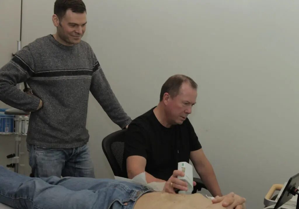
John Marino was somewhat skeptical when he came to see Dr. David Rosenblum about his posterior knee pain. Previous treatments including knee surgery hadn’t delivered the results he had hoped for. But he sounded optimistic when we spoke to him shortly after an ultrasound-guided pain injection by Dr. Rosenblum.
This was quick. Nothing I’ve done before was done on the same day, including imaging my knee. Right now, my knees don’t feel any pain,” he says after the treatment.
Dr. Rosenblum chose to perform an iPACK (infiltration Between the Popliteal Artery and Capsule of the Knee) block on John. Watch the following 2-minute video to see how he locates the popliteal artery and precisely guides his needle using the Clarius L7 HD3 wireless ultrasound scanner.
The Clarius ultrasound gives me a lot of confidence when performing procedures. It also inspires confidence in my patients,” says Dr. Rosenblum. “My patients see that I’m using a newer piece of tech, not using an archaic machine that some of the other physicians are using and it makes them feel more comfortable because they know I have the best technology to help me treat my patients. They tend to look at what I’m doing as something cutting-edge and high-end.”
Watch this 1-minute video to see why John thinks his treatment was successful.
Take the Guesswork out of Pain Management with Clarius Ultrasound
Affordable and easy to learn and use, Clarius wireless ultrasound scanners are the leading choice for pain management specialists looking for the best image quality.
If you’re interested in joining our growing community of users, please visit our wireless ultrasound for pain management page to learn more about which Clarius scanner is right for your practice. Or contact us today for a virtual demo to help you select the best Clarius scanner for your practice.
[WEBINAR] Ultrasound-Guided Techniques for Rapidly Dissolving Misplaced Facial Fillers
Facial fillers have become increasingly popular in aesthetic medicine, but complications can arise when they are misplaced or have migrated. Fortunately, ultrasound technology has revolutionized the way practitioners identify and treat these issues, offering a safer and more efficient approach to filler dissolution.
Dr. MJ Rowland-Warman, founder of Smileworks Hub and clinician director at Smileworks in Liverpool, considers ultrasound the most groundbreaking development in aesthetic medicine since the invention of filler. She practices non-surgical aesthetics with a special interest in the management of dermal filler complications using ultrasound.
According to Dr. MJ, ultrasound has emerged as a game-changing tool in the field of aesthetic medicine. It allows practitioners to visualize facial anatomy beneath the skin, identify misplaced fillers, and guide treatment with unprecedented precision and improved safety. This technology is particularly valuable for addressing one of the most common complications in aesthetic procedures: misplaced facial fillers.
Understanding Misplaced Fillers
Misplaced fillers occur when the product is injected into an incorrect plane or location, or when it interferes with adjacent structures. This can happen immediately during treatment or develop over time due to tissue movement. While some practitioners may overlook minor misplacements, these issues can cause significant distress for patients and potentially damage the reputation of the aesthetic industry.
2. Targeted Treatment: With ultrasound guidance, hyaluronidase can be injected directly into the filler deposit.
3. Reduced Hyaluronidase Usage: Ultrasound-guided techniques typically require less hyaluronidase compared to traditional “flooding” methods, minimizing potential side effects.
4. Real-Time Visualization: Practitioners can observe the dissolution process in real-time, ensuring complete treatment.
5. Improved Patient Outcomes: Patients experience fewer interventions, faster resolution of complications, and better overall results.
The Dissolution Process
When performing ultrasound-guided filler dissolution, practitioners typically follow these steps:
1. Scan the Area: Use the ultrasound to identify the misplaced filler and surrounding anatomy.
2. Prepare for Injection: Utilize an in-plane technique, aligning the needle with the ultrasound probe for optimal visibility.
3. Inject Hyaluronidase: Administer small, targeted doses of hyaluronidase directly into the filler deposit.
4. Monitor Dissolution: Observe the filler taking on a characteristic “cotton wool” appearance as it dissolves.
5. Reassess and Repeat: If necessary, administer additional doses until the filler is completely dissolved.
Case Studies
Dr. MJ Rowland-Warmann, a renowned expert in aesthetic medicine, shared two compelling cases demonstrating the effectiveness of ultrasound-guided filler dissolution:
Tear Trough Complication
A patient presented with bilateral interorbital swelling 18 months after tear trough filler treatment. Ultrasound revealed misplaced filler and associated hypervascularity. Targeted dissolution provided swift resolution. Watch this video for her case review and demonstration.
Forehead Filler Complication
A patient experienced severe symptoms after receiving forehead filler from an unlicensed practitioner. Ultrasound-guided dissolution offered relief when previous attempts had failed. Watch this video for her case review and detailed demonstration.
Clarius Handheld Ultrasound: The Leading Choice for Aesthetic Professionals
As Dr. MJ demonstrates during the webinar, ultrasound-guided techniques for dissolving misplaced facial fillers represent a significant advancement in aesthetic medicine. She has been using the Clarius L20 HD3 handheld ultrasound at her practice for three years and says using it for filler treatment “is a godsend, whether that be before treatment to vascular map, during treatment to place filler using ultrasound guidance, or after treatment to confirm placement.
Wireless and pocket-sized, Clarius handheld ultrasound scanners deliver the high-definition imaging and performance of traditional ultrasound systems for a small fraction of the cost. They are the leading choice for plastic surgeons and aesthetics practitioners performing ultrasound-guided procedures to ensure patient safety.
New T-Mode™AI by Clarius is a groundbreaking educational technology to help clinicians new to ultrasound advance their image interpretation skills using Clarius handheld scanners.
Visit our aesthetics page to learn more or request a virtual ultrasound demo today to learn how high-definition ultrasound imaging with voice controls can improve safety and deliver consistent patient outcomes at your aesthetic practice!
[VIDEO] Expert Insights: Dr. Steven Weiner on Using Ultrasound for Aesthetic Injections
Dr. Steven Weiner, a facial plastic surgeon from Santa Rosa Beach, Florida has been a proponent of ultrasound use for safer procedures in the aesthetics community since he experienced the benefits in 2019. We were delighted to have the opportunity to interview him during this year’s Aesthetics Meeting about his journey. Read on for highlights of our conversation or watch this 5 minute video.
How has your use of ultrasound evolved?
I was introduced to ultrasound when I went to Amsterdam and learned from my mentor, Leonie Schelke, and the Cutaneous group. I did that in 2019, so I’ve been using ultrasound for about five years. I use it for pre-mapping, finding where the arteries are or aren’t, prior to injecting in the temples, in the deep piriform area. And I also use it to look at patients beforehand, that have had previous fillers to see what fillers they had and where they’re placed. But I also use it a lot for complications. Swelling around the eyes, lumps and bumps, pain, discomfort. And last but not least, I use it a lot for vascular occlusions where the patient is in distress because the artery has been injected with filler. ”
When did you start using Clarius ultrasound?
I learned about the L20 in 2020 from Dr. Marc Salzman, I think he was doing a webinar. I thought it was a great tool for the average injector. It wasn’t too expensive, it was very easy to learn because a lot of the knobology, the changes in the controls were very similar to the iPhone. And it’s on an iPad and the photos are very interchangeable with your iPhone and Mac. They make it really easy for a new user. And then, obviously, with the new AI, it’s made it even easier for the person who has no experience. ”
“I didn’t have any experience using ultrasound in 2019. And yeah, it takes a long time. In fact, I’m still learning, but the Clarius kind of dumbs it down a little bit, so that almost everyone can at least use it. And in my trainings, I tell people… And I do the trainings almost exclusively with Clarius, I tell them that you’re not going to be an expert by the time this day’s over, but I can tell you you’ll be able to use the Clarius in your office by the time you’re done. And it’s an eight-hour course, and by God, most can use it.”
Any thoughts on the Clarius T-Mode AI for helping clinicians who are new to ultrasound?
The new T-Mode is really good. It’s actually very accurate, which was surprising. And it’s particularly good in the temple anatomy because there’s 10 layers of the temple and it’s very difficult for people to understand that. But with the T-Mode, you throw it on the temple and you automatically see the layers, it even labels them for you if you press the freeze button, pause button. It’s also great for lips and cheeks. So, it’s a great learning tool. I think that when you’re using it on a patient, patients love to see first, that you care about their safety, but number two, that you can teach the patient too, about the different layers. And they’re intrigued with that.”
“And you can show them abnormalities or variations in anatomy of them versus what normally exists as well. And then you can show them the filler before and after, what filler she had before and then your fillers afterwards. That’s very interesting to them and they appreciate the extra time that you take. It doesn’t take a lot of extra time, but if you have a few moments, particularly your well-educated patients or your long-term patients, they want to see that and they really understand that you have taken a lot of time and effort to learn, to purchase a device, and I think it just takes your practice to a different level. ”
What do you like best about using Clarius ultrasound?
Keeping with the iPhone type of mantra, it’s just intuitive. You want to enlarge it, you do this (pinch out), you want to change the depth, you just slide. One of the advantages that really isn’t talked about much, is that since it is very compatible with the iPad, you can do a screen recording. So instead of hooking up a whole different graphics card when I’m using some other device, you can just screen record on your iPad and you get 15, 30 minutes of recording. Let’s say you’re taking care of a guided injection of a complication, or so forth. On the other devices, without that, you only get around 15 second increments, but if you screen record, you can get 15, 30 minutes.”
“They’re also basically indestructible. You could throw it into a fish tank for 48 hours and then pull it out and use it. So that’s a pretty neat feature too. And you can wipe it with anything and you’re not going to destroy it. I’ve been carrying it around, through the airports, in my backpack, and everything has worked fine. They’re indestructible. The ones that you get in your office are much more delicate and you have to watch what you clean them with, and if you drop it, they don’t function. I haven’t dropped the Clarius, but I’m sure it would do fine.”
Watch this Free On-Demand Webinar to Learn Dr. Weiner’s Ultrasound Techniques for Safer Injections
We invite you to watch a popular webinar from our archives during which Dr. Weiner demonstrates step-by-step techniques for performing safer filler injections by using ultrasound to limit vascular complications. You’ll find the webinar here.
A renowned educator, Dr. Weiner is the founder of Sonosthetics, an ultrasound training course for aesthetic injectors. He is a board member of CMAC (Complications in Medical Aesthetics Collaborative) which educates on the prevention and management of filler complications. Dr. Weiner is also the Ultrasound Director of the Academy for Injection Anatomy, a monthly 2-day course on cadaver and ultrasound anatomy and injection techniques.
Improve Patient Safety with High-Definition Ultrasound
Dr. Weiner uses the Clarius L20 HD3 ultra-high frequency wireless handheld ultrasound scanner for facial aesthetic procedures. Designed to provide extremely high image quality in the near field, from the skin line to 4 cm, the Clarius L20 HD3 is ideal for a variety of clinical settings requiring superior superficial imaging. It is the only handheld ultrasound with ultra-high frequency of 20 MHz. Wireless and affordable, it delivers exceptional superficial imaging with an easy-to-use app for your iOS or Android device.
[VIDEO] Why Dr. Pazmiño Chooses Clarius Ultrasound: Five Key Benefits for Plastic Surgery
Dr. Pat Pazmiño is nationally renowned for inventing new procedures that combine the latest techniques in aesthetic surgery with ultrasound, such as the ultraBBL™, for safe Brazilian Butt Lift (BBL) procedures. A vocal fan of Clarius ultrasound, he recently sat down with us to discuss why he prefers to use and recommend Clarius for plastic surgery. Watch this 4-minute video or scroll for highlights.
1. AI-Powered Clarius Is Simple to Use for New and Experienced Users
One of the reservations that surgeons sometimes have about ultrasound is that they feel that it’s an unknown technology. They feel that there’s a big learning curve, and as surgeons, we just want to focus on the patient.”
“One of the strengths about the Clarius system is that it’s not just ultrasound, it’s smart ultrasound. It allows us to go ahead and use that technology to optimize the settings to get the best image possible so that way we can really focus. And I think that’s been one of the big strengths with the Clarius system, that with the artificial intelligence that they use in their software, it helps optimize the image. So that way surgeons, we can really focus on the patient, on the procedure, and not worry so much about playing with different settings, turning knobs, it really makes it much more effortless.”
2. Clarius Makes Learning Ultrasound Easier
Some surgeons find ultrasound intimidating; they don’t understand what they’re seeing. And Clarius has done an incredible job with the T-mode, which helps with education because for the first time in real time, you can actually see a color-coded image of what you’re looking at on the screen.”
“I have used the T-mode in training, and it really is eye-opening for the doctors because when they see it, it clicks. They finally understand what is on the other image, which can look like a TV full of static. But the T-mode really helps identify the anatomy and it really puts surgeons at ease.”
3. Clarius Tailors Its Systems for Specialty Applications
I consider Clarius a leader in medical imaging, especially for plastic surgeons. I think that they have a high-quality piece of equipment, but more importantly, the team is very responsive; they understand what our needs are, and they’ve really worked very diligently at meeting them. Years ago, I worked with Clarius to create the BBL mode, and that was the first time I had seen that happen in medical imaging. It was a mode specifically for a procedure that was designed to be performed with ultrasound. And because of that, it allows surgeons to do this safer, more effectively, and faster.”
“There are systems that are very expensive, fifty thousand to a hundred-thousand dollars. But none of them are portable and none of them have a great image. And those systems do not have the modes that specifically help us in surgery, help us identify and understand the images.”
4. I Can Carry Wireless Ultrasound Everywhere
A wireless system is very important for surgeons because number one, when I’m in the clinic, I can keep it in my pocket and I can pair it with my iPhone, with an iPad, so it goes everywhere with me. I can bring it up easily in the middle of a conversation with a patient, and it helps me get information so I can make decisions and start treating them.”
“Of course, there are some cart-based systems that have better image quality for certain applications. But the best ultrasound is the one that is with you and that you can use at any time. And I think that’s the strength of Clarius.”
5. I Can Enclose It in a Sterile Bag for Surgery
A wireless system is even more important in the operating room because in the operating room, we can put a Clarius wireless system completely into a sterile bag. And what that allows us to do for the first time, we can bring ultrasound onto the operative field. We can bring ultrasound into the OR, and it allows a surgeon for the first time to see under the skin while they are working.”
Clarius Ultrasound for Safer Plastic Surgeries
Wireless and pocket-sized, Clarius delivers high-definition imaging devices with an easy-to-use app that’s powered by artificial intelligence. Our Advanced Aesthetics package, included with membership, offers customized workflows for confident plastic surgery examinations and procedural guidance.