After following a few practitioners, I decided to choose Dr. Zainab because she seemed like an incredibly knowledgeable specialist, and also because she was using ultrasound to make every single procedure very safe,” says Ruxandra.
Dr. Zainab has a decade of experience in facial aesthetics, which she added to her aesthetic dentistry practice. She has been using Clarius ultrasound since 2021 and uses the Clarius L20 HD3 ultra-high frequency scanner.
I feel a lot more confident and comfortable with the practitioner knowing that they’re using an ultrasound before the filler procedure to understand where they need to inject. I am very happy with the results of the treatments.”
Watch this 3-minute video to see Dr. Zainab in action and to learn more about Ruxandra’s procedure.
Confirming Filler Placement Post-Injection
Dr. Zainab scans her patients to confirm safe filler placement after a procedure. Watch this 2-minute video to see her scan the filler in Ruxandra’s nose.
On-Demand Webinar: Dr. Zainab’s Technique for the Ultrasound-Guided Nonsurgical Rhinoplasty
Dr. Zainab recently presented a 1-hour webinar where she shared her technique for planning safe injections in the nose by identifying anatomical structures, including blood vessels in advance of the procedure. Watch the webinar at your convenience to learn her safer approach to nonsurgical rhinoplasty.
Free Webinar
Ultrasound for Nonsurgical Rhinoplasty: Avoiding Vascular Complications and Improving Results
New to Ultrasound? Gain Confidence with Clarius T-ModeTM AI
The new Clarius T-ModeTM AI is a groundbreaking educational technology to help aesthetic clinicians advance ultrasound interpretation skills using Clarius wireless handheld scanners. Powered by artificial intelligence (AI), T-Mode AI enhances a grayscale image in real time using distinctive colors, patterns, and labels, reminiscent of illustrations found in anatomical textbooks, so you’ll quickly learn how to identify anatomical tissues and structures during an ultrasound exam.
[VIDEO] Patient Perspective on Aesthetic Ultrasound: “It Gives You the Extra Safety Blanket”
As the head of a social media agency in the medical and dental industry, Chelsea Stewart had heard good things about Dr. Zainab Al-Mukhtar before she booked her first appointment for aesthetic treatments three years ago at Harrow on The Hill Dental and Facial Aesthetics where Dr. Zainab is the clinical lead in facial aesthetics.
Chelsea sees Dr. Zainab once or twice yearly for “tweaks to refresh and rejuvenate and hydrate my face.” During Chelsea’s recent visits, Dr. Zainab has been using ultrasound.
She has always got the latest equipment and has been using ultrasound, which I think improves the treatment because it’s a safer approach,” Chelsea explains. “Working within the medical field, I’ve seen cases of other patients in other practices where people have had necrosis of the nose and that is a big fear of mine is something going wrong where it starts to erode at your skin and can leave further complications down the line. So, having that ultrasound, it gives you that extra safety blanket.”
Watch this 3-minute video to learn more about Chelsea’s experience.
Pre-Procedure Vascular Mapping of Chelsea’s Nose
Dr. Zainab performed a successful nonsurgical rhinoplasty for Chelsea during her last visit. Watch this 3-minute video to see Dr. Zainab confirm the presence and location of vascular structures in the radix of the nose prior to injection, using high-resolution ultrasound and color Doppler.
Free On-Demand Webinar: Learn Dr. Zainab’s Technique for Ultrasound-Guided Nonsurgical Rhinoplasty
Dr. Zainab recently presented a 1-hour webinar where she shared her technique for planning safe injections in the nose by identifying anatomical structures, including blood vessels. Watch the webinar to learn her safer approach to nonsurgical rhinoplasty.
Free Webinar
Ultrasound for Nonsurgical Rhinoplasty: Avoiding Vascular Complications and Improving Results
Get Better Results with Ultra-high-definition Ultrasound for Facial Aesthetics
Dr. Zainab uses the Clarius L20 HD3 handheld wireless ultrasound scanner to map high-risk areas before a procedure and for ultrasound-guided injections for high-risk procedures. It’s the only specialty-designed handheld ultrasound with an ultra-high frequency of 20 MHz. Wireless and affordable, it delivers exceptional superficial imaging to 4 cm with an easy-to-use app for your iOS or Android device.
If you’re new to using ultrasound, gain confidence in your scanning skills with Clarius artificial intelligence (AI) tools such as the new Clarius T-Mode AITM. It’s a groundbreaking educational technology designed to help aesthetic clinicians advance ultrasound interpretation skills using Clarius wireless handheld scanners. Clarius T-Mode AI enhances a grayscale image in real time using distinctive colors, patterns, and labels, reminiscent of illustrations found in anatomical textbooks, so you’ll quickly learn how to identify anatomical tissues and structures during an ultrasound exam.
[VIDEO] Why an Aesthetic Clinician Chose Not to Inject Her Patient Following a Pre-Procedure Aesthetic Ultrasound Exam
28-year-old Mimoza, born and bred in London, UK, has never liked her nose and was excited about having a nonsurgical rhinoplasty.
Dr. Zainab used an ultrasound during my consultation and proved that the veins at the side of my nose are not in the way and she could perform the nonsurgical rhinoplasty whereas other aestheticians that didn’t use ultrasound immediately said no,” explains Mimoza. “She likes my nose the way it is, which we agreed to disagree on because I’ve always been self-conscious of my nose. I feel very confident and comfortable getting this treatment done by Dr. Zainab because she can see exactly what’s under the skin.”
Pre-Procedure Vessel Mapping Identifies Danger Zone
Unfortunately, Mimoza’s hopes were dashed during her second visit to have the non-surgical rhinoplasty performed. Watch this video to see why Dr. Zainab decided it wasn’t safe to proceed after a detailed pre-procedure scan to map the vascularity of Mimoza’s nose.
Patient Perspective on Ultrasound for Aesthetics
Watch this 1-minute video to hear why Mimoza chose to see Dr. Zainab for her treatment.
Learn Dr. Zainab’s Technique for the Ultrasound-Guided Nonsurgical Rhinoplasty
Trained as a dentist, Dr. Zainab Al Mukhtar practiced aesthetic dentistry for 13 years and has been focused on facial aesthetics for the past 10 years. She is a national trainer in aesthetic injectables and owns a clinic called Harrow on The Hill Dental and Facial Aesthetics where she is the clinical lead in facial aesthetics.
Dr. Zainab recently presented a 1-hour webinar where she shared her technique for planning safe injections in the nose by identifying anatomical structures, including blood vessels. Watch the webinar at your convenience to learn her safer approach to nonsurgical rhinoplasty.
Improve Safety with Ultra-high-definition Ultrasound for Facial Aesthetics
Dr. Zainab uses the Clarius L20 handheld wireless ultrasound scanner to map high-risk areas before a procedure and uses it for ultrasound-guided injections for high-risk procedures. It’s the only specialty-designed handheld ultrasound with an ultra-high frequency of 20 MHz. Wireless and affordable, it delivers exceptional superficial imaging to 4 cm with an easy-to-use app for your iOS or Android device.
If you’re new to using ultrasound, gain confidence with the new Clarius T-ModeTM AI, a groundbreaking educational technology to help aesthetic clinicians advance ultrasound interpretation skills using Clarius wireless handheld scanners. Powered by artificial intelligence (AI), Clarius T-ModeTM AI enhances a grayscale image in real time using distinctive colors, patterns, and labels, reminiscent of illustrations found in anatomical textbooks, so you’ll quickly learn how to identify anatomical tissues and structures during an ultrasound exam.
[VIDEO] Google Uses Clarius Open-source Platform to Research AI for Prenatal Ultrasound Exams
Handheld, portable ultrasound devices are changing the way ultrasounds are delivered, bringing this technology to more patients and in more care settings.
When Google initiated a global health initiative to develop technology that would enable more caregivers to perform prenatal ultrasound exams, their development team went searching for handheld ultrasound scanners with Android-compatible software they could work with.
According to Kris Dickie, Clarius’ Chief Technology Officer, although the original Clarius research SDK didn’t meet Google’s specifications, they quickly began working on a more flexible platform.
We developed an SDK that ran specifically on Android that would allow them to build software that runs alongside the Clarius App,” he explains.
Once the Android SDK was available, the Google team purchased the Clarius C3 curvilinear scanner with the Clarius Clinical Research package, which provides access to raw data collected internally and custom software for real-time analysis.
Google’s research was recently featured in a BBC report filmed in Kenya, which highlighted how Google’s ultrasound AI can be used to enable more care givers in remote and under-resourced areas to perform critical prenatal ultrasound exams. Only 10% of mothers in rural Kenya currently have access to ultrasound services.
Google’s AI uses the SDK to access ultrasound captured by the Clarius scanner and transferred to a tablet or smartphone.
Expanding the Clarius SDK Empowers More AI-Powered Ultrasound Innovation
Developing a new and improved SDK spawned additional benefits for Clarius beyond being helpful to Google.
Our expanded SDK has enabled many other companies to develop AI solutions that are improving access to medical imaging for patients including ThinkSono AI and DESKi’s HeartFocus.”
In 2022, Clarius announced Clarius Marketplace, which enables ultrasound innovators to bring their AI-powered software solutions to market faster. The Clarius SDK provides developers with a set of tools to quickly take their solution from code directly to the clinician’s hand without creating custom hardware.
Now, through the Clarius Marketplace, clinicians who purchase a Clarius scanner with the Clarius Membership have access to new third-party ultrasound innovations, designed to improve workflows, streamline training, help with faster diagnoses, and automate reporting.
Get An Instant Window into the Health of Mothers and Their Fetuses with Clarius Ultrasound
Point-of-care ultrasound is recommended as a “routine extension of practice for most OB/GYN clinicians.” Ideal for first visits, quick check-ups, and when you need to investigate pelvic pain, pre-term labor symptoms, and fetal viability, the Clarius C3 HD3 convex and EC7 HD3 endocavity scanners help deliver the image quality you need for a confident assessment.
[VIDEO] Why an Experienced Rheumatologist Calls Ultrasound His “Truth Machine”
Ultrasound has been a passion for Dr. Mohammed Bardi, a rheumatologist practicing in Vancouver for seven years. After completing his rheumatology training at the University of British Columbia, he pursued additional training with the American College of Rheumatology and then trained in Norway and the UK. Now, he considers ultrasound a natural extension of his physical exam.
Some people have an ultrasound day or a clinic, but the way I’ve set up my practices, my ultrasound is like my stethoscope,” he explains. “It makes practice more rewarding. I sometimes call the ultrasound the truth machine. It’s like the tiebreaker when sometimes if I’m not sure about my diagnosis.”
Beyond the ability to assess anatomy and confirm diagnosis, Dr. Bardi is excited about using ultrasound to improve success with patient adherence to therapy by clearly showing progress using ultrasound clips captured before and after therapy.
We say a picture is worth a thousand words. When I can show patients their old images and their new images and they can see the difference, it really helps one with patient adherence.”
Watch our 4-minute video to learn what he likes about Clarius L15 HD3 wireless ultrasound for rheumatology.
See the Fine Details You Need with Clarius High-Definition Handheld Ultrasound
Delivering the same high-powered imaging as the best cart-based systems, Clarius HD3 ultrasound shows you the details you need to quickly investigate an area of concern for an accurate diagnosis. Watch patient engagement soar with real-time imaging of their progress.
Visit our rheumatology page for more information. Or book a virtual demonstration with a Clarius expert to learn which Clarius ultrasound is right for your practice.
[VIDEO] Physical Therapist Unveils His Patients’ Specific Source of Pain and Tracks Outcomes with Handheld Ultrasound
Having been through residency and fellowship training and annual stints in the cadaver lab, physical therapist Dr. James Escaloni, RMSK, thought he knew anatomy. He only realized what he had been missing when he started incorporating musculoskeletal sonography into his regular practice.
Now that I have this information, I’ve got a greater depth of understanding, so I feel more confident as a clinician, and especially as an educator,” says Dr. Escaloni.
After his interest in sonography was piqued while watching an ultrasound-guided treatment about five years ago, Dr. Escaloni dedicated himself to achieving his RMSK (Registered in Musculoskeletal sonography) certification and now uses ultrasound daily at his chronic pain management practice “to provide the specificity that’s missing from my clinical exam.”
He relies on the real-time, dynamic imaging of ultrasound instead of MRI and X-rays to reveal a patient’s specific cause of pain. “The benefit of ultrasound is that you can actually have the person who you just tested, in front of you; you’ve just poked them on that spot, and they say, « That is the pain. That’s the spot. »”
In addition to identifying the cause of chronic pain, Dr. Escaloni uses ultrasound to guide his dry-needling procedures and monitor progress over weeks of treatment.
There’s been a revolution in physical therapy with dry needling,” he says. “It started as a fringe thing, nobody was doing it, and then all these people were getting these amazing outcomes, and then, all of a sudden, 10 years later, it’s taught in all the PT (physical therapy) schools. Everybody does it, and if you’re not doing that as part of your practice, then you’re not the best clinician.”
Although he had access to a cart-based system at his clinic, James primarily uses his Clarius HD3 wireless ultrasound scanner, especially during a busy day when he carries it in his pocket to use with his phone.
The Clarius system has got a very good image, especially for more superficial tissues and the Clarius voice control features are a fantastic thing for me in the office. It helps me maintain a clean area; I can use voice-activated settings to change the gain and needle mode as I need it. Clarius ultrasound has helped me deliver the best patient care, because I’m able to track outcomes with my patients and show them we’re making progress.”
For those interested in the rigorous scientific research and high-quality randomized controlled trials (RCTs) that Dr. Escaloni and his team have accomplished, you can explore their findings here. Watch this 4-minute video to learn more about Dr. Escaloni’s experience with Clarius.
Watch Webinar for Practical Tips from Two Physiotherapists
FREE WEBINAR
Ultrasound-Guided Dry Needling: Enhancing Precision and Outcomes
Join us for a one-hour webinar with Dr. James Escaloni and Dr. Ian Young to learn their practical tips for using ultrasound to improve physical therapy outcomes.
Dr. Ian Young, DSc, PT, Dip. Osteopractic, is a physical therapist & founder and owner of Tybee Wellness & Osteopractic in Tybee Island, GA. He has been practicing full-time in orthopedics and sports for the last 27 years. Dr. Young is senior faculty within the American Academy of Manipulative Therapy (AAMT) Fellowships in Orthopaedic Manual Physical Therapy & Musculoskeletal Ultrasonography.
Dr. James Escaloni, DPT, FAAOMPT, Dip. Osteopractic, serves as a physical therapist and movement specialist at Wellward Regenerative Medicine in Lexington, KY. He is board-certified, residency, and fellowship trained in orthopedics. In addition to identifying the cause of pain in the most complex patients, Dr. Escaloni teaches around the world on topics like differential diagnosis and manual therapies.
2-Minute Video: USG Dry Needling of the Supraspinatus
Watch Dr. Escaloni’s technique for dry-needling the supraspinatus tendon using the Clarius L15 HD3.
Take the Guesswork Out of Pain management with Clarius Ultrasound
Recognized for the best imaging quality among handheld systems, Clarius HD3 is affordable and easy to use. Now with new features like T-Mode AI and MSK AI, Clarius ultrasound is even easier to learn.
Enhance your skills by attending in-person courses offered by the American Academy of Manipulative Therapy, where Clarius scanners are used to teach diagnostic MSK ultrasonography and ultrasound-guided dry-needling
[VIDEO] Why Handheld Ultrasound Is “the Next Big Thing” for Physical Therapy
During most of his time as a physical therapist, Dr. Ian Young, DSc, PT, Dip. Osteopractic, RMSK relied on palpation, landmarking, and MRI reports to determine and carry out a treatment plan for his patients. Then he had an ultrasound-guided knee injection in an orthopedic office and saw the possibilities for his practice.
I thought this is really cool; I can see the needle go in and where the fluid’s going in and the patella tendon and I can see the joint. It really opened my eyes to the possibility that this was something that I could get into because we do a lot of dry needling and soft tissue treatments that we could monitor before and after,” Ian explains.
He began his ultrasound training journey about seven years ago and after lots of courses and practice, he and his business partners use ultrasound every day on every patient and to teach other physiotherapists.
As you move along over the years and you get the right training, ultrasound is imperative, imperative to my clinical practice. But it was definitely a challenging road to get here. I think that happens with most clinicians when they are just getting started. But once you get it, it’s going to be part of your practice no matter what.”
Dr. Young uses the Clarius L15 HD3 and C3 HD3 wireless handheld ultrasound systems as well as a cart-based system at his cash practice in Savannah, Georgia where he sees 10-15 patients per week for longer and more expensive visits using ultrasound for diagnosis and guided needling. He also uses Clarius scanners for ultrasound courses where he and his team teach 15-45 physiotherapists ultrasound-guided dry needling, musculoskeletal ultrasound, diagnostic musculoskeletal ultrasound for the upper extremity, lower extremity spine, and some regenerative processes.
It’s really new for a lot of the physical therapists around the country, really around the world,” Ian says. “And it’s become an exciting thing because we really think it’s the next big thing for physical therapy, and we want to make it almost just a normal thing to have in your clinic. If you don’t have ultrasound and you’re not doing diagnostic ultrasound, then you’re way behind.”
For those interested in the rigorous scientific research and high-quality randomized controlled trials (RCTs) that Dr. Young and his team have accomplished, you can explore their findings here. Watch this 5-minute video to see what Dr. Young says about his experience using the Clarius wireless handheld ultrasound system and how it compares to his cart-based system.
Watch Webinar for Practical Tips from Two Physiotherapists
FREE WEBINAR
Ultrasound-Guided Dry Needling: Enhancing Precision and Outcomes
Join us for a one-hour webinar with Dr. James Escaloni and Dr. Ian Young to learn their practical tips for using ultrasound to improve physical therapy outcomes.
Dr. Ian Young, DSc, PT, Dip. Osteopractic, is a physical therapist & founder and owner of Tybee Wellness & Osteopractic in Tybee Island, GA. He has been practicing full-time in orthopedics and sports for the last 27 years. Dr. Young is senior faculty within the American Academy of Manipulative Therapy (AAMT) Fellowships in Orthopaedic Manual Physical Therapy & Musculoskeletal Ultrasonography.
Dr. James Escaloni, DPT, FAAOMPT, Dip. Osteopractic, serves as a physical therapist and movement specialist at Wellward Regenerative Medicine in Lexington, KY. He is board-certified, residency, and fellowship trained in orthopedics. In addition to identifying the cause of pain in the most complex patients, Dr. Escaloni teaches around the world on topics like differential diagnosis and manual therapies.
2-Minute Video: USG Dry Needling of the Lateral Elbow
Watch Dr. Young’s technique for guiding his needle to the lateral epicondyle using a Clarius L15 HD3.
Eliminate Guesswork with Clarius High-Definition Ultrasound
Recognized for the best imaging quality among handheld systems, Clarius HD3 is affordable and easy to use. Now with new features like T-Mode AI and MSK AI, Clarius ultrasound is even easier to learn.
Enhance your skills by attending in-person courses offered by the American Academy of Manipulative Therapy, where Clarius scanners are used to teach diagnostic MSK ultrasonography and ultrasound-guided dry-needling
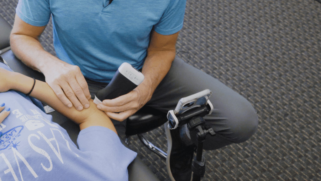
[VIDEO] How Portable Ultrasound Has Elevated Medical Aesthetics
As one of the first injectors in the United Kingdom to adopt ultrasound for facial aesthetics, Dr. Zainab Al-Mukhtar has much to say about how portable ultrasound has revolutionized her field.
Ultrasound has been around in medicine for many, many years but missing in aesthetics because there was no portable option,” she explains when we recently spoke to her at her clinic in London, England.
“The introduction of the portable ultrasounds completely changed that. It means that now within an affordable price, medical aesthetic practitioners can purchase a portable device and are in a fortunate position where there is more training available. I think that it has elevated aesthetic practice a lot. There is an increase in use as a precautionary measure prior to injections. There is an increased use of ultrasound-guided injections where necessary or where it is deemed helpful. There is an increased predictability in the way that we manage complications as an industry and there is also a greater understanding of dynamic anatomy as a result of being able to see the anatomy in real-time.”
A Clarius ultrasound user since 2021, Dr. Zainab recently upgraded to the Clarius L20 HD3 ultra-high frequency scanner and is particularly enthusiastic about Clarius Voice Control, which she claims even impresses her patients.
The voice control is definitely my favorite. I don’t need to have somebody present in the room at all times. I don’t need to have a glove-less hand to press buttons with. I can focus completely on the patient without having to make a mess trying to change things on the screen with my fingers. It’s brilliant!”
Watch our 4-minute video interview with Dr. Zainab to learn why she is a big fan of the Clarius L20 HD3 for facial aesthetics.
Learn Dr. Zainab’s Techniques for Avoiding Complications and Improving Results of Nonsurgical Rhinoplasty
Due to the anatomical nature of the nose, vascular compromise is a potential complication during nonsurgical rhinoplasty procedures. Practitioners like Dr. Zainab use high-resolution ultrasound to gain valuable insight into patients’ vascular anatomy in real-time, making injections safer and more accurate.
Join us for a one-hour webinar with Dr. Zainab Al-Mukhtar to learn her technique for planning safe injections in the nose by identifying anatomical structures, including blood vessels.
Free Webinar
Ultrasound for Nonsurgical Rhinoplasty: Avoiding Vascular Complications and Improving Results
Avoid Complications with Ultra-high-definition Ultrasound for Facial Aesthetics
Medical aesthetic practitioners looking to improve patient safety rely on the Clarius L20 HD3 – the world’s only ultra-high frequency ultrasound in a wireless scanner. Impress your patients with clear imaging of the skin, muscles, vessels, and fascia for safe, consistent outcomes.
[WEBINAR] Guided Treatment for Managing Permanent Facial Filler Complications
A specialist in the management of dermal filler complications, Dr. MJ Rowland-Warmann sees complication cases from other injectors nearly daily and has become a vocal advocate for using ultrasound to improve the safety and effectiveness of aesthetic procedures.
Filler complications are out of control,” says Dr. MJ at the start of the webinar. “Not a week goes by when we don’t see a horror story in the news or on social media about someone’s awful experience with fillers. And these cases are difficult when patients are injured and upset and a resolution is needed quickly. I couldn’t be without ultrasound. My Clarius L20 is a staple in my practice, and I’ve been using it for around two years. It’s an absolute marvel.”
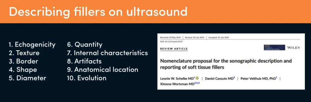
Types of Dermal Fillers and What They Look Like with Ultrasound
There are hundreds of different filler products out there, different types, different brands. Some countries have more products on their market. Some like the US with stricter licensing laws, they have less. On the UK market, there’s an estimated 500 plus filler products. Some are good, some not so much. To be approved medically, the ideal cosmetic filler should be non-toxic, non-carcinogenic, non-immunogenic, and to achieve excellent aesthetic results.
This paper by Schelke and others from 2019 is really helpful because it describes the ultrasound characteristics of dermal fillers using 10 parameters.”
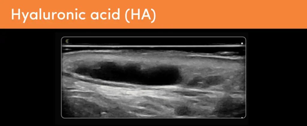
Hyaluronic Acid Filler
Hyaluronic acid filler is undoubtedly the most commonly used type of filler due to its safety and efficacy. Net accounts for about 80% of fillers use worldwide. It’s what you see in your patient’s faces most of the time.
This is HA, it’s anechoic and homogeneous with a well-defined border. HA is usually round or oval in shape. The diameter varies according to the size of the deposits, but generally, per deposit we expect less than 0.5 to a ml, given the size of syringes on the markets. There are no internal reflections. And this tells us the product is liquid or gel-like.
There is a strong posterior enhancement due to the liquid contents, and this amplifies sound waves passing through the filler to the tissues underneath, making them appear whiter. And that’s what you could see there by the white band underneath the filler deposit. This is the classic presentation of HA in tissues. HA filler is extremely consistent over a long period in tissues even years.”
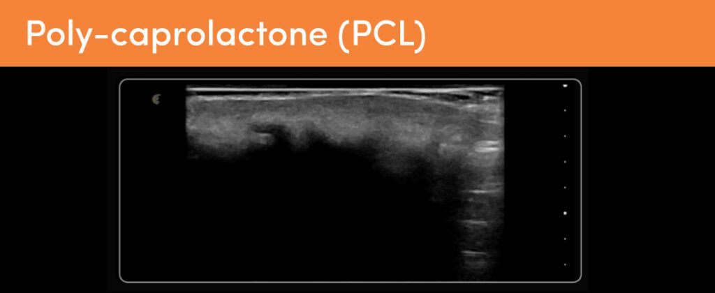
Poly-caprolactone Filler
This is polycaprolactone, often marketed under the trade name Ellansé. It’s a hydrophobic filler that is hypoechoic on ultrasound. It appears homogenous. It’s got a poorly defined border and a band-like shape. PCL creates a significant posterior shadow. It blocks out the tissues underneath. This is the jaw line, and the tissues including the bone under the filler have been completely shadowed. Ellansé is a popular bio stimulator in South America and in Europe, so you will likely see this if you work there. It’s injected in a CMC carrier gel, which resorbs. And the results of PCL last about two to four years. It’s not reversible, but it does degrade over time.”
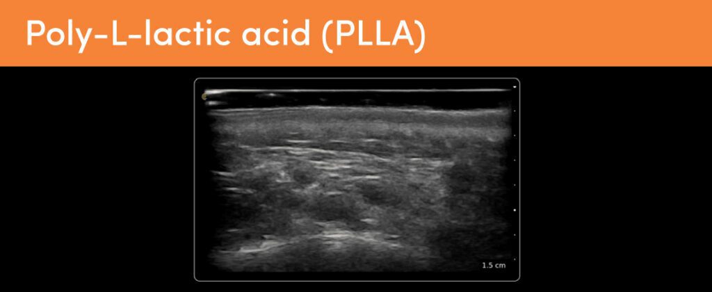
Poly-L-lactic acid (PLLA)
Here we see poly-L-lactic acid. We know it as Sculptra. It’s a hydrophobic bio stimulator. It causes fibrosis, and on ultrasound it’s characterized by widespread hyperechoic patches with slight posterior shadowing, making this diffuse sort of dappled appearance. PLLA is not retained within the tissues at the filler deposit, but instead it causes this fibrotic tissue response. And these kinds of hyperechoic patches are only often really evident when you are comparing it to normal isoechoic fat tissue.”
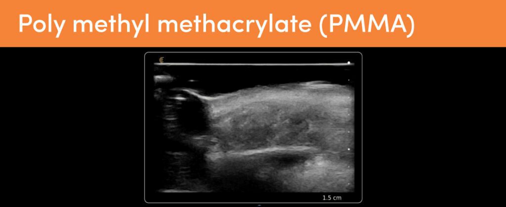
Polymethyl methacrylate (PMMA)
An uncommon synesthetic filler, this is PMMA or poly methyl methacrylate.This patient had cheek filler about 10 years prior, which over time displaced infraorbitally. And here you can see it underneath the orbicularis oculi as an ill-defined hyperechoic mass-like deposit with mini comet-tail artifacts. Those are those small hyperechoic dots that you can see within the filler deposit and which are typical of PMMA. PMMA microspheres are injected in a carrier gel like hyaluronic acid, which is why we’ve got that anechoic deposit right there. And it’s largely fallen out of favor as it’s permanent and it hardens with time. It is still used in South America, and this is where this patient had it placed. So, make sure you take a good history if you suspect PMMA.”
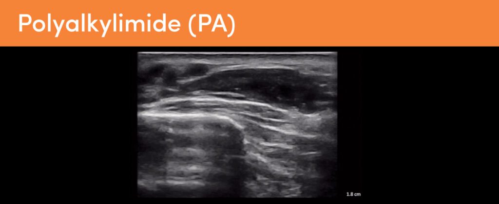
Video Case Review: Woman with Permanent Filler Complication
In this 5-minute video, Dr. MJ describes the case of a 57-year-old lady suffering from significant aesthetic disfigurement due to historic filler treatment. The midface is overfilled and lumpy. Dr. MJ demonstrates the ultrasound appearance of polyalkylimide permanent filler and discusses options for treatment. Watch the webinar for more details.
Affordable, High-Definition Ultrasound for Aesthetics
Wireless and pocket-sized, Clarius handheld ultrasound scanners deliver the high-definition imaging and performance of traditional ultrasound systems for a small fraction of the cost. They are the leading choice for plastic surgeons and aesthetics practitioners performing ultrasound-guided procedures to ensure patient safety.
New T-Mode™ AI by Clarius is a groundbreaking educational technology to help clinicians new to ultrasound advance their image interpretation skills using Clarius handheld scanners.
Visit our aesthetics page to learn more or request a virtual ultrasound demo today to learn how high-definition ultrasound imaging with voice controls can improve safety and deliver consistent patient outcomes at your aesthetic practice!
[WEBINAR] Learn the Basics of Treating Misplaced Filler Under Ultrasound Guidance
Dr. MJ shifted from general dentistry to complete her master’s in orthodontics and aesthetic medicine. Today, she practices non-surgical aesthetics with a special interest in managing dermal filler complications using ultrasound. Overcorrection or misplaced filler is one of the most common complications she sees in her practice. Besides being aesthetically disappointing, this can cause physical or psychological issues for patients.
Why Use Ultrasound for Aesthetic Medicine
Dr. MJ has been using the Clarius L20 handheld ultrasound for her practice for two years and says using it for filler treatment “is a godsend, whether that be before treatment to vascular map, during treatment to place filler using ultrasound guidance, or after treatment to confirm placement.
It is key to predictable outcomes. We can check up on our filler over time, and decide what’s hidden in our patients’ faces if we come across historic filler, and we can learn a huge amount of anatomy using it in our daily practice, but one major use is in dermal filler complications. Diagnosing and managing complications has arguably taken a giant leap forward since ultrasound. Being able to see beneath the surface of the skin to identify problems from filler is revolutionary. Gone are the days when we’re left guessing what the problem is or even where the problem is. Now, this simple two-minute diagnostic can help us resolve problems swiftly and effectively.”
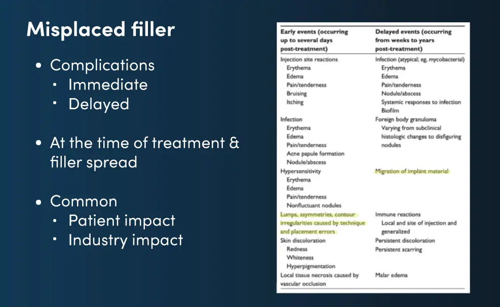
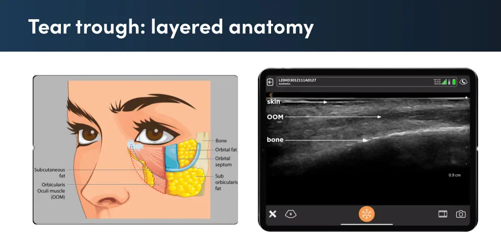
Overview of Adverse Reactions of Misplaced Fillers
This patient presented with bilateral interorbital swellings, which have been there since she had tear trough filler 18 months ago. They’re not painful. They don’t fluctuate, but they’re unaesthetic. She was told by her injector that the filler had gone after all this time. But as we know from recent studies, like the 2021 by Mobin Master, filler sticks around for years if not decades. So, this is a classic case of an immediate complication not being handled and turning up delayed.
Dr. MJ provides a detailed review of the case and tear trough anatomy during the webinar. Watch this video to see her technique for dissolving tear trough filler. Instead of using a “shotgun approach” that floods the area with 1500 units of hyaluronidase, she pinpoints the area with ultrasound and uses a much lower dose with better results.
Q&A: Which needles are you using when injecting hyaluronidase?
Dr. MJ Rowland-Warmann: I don’t use a cannula. I use a needle. I use a 27-gauge, one- and three-quarter-inch standard needle, not anything highly reflective. You can see that perfectly well on the Clarius. I would say the imaging that I get is great. The problem that you’ll have is lining it up. You’ve got to make sure that the needle is directly in line with your transducer, and that it doesn’t deviate. That’s where all of the problems come from. So, you’ve got to develop an eye for keeping that needle in line with the transducer, and you will get brilliant results even with a 27-gauge needle like I use.
Q&A: Are you using lidocaine for your injection entries?
Dr. MJ Rowland-Warmann: Yes, I sometimes do. I often dilute the hyaluronidase itself with four parts saline, and one part lidocaine so that it’s less painful for the patient. I found that works quite well. I use in total a five-milliliter dilution of 1,500 units of hyaluronidase.
Q&A: Would you say all misplaced filler remnants can be dissolved? In essence, can a patient gain back absolute original features?
Dr. MJ Rowland-Warmann: Are we going to put them back to a time ago before they had filler? The answer is no and never. There are multiple factors that contribute to a complication, and the filler itself is just one of them. If we remove the filler, the filler might be gone, but the complication itself has effects on the tissue, so there may be an adaptive response, and some of this adaptive response is hypervascularity that you see. Some of it may be edema of the tissues. Sometimes around the tear trough, that doesn’t completely go.
We also have two other factors. One of them is time. People age. If you’ve had a longstanding complication, me removing that filler is not going to put you back to where you once were. That has to be very carefully managed, and patients need to be instructed on that.
The last is perception drift. People always seem to remember themselves as better than they were. Objects in your memories are always seen with rose-tinted spectacles. So, the phenomenon of perception drift is patients always perceive themselves as complete before the complication.
When you remove that hyaluronic acid, and you leave them with the effects of time and potentially with the effects even minor of the complication, sometimes they’ve become quite despondent. So, management is very crucial at the start that that is done properly, but the actual hyaluronic acid filler itself, yes, can be removed in most cases. There are some rogue fillers out there on the market, especially in Europe where we are a little bit less licensed, that are much more difficult to remove. Obviously, permanent fillers cannot be removed with hyaluronidase.
Affordable, High-Definition Ultrasound for Aesthetics
Wireless and pocket-sized, Clarius handheld ultrasound scanners deliver the high-definition imaging and performance of traditional ultrasound systems for a small fraction of the cost. They are the leading choice for plastic surgeons and aesthetics practitioners performing ultrasound-guided procedures to ensure patient safety.
New T-Mode™AI by Clarius is a groundbreaking educational technology to help clinicians new to ultrasound advance their image interpretation skills using Clarius handheld scanners.
Visit our aesthetics page to learn more or request a virtual ultrasound demo today to learn how high-definition ultrasound imaging with voice controls can improve safety and deliver consistent patient outcomes at your aesthetic practice!
Réservez une démonstration virtuelle avec un expert en échographie!
Voyez Clarius HD3 en action pour découvrir combien il est rapide, facile et abordable d'ajouter l'échographie haute définition à votre cabinet.