

Ultrasound lets me get quick answers to specific clinical questions to support the right course of treatment for my patients,” says Melbourne-based podiatrist James Ferrie, B.Pod, M.A. Pod.A.
James is one among many physicians treating patients for musculoskeletal (MSK) ailments who have recently incorporated Clarius point-of-care ultrasound into their practice to deliver better patient care.
As clinicians, we listen and take a comprehensive history. Before physically assessing the patient, we have already comprised a provisional diagnosis and several differential diagnoses. The integration of point-of-care diagnostic ultrasound during a clinical examination adds a sixth sense, the ability to see below the skin, it helps to confirm or disconfirm my provisional diagnosis quickly and accurately,” James explains.
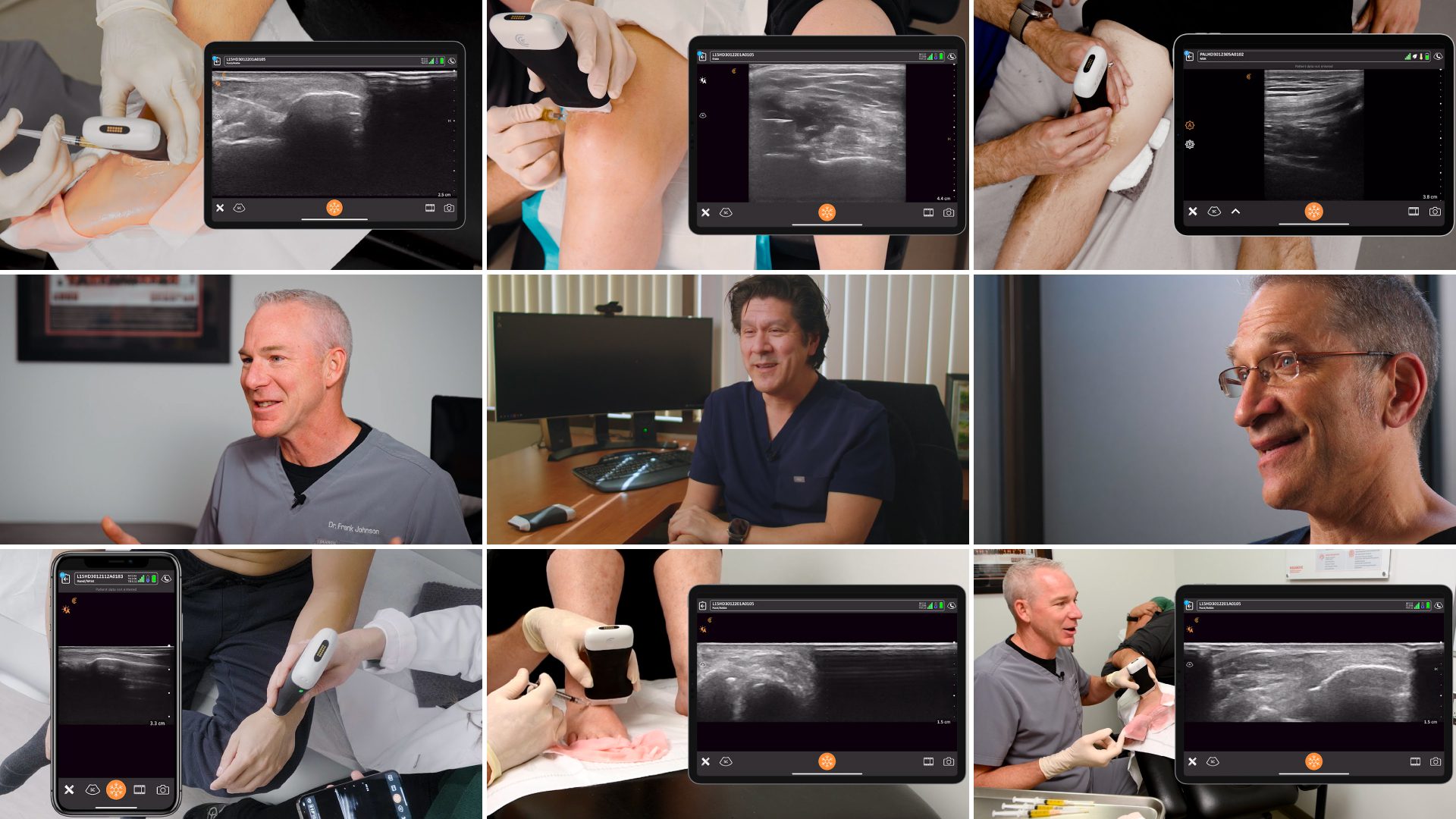
While James was experienced with ultrasound before choosing Clarius, many of our users are relatively new to ultrasound. That’s why we collaborated with Dr. Oron Frenkel to put together a series of brief educational videos that are easily accessible in our Clarius Classroom and in the Clarius App. Following is a series of videos on how to scan the lower extremities and videos showing common pathologies.
Learn How to Scan the Knee with Clarius Ultrasound
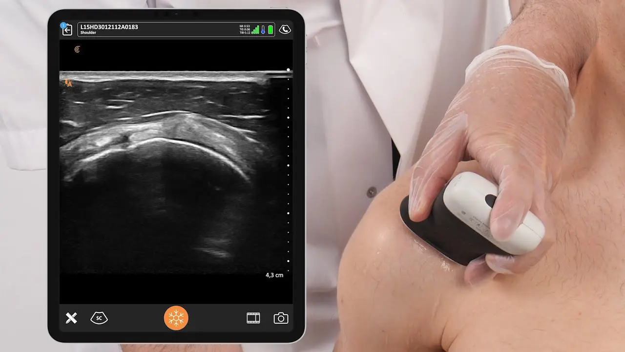
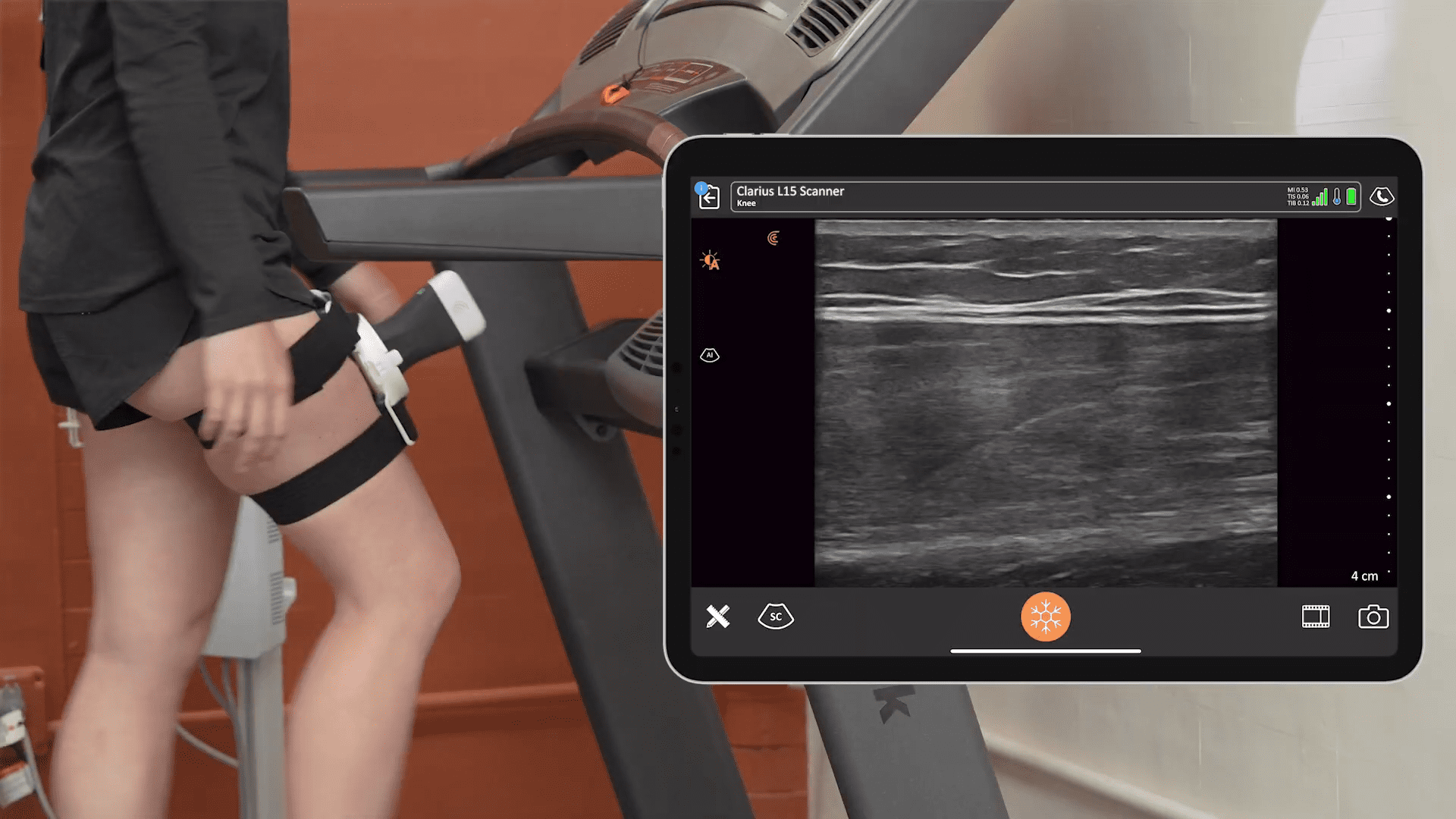
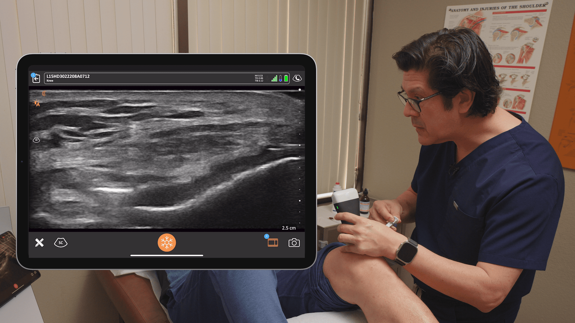
For a patient presenting with knee pain, a quick scan of the knee can rapidly identify pathology with more specificity than a physical exam. In this video, Dr. Frenkel uses the Clarius L15HD high-frequency ultrasound scanner to identify any tendinopathy or tears, which he doesn’t find on this healthy model. See what a knee with pathology looks like in the video shown below.
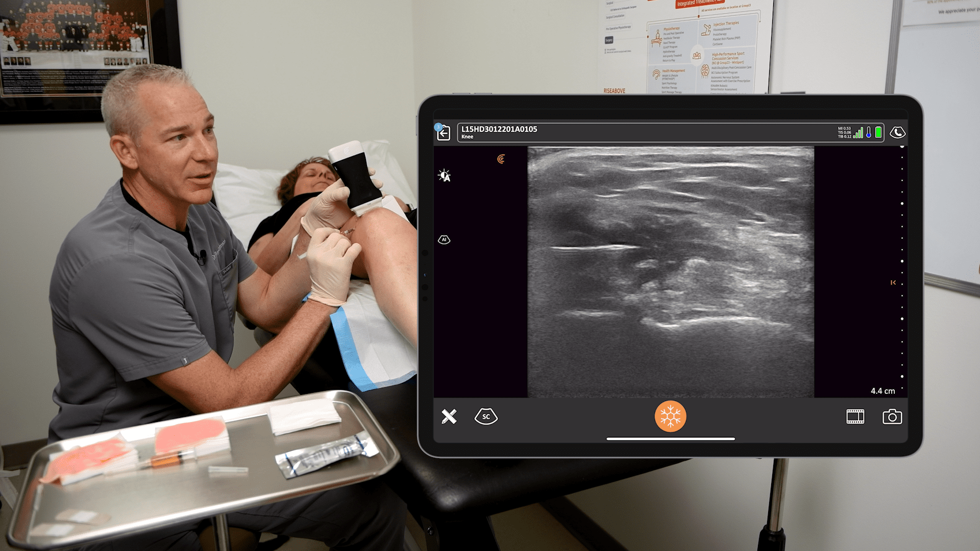
See How to Identify a Knee Joint Effusion on Ultrasound
In this video, Dr. Frenkel identifies a knee joint effusion in a longitudinal scan of the knee captured with the Clarius linear scanner. The patient presented with a swollen and painful knee.
Learn How to Scan the Heel with Clarius Ultrasound
Dr. Frenkel demonstrates how to quickly scan a patient’s foot to identify signs of plantar fasciitis. He uses the plantar present on the advanced MSK preset package that is available with Clarius HD linear ultrasound scanners. Watch the following video to see what a case of plantar fasciitis looks like with ultrasound.
See How to Identify a Case of Plantar Fasciitis Using Ultrasound
In this video, you’ll see an ultrasound scan of thickened plantar fascia, which is best identified in the longitudinal axis from its insertion into calcaneus. This is essentially diagnostic of plantar fasciitis. Visit Clarius Classroom to see videos of Achilles tendinopathy.
Learn How to Scan the Hip with Clarius Ultrasound
For patients presenting with hip pain, a quick ultrasound scan of the deep joint can identify basic and useful clinical information such as evidence of joint effusion or trochanteric bursitis. In this short video, Dr. Frenkel uses the Clarius L7 HD scanner to perform an ultrasound exam of the hip where he finds no obvious evidence of pathology.
Learn How to Scan Small Joints with Clarius Ultrasound
Dr. Frenkel uses the Clarius HD 15 high-frequency linear scanner to examine small joints in a healthy patient. This ultrasound exam is used to identify effusions and fractures in the phalanges and associated joints in the hands and feet.
See What Fluid Looks like in the 1st Metatarsophalangeal Space Using Ultrasound
In this video you can see a hyperechoic fluid collection in the first metatarsophalangeal space suggesting a joint effusion. Turning on power doppler we see inflammation, which could indicate gouty arthropathy, a septic joint or arthritis.
Learn How to Scan the Achilles Tendon
Watch this video to quickly and confidently scan the Achilles tendon to identify a rupture, tendinopathy, or tears of the gastrocnemius muscle.
Clarius offers both live and on-demand webinars for clinicians looking to hone their ultrasound scanning and interpretation skills. Learn from renowned SonoSkills educator Marc Schmitz as he teaches anatomy, sonoanatomy, technical scanning skills and pathologies common to the shoulder, elbow and knee in the free webinar «Pragmatic MSK Ultrasound: Scanning the Rotator Interval, Common Extensor Tendon and Patellar Tendon.»
Handheld Ultrasound Designed for MSK Clinicians
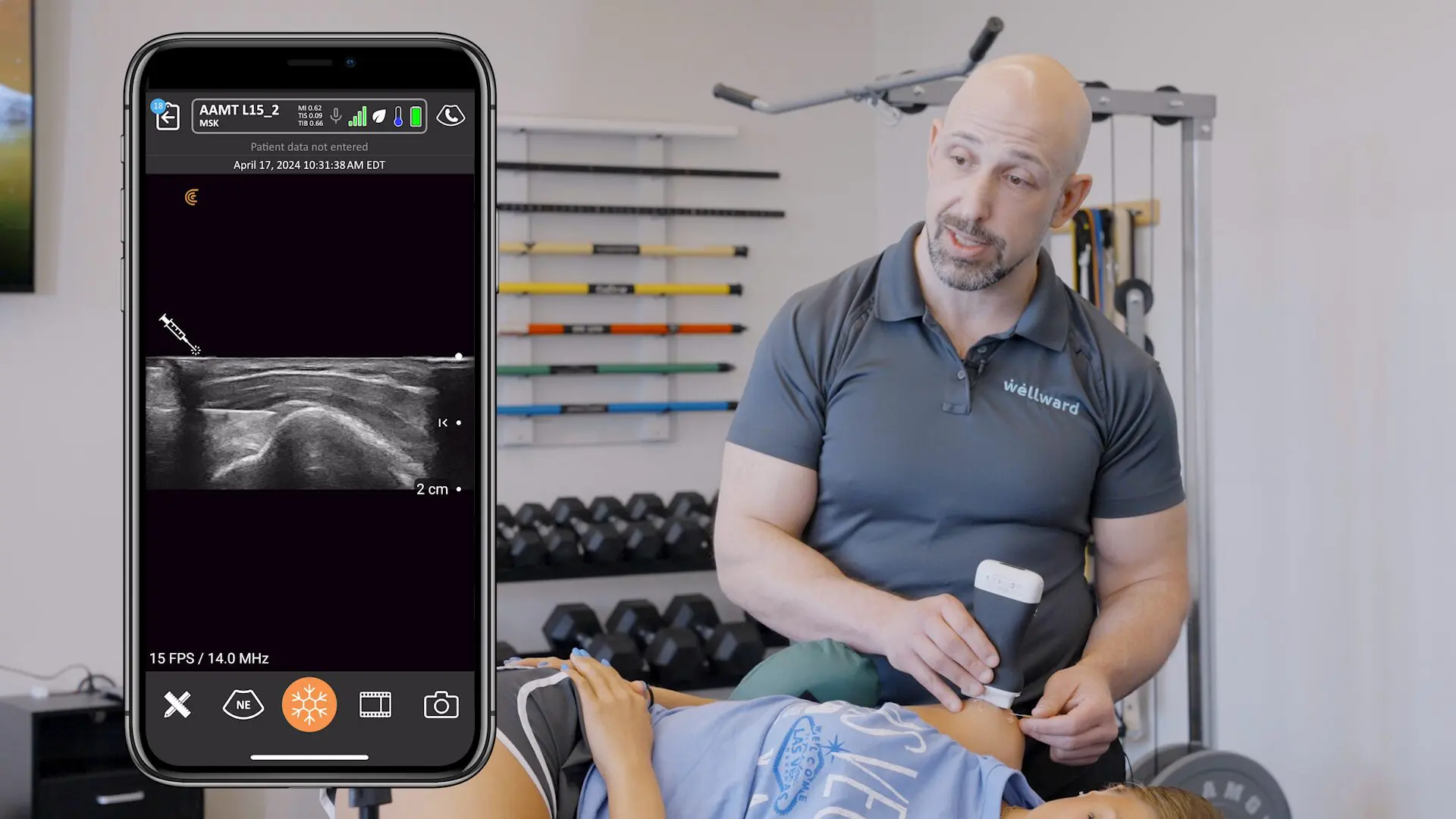
Getting detailed ultrasound images of musculoskeletal anatomy to make a confident diagnosis has never been easier. Ideal for MSK anatomy down to 7 cm, the Clarius L15 HD high-frequency ultrasound scanner is available with an optional Advanced MSK Package for diagnostic and interventional procedures.
Clarius offers four ultrasound scanners that are suitable for MSK applications. Learn more about which Clarius Handheld Ultrasound scanner is right for your practice. Or contact us today to request an ultrasound demo.


