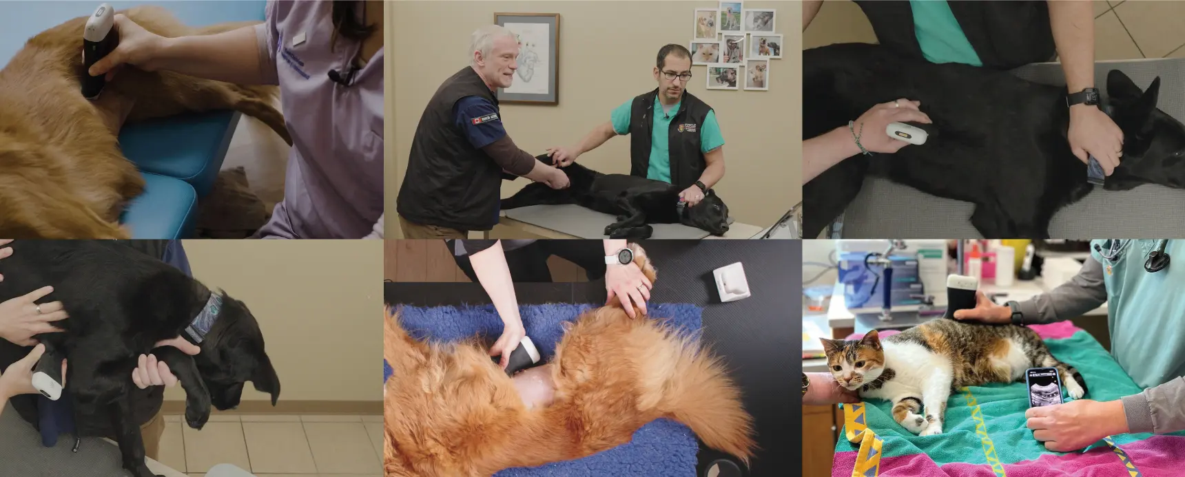
After years of working as an emergency and critical care veterinarian, Dr. Camilla Edwards, DVM, CertAVP, MRCV, is now dedicated to helping small animal vets with ultrasound. When we learned about her interest in Clarius ultrasound for veterinarians, we were quick to engage Dr. Edwards to help us build our library of veterinary ultrasound videos.
We recently recorded a free one-hour webinar with Dr. Edwards, which we invite you to watch: Practical Small Animal Ultrasound: Diagnosing Pathology with Intestinal, Gallbladder & Spleen Exams. Read on for highlights about splenic ultrasound from the webinar from Dr. Edwards.
Splenic Ultrasound Anatomy in Cats and Dogs
“The spleen is an elongated flattened organ, which is triangular in cross section in dogs and ovoid in cats. It’s located near the stomach on the left side and it goes under the ribcage cranially. The dorsal head is close to the stomach and fixed in position. Because the tail ventrally is very mobile, it can point towards the urinary bladder or down. You have to look for where it’s headed in each animal.

Indications for Scanning the Spleen
In general practice, we would examine the spleen with ultrasound when we see cranial abdominal organomegaly or if we have a hemoperitoneum.
A classic case will be a lethargic, pale dog with an enlarged abdomen. When we stick the probe on the abdomen, we’ll see some fluid. Then we need to have a look at the spleen to assess whether we have a bleeding hemangiosarcoma on the spleen.
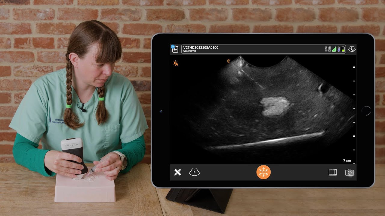
Besides looking for pathology, ultrasound is useful when getting a sample of fluid in the belly to determine if it’s blood or something else. We don’t want to hit a blood vessel or the intestines, so ultrasound-guided sampling is a really useful tool. It makes procedures a lot safer without blindly sticking a needle inside the animal.
Normal Views of the Spleen
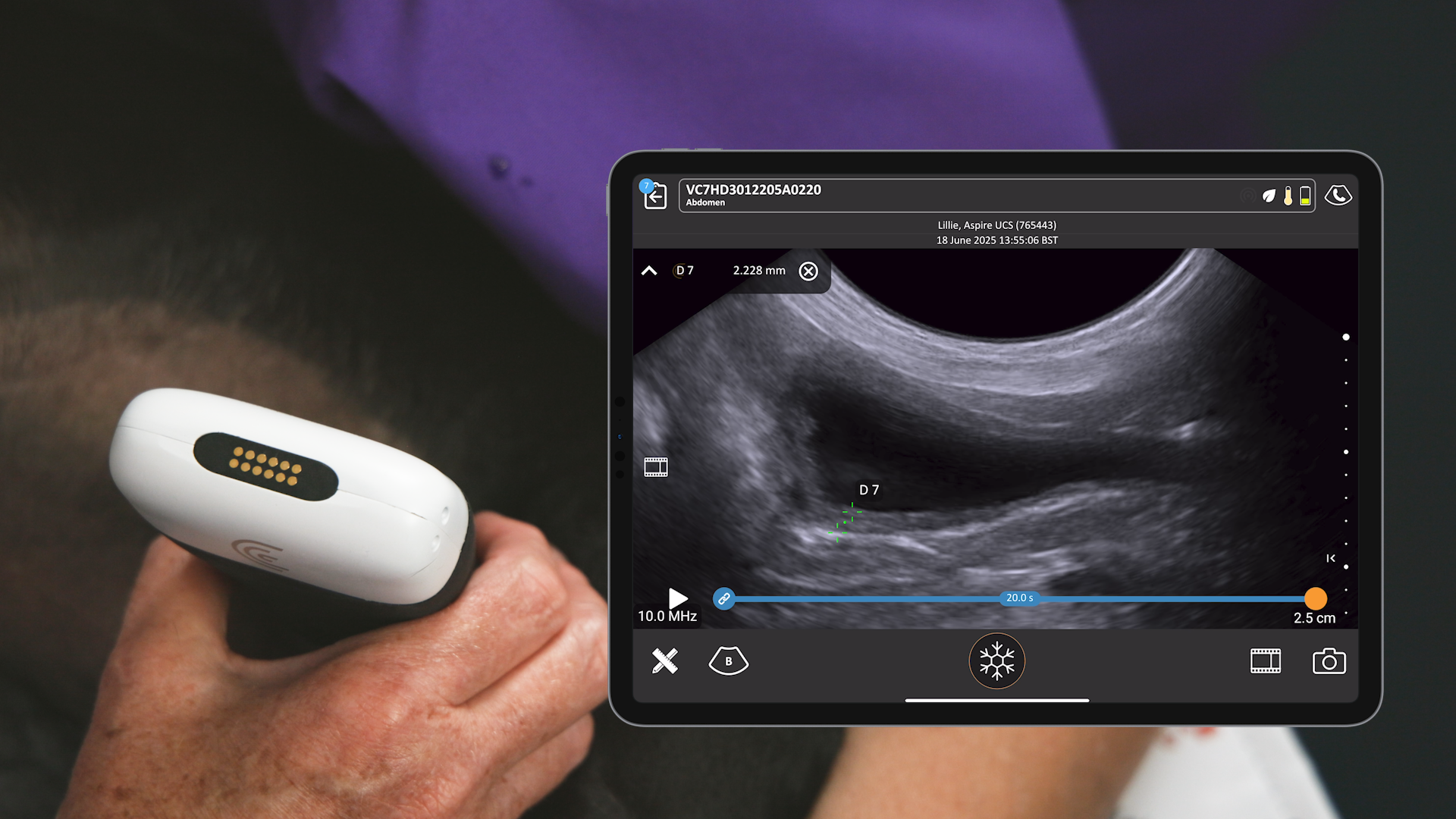
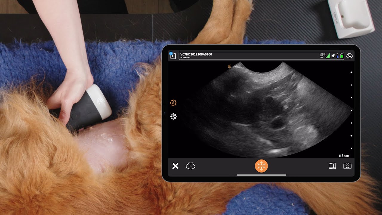
In the ultrasound video below, we see the splenic head underneath the rib cage, the stomach cranially and the left kidney up on the right of the image. Then we aim the ultrasound probe under the ribcage and we move down to the splenic body.
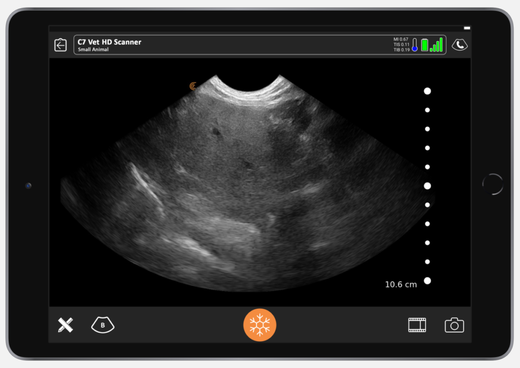
At the start of the next video you see the hilus, there are blood vessels entering the spleen, and then we follow it all the way down to the tail. It’s really important to follow it to look at the tail and the head because there can be a nodule or a mass right on the tip. We should see a uniform homogenous organ in a healthy spleen.
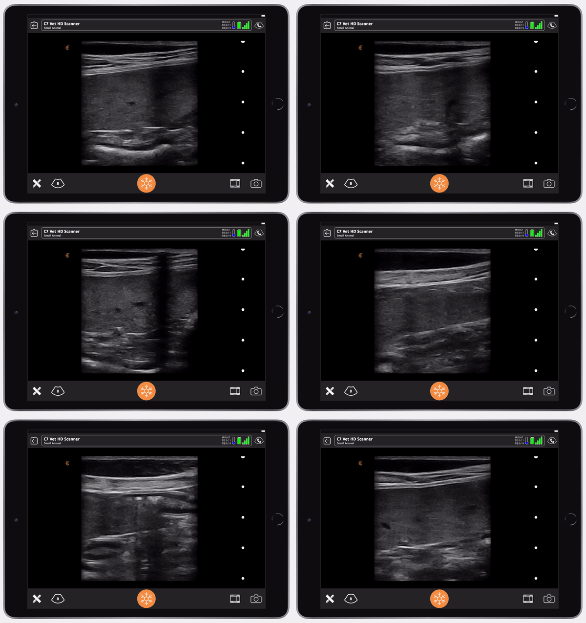
In dogs, a normal spleen can vary enormously in size. But in cats, it should never be more than one centimeter thick at the hilus where the blood vessels enter, and it should never be folded. If these are your findings, then you’ve got an enlarged spleen.
How to Scan a Spleen on a Dog Using Ultrasound with the Clarius C7 HD Vet
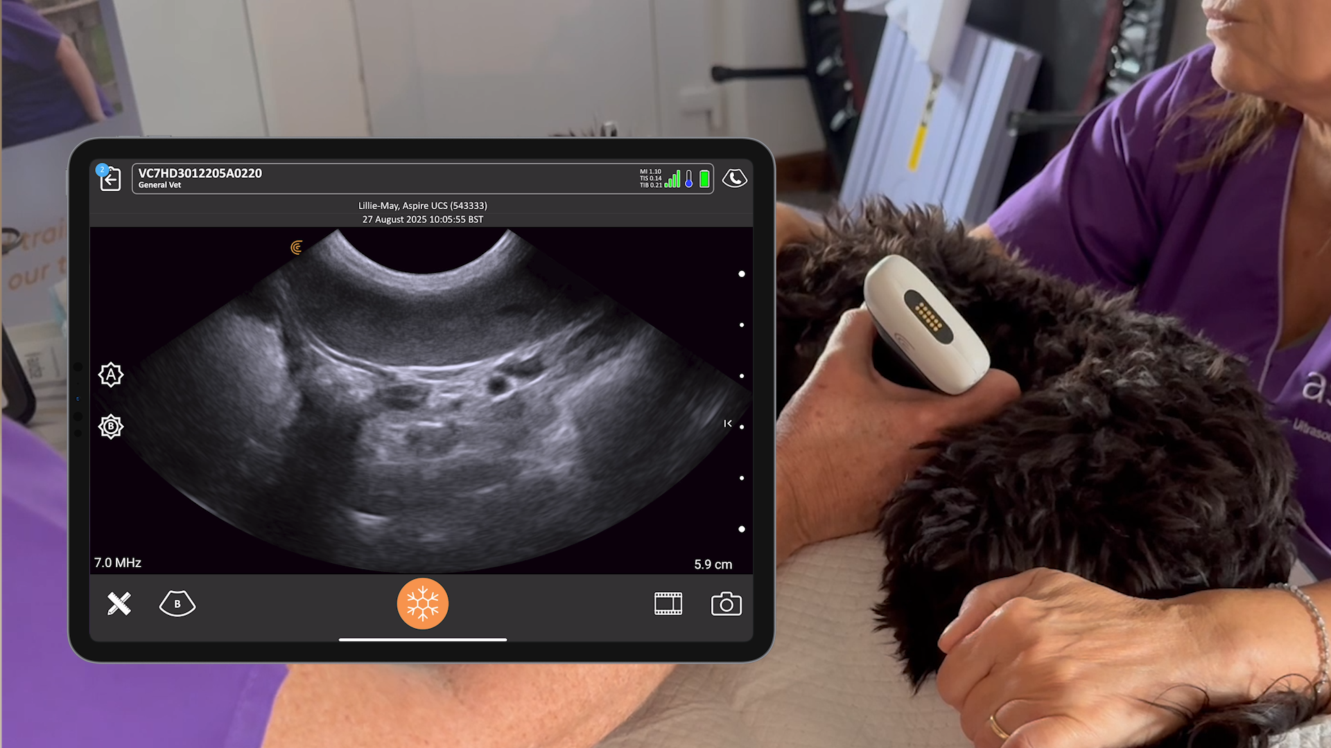
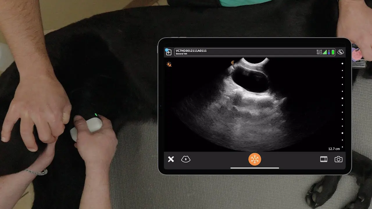
I scanned my dog Pippy for this next tutorial. We start in the same position for the liver: the xiphisternum. Then we follow the costal arch up. The first organ we come across after the liver is the stomach. In the video, we can see some of the stomach and we can see some spleen at the top. We get the left kidney coming into view, and then we fan underneath the rib cage to see all of the spleen that we wouldn’t otherwise see.
Since you can’t palpate the area around the spleen, ultrasound is really a brilliant way of having a look.
And it’s important because there’s a big chunk of the spleen under there. Once we find the head of the spleen, we lift the probe and rotate it to aim longitudinally down and in the direction of the spleen.
Sometimes that can be a little difficult and we need to make small adjustments. The principle is to slide and fan, slide and fan, slide and fan until we get to the tail of the spleen. Sometimes that involves going around to the other side of the body.
When I captured this video, I had been scanning Pippy for quite a while by this point. The spleen grows with massage and hers had grown quite a lot during the exam. That’s why I needed to extend around to the other side of the abdomen to get to the tail that we can see there. One we reach the tail, we turn and rotate the probe again, 90 degrees, and bring it all the way back up to the ribcage. In this video we’ve looked at the whole spleen in two different directions: transverse and longitudinal.
Common Pathology Seen in the Spleen

As mentioned before, it’s important to know that the size of the spleen varies a lot. It’s normal for the spleen to grow and get small. So, we need to look closely at the spleen to identify pathology, which can include congestion and sedation or lymphatic fluid hyperplasia, extramedullary, hematopoiesis, infiltrative disease and splenic torsion.
Use ultrasound to check to see if the margins are regular or irregular. Look for nodules and masses. If you see a hyper echoic nodule, it’s often a myelolipoma, which is a fatty, benign tumour. You will need to sample to know for sure. We might find hypoechoic nodules or mixed echogenicity, and then we need to look at their distribution.
There are a few things that are quite characteristic that we might find on ultrasound. One of them is a lacey spleen, where we see sort of hypoechoic (darker) hacks in the spleen. This can indicate a splenic torsion, which is a real emergency. With a splenic torsion, the dog is in a lot of pain and you know it’s coming from the abdomen somewhere.
The other one that we might see is a Swiss cheese spleen, where there are hypoechoic holes in the spleen. This is commonly seen with lymphoma. And again, we think about the distribution of the pathology.
Splenic Mass in a Beagle
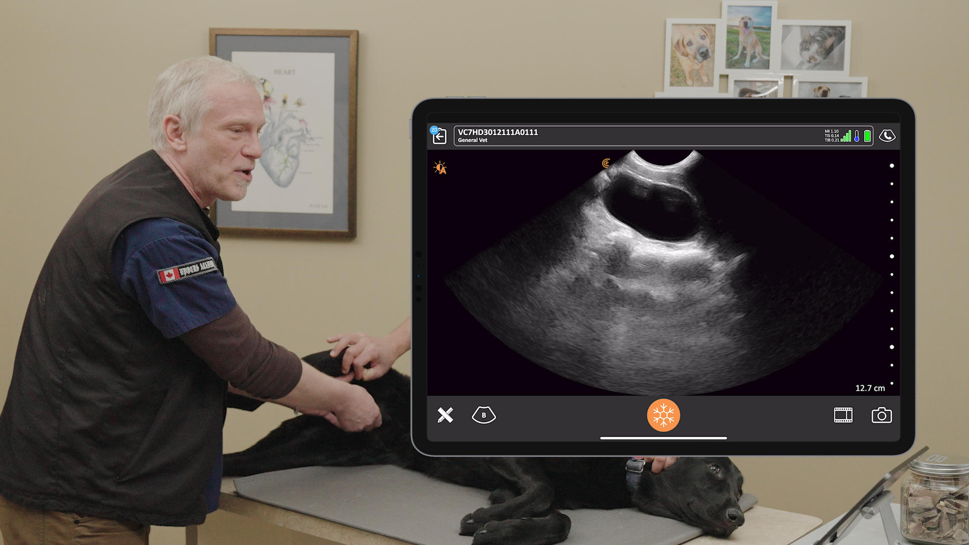
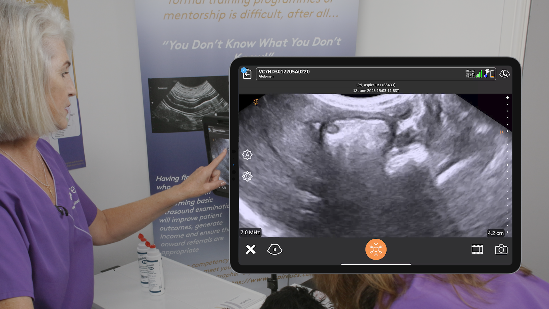
The ultrasound image below shows pathology we found in a beagle. The splenic head looks quite normal. But we see a mixed echogenicity mass.
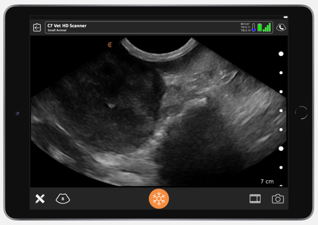
There could be so many differentials. The diagnosis becomes more obvious if there’s also bleeding and then we are more worried that it is hemangiosarcoma. To confirm diagnosis, we need to get a sample. In this case, a sample unfortunately confirmed a hemangiosarcoma.

Being able to use an ultrasound probe is really useful so you can treat it right away instead of sending the animal somewhere else for imaging.”
Interested in Learning More About Veterinary Ultrasound?
Watch our fun, fast-paced webinar “Veterinary Point-of-Care Pleural Space and Lunch Ultrasound (PLUS) for Everyday Practice” featuring educators Dr. Soren Boysen, DVM, DACVECC and Dr. Serge Chalhoub, BSc, DVM, DACVIM (SAIM).
About the Clarius HD Vet Handheld Ultrasound Scanners
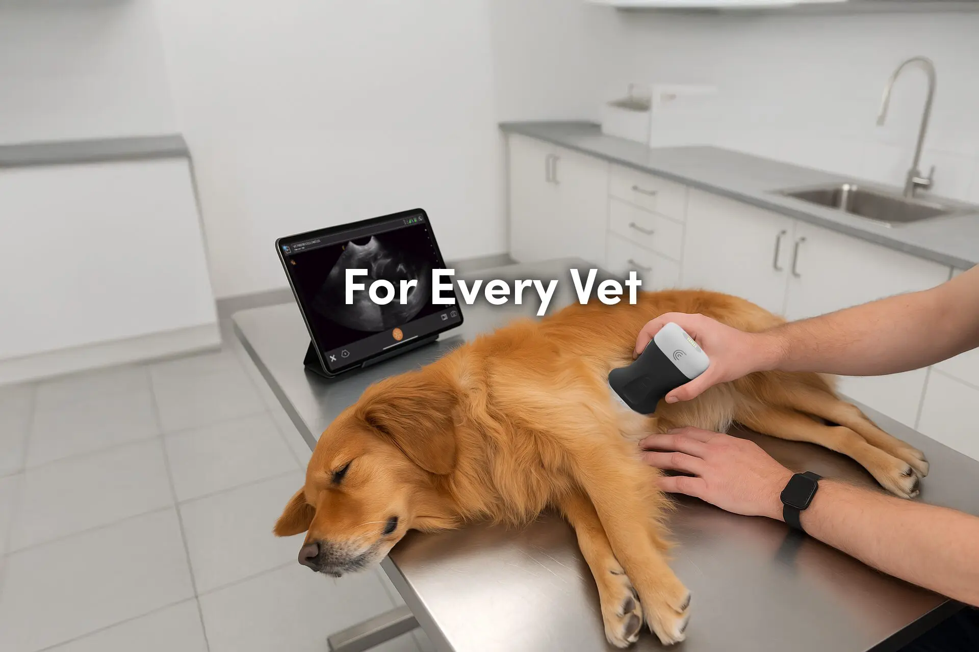
Affordable and easy-to-learn and use, Clarius handheld ultrasound is making it possible for veterinarians to quickly diagnose and treat animals in pain instead of sending patients away for diagnostic imaging. Our customers report increased word-of-mouth referrals and better client satisfaction after bringing ultrasound in-house.
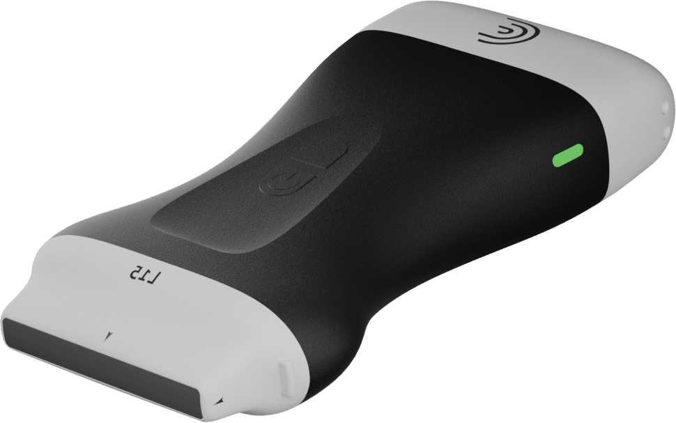
The images that Dr. Edwards showed in this article were captured with the Clarius C7 microconvex vet scanner, which is specifically designed for clinical imaging of small and medium-size animals. Our range of ultrasound scanners for veterinarians includes the C3 HD Vet convex scanner for large animals, and the L7 HD Vet linear scanner for equine musculoskeletal imaging.
To learn about how easy and affordable it is to add Clarius handheld ultrasound to your veterinary practice, visit our Clarius veterinary ultrasound page for a video demonstration and product details. Or contact us today to discuss which scanner is right for your veterinarian practice.
If you’re interested in alerts for our new Clarius Classroom video tutorials, please subscribe to our Facebook, Twitter, LinkedIn, Instagram or YouTube channel.
You can also watch a review by Dr. Edwards of the Clarius C7 HD Vet on her website.
