

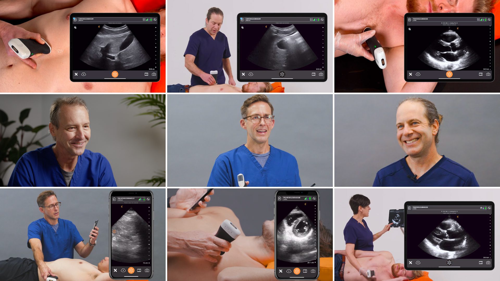
You could say that Tom Cook, MD, an emergency physician based in South Carolina, is an ultrasound fanatic. He has been fascinated with the technology’s potential to help save lives since he touched his first mechanical probe more than 25 years ago. When ultrasound was miniaturized to the size of his hand, he stopped carrying a stethoscope.
I was by sheer luck pushed into learning about ultrasound technology and it’s done so many amazing things for me personally, but also for the patients I take care of and the residents I train,” says Dr. Cook. “Many physicians think it’s a fantasy to be able to walk up to a patient and immediately look inside them and know what’s wrong with them. But it’s not a fantasy. That’s exactly what ultrasound technology does today.“
We recently had an opportunity to interview Dr. Cook about how he uses ultrasound to make better decisions about caring for his patients. Watch his video interview to learn what he had to say. Or read on for highlights.
POCUS Is a Safe and Efficient Way to Determine a Patient Management Strategy
Before we had ultrasound, it really was a bit of a guessing game. We could come up with a differential, and we would try and eliminate the possibilities in that differential based on a variety of different laboratory studies and physical findings.
What I point out to people now is that you can walk into a patient’s room with a differential in mind, do an ultrasound exam, and immediately eliminate several pathologies or even make the diagnosis.
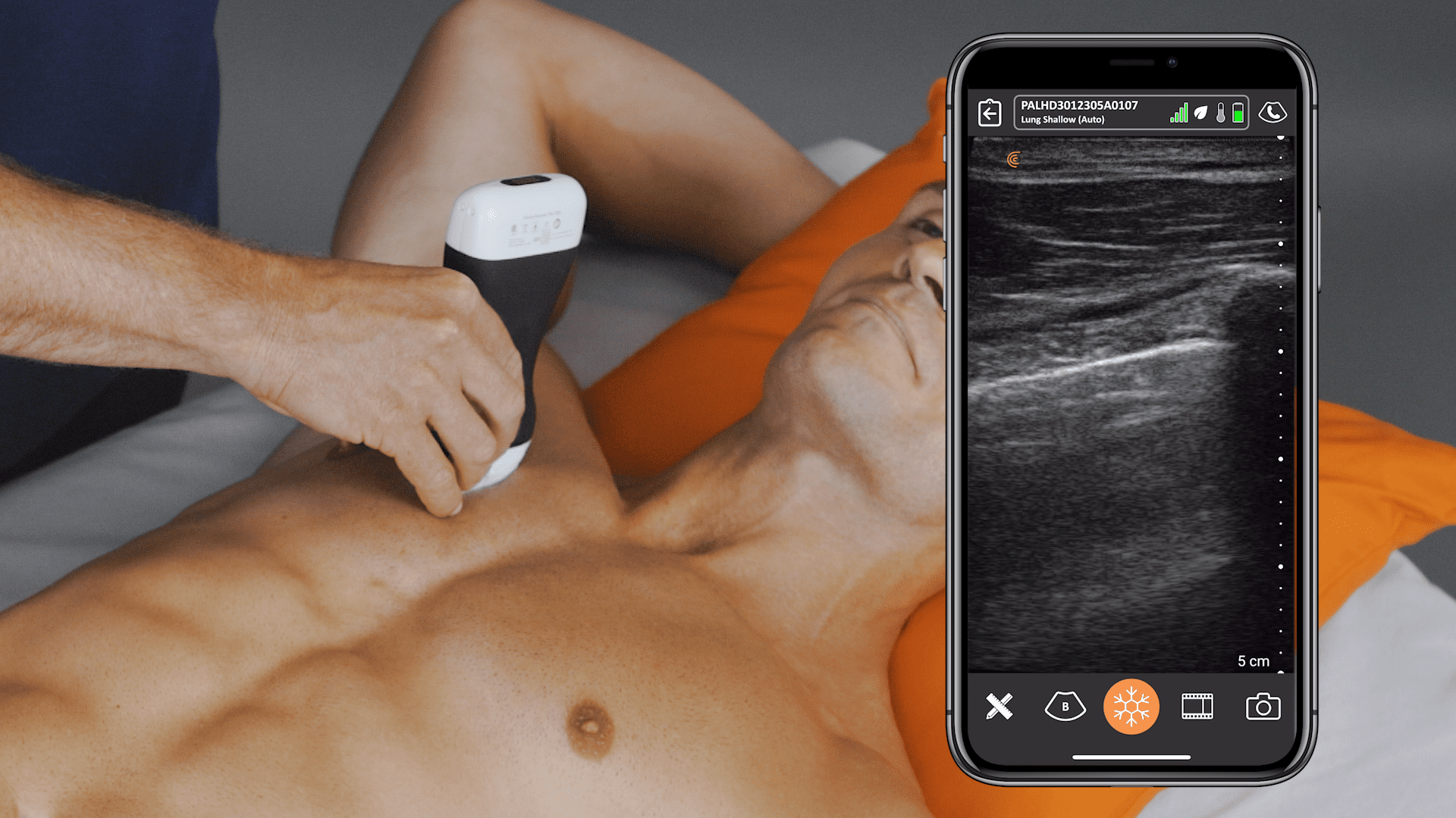
An example would be a patient comes in with chest pain and shortness of breath. You walk in the room, perform a cardiac ultrasound, and immediately ask yourself, „Does this guy have heart failure, a pericardial effusion, a pulmonary embolus, or a valvular problem?“ Then, you examine the lungs. „Does he have pulmonary edema or pleural effusions?“
As you walk out of the room you are thinking, „I just crossed off six items from my differential. My original differential had ten possibilities, but I’ve whittled it down to three or four pathologies from my ultrasound examination alone. And, I did it within a couple of minutes. That is real powerful technology.”
POCUS Webinar Series with Dr. Tom Cook
In addition to practicing as an emergency physician, Dr. Cook has spent his career encouraging other clinicians to use ultrasound in daily practice through the ultrasound education company he founded in 1998, 3rd Rock Ultrasound. We have the privilege of hosting a series of four POCUS webinars with Dr. Cook in the coming months.
Join us for the first webinar, Cardiac POCUS: Techniques for Assessing Left Ventricular Function, to learn how to use handheld ultrasound to accurately assess left ventricular function in your patients presenting with chest pain, dyspnea, or unstable vital signs.
During the free one-hour webinar on October 25th, Dr. Cook will give you four easy tools to quickly and reliably determine whether the left ventricle is pumping adequately.
- Visual estimation of myocardial contractility
- E-Point Septel Separation (EPSS)
- Fractional Shortening
- Mitral Annular Plane Systolic Excursion (MAPSE)
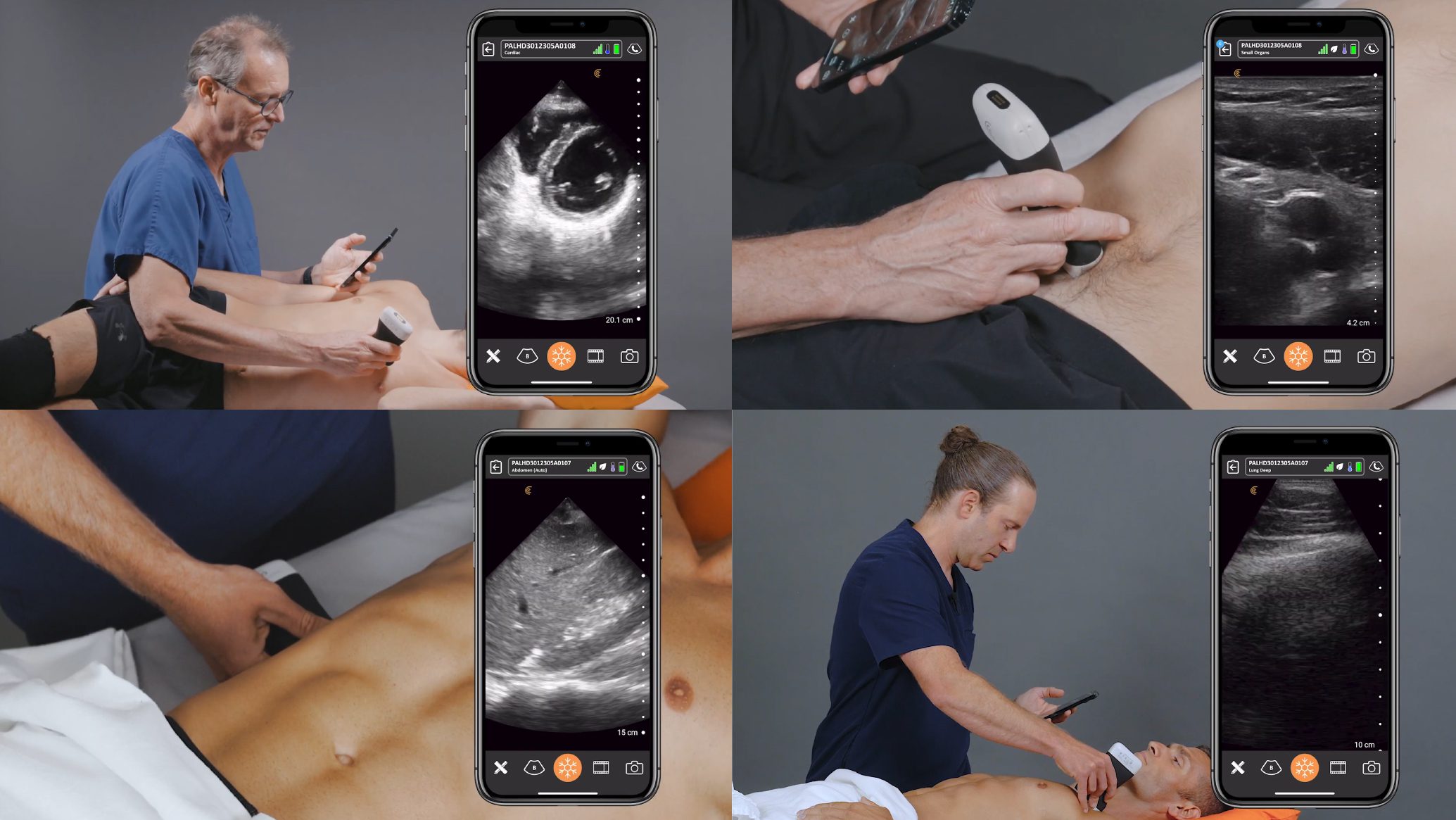
Clarius for Emergency Medicine: Saving Lives by Expediting Triage and Treatment
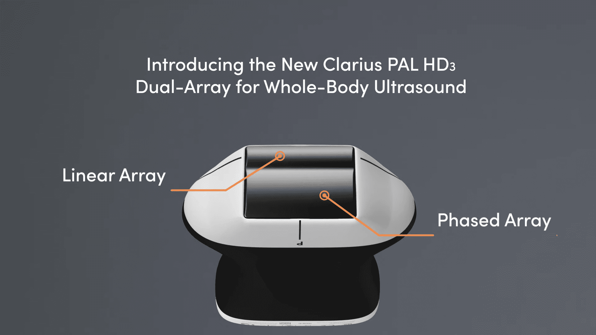
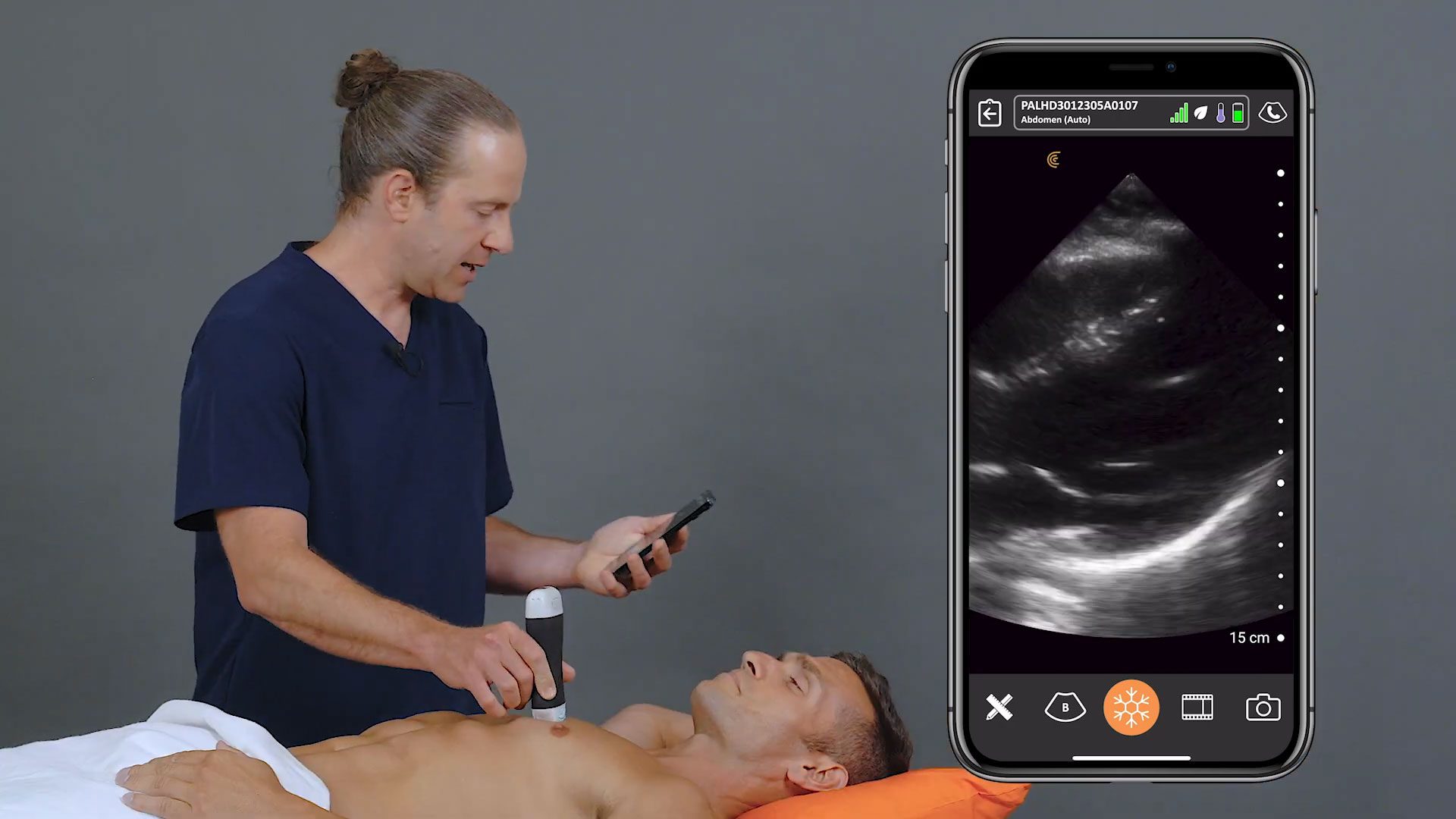
Dr. Cook primarily uses the Clarius PA HD3. It’s one of three Clarius scanners suitable for emergency medicine. Wireless and app-based, Clarius ultrasound delivers fast imaging and sharp detail at the bedside. Visit our emergency medicine page to learn more about which scanner is suitable for your practice. Or book a virtual demo with a Clarius expert in your region.


