

Veterinary abdominal point-of-care ultrasound has evolved from the abdominal FAST exam, which looked for free fluid in the abdomen. Now, many veterinarians are using the 5-point abdominal point-of-care ultrasound (APOCUS™) exam for real-time assessment of fluid and multiple abdominal organ abnormalities in ill small animal patients.
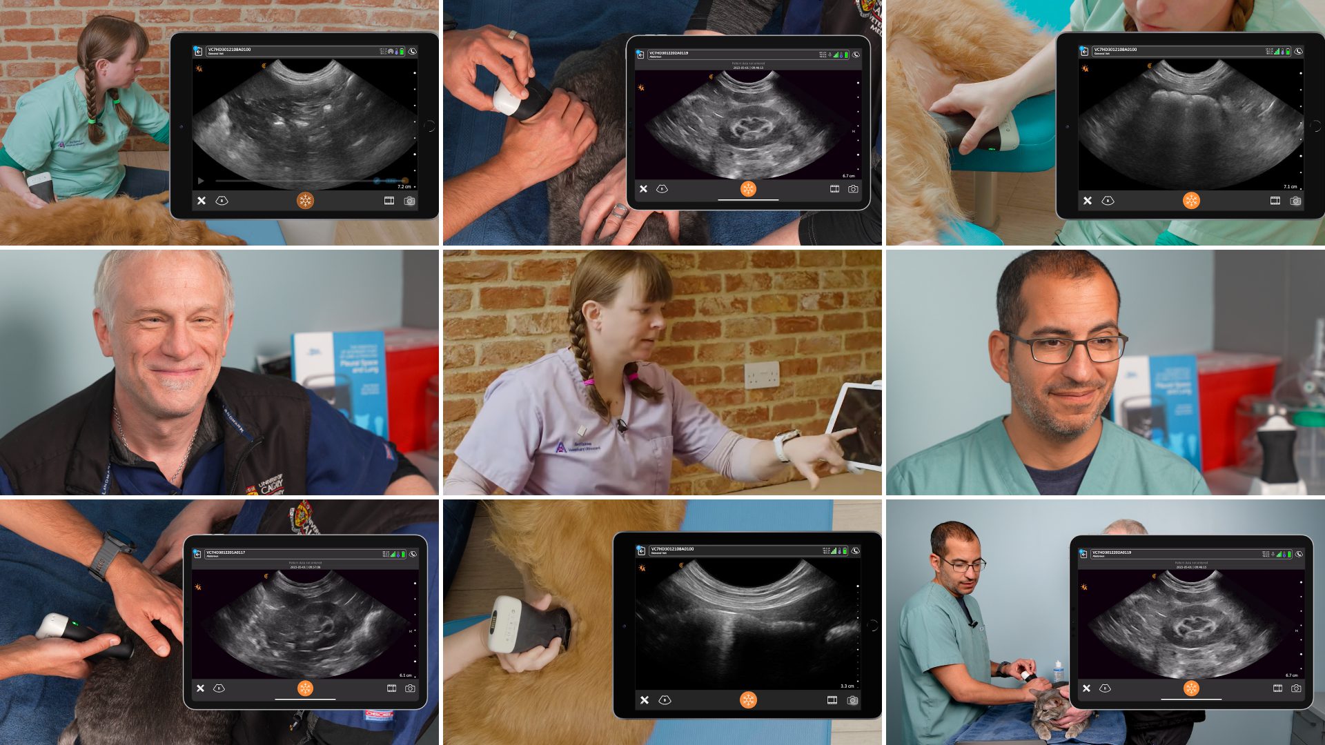
We recently presented a webinar with Dr. Soren Boysen, a specialist in veterinary emergency medicine and critical care, and Dr. Serge Chalhoub, a specialist in veterinary internal medicine in small animals during which they shared their techniques for performing a 5-point APOCUS™ exam. Read on for highlights or watch the free webinar, « Veterinary POCUS: Assessing Acute Abdominal Conditions Using the Rapid 5-Point Abdominal POCUS Exam« , at your convenience.
Drs. Boysen and Chalhoub are considered pioneers in veterinary point-of-care ultrasound, and have a combined total of over 25 peer-reviewed articles and books on the subject. During the webinar, they demonstrate how the 5-point APOCUS™ exam is performed and describe how point-of-care ultrasound is applied based on the clinical setting using an actual case example.
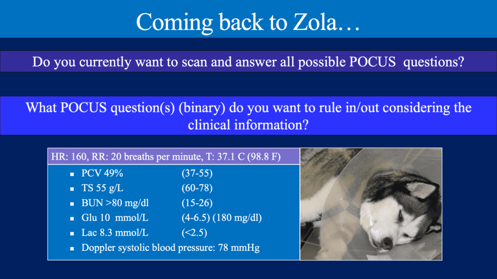
Case Study: Zola
- 3-year old male neutered Husky
- Landed on the tailgate of a truck 48 hours ago
- Seemed fine immediately afterwards
- Vomiting, not eating the past 24 hours
- Does not want to get up or walk
Physical Exam Findings:
All aspects of point-of-care ultrasound are not always indicated in every single patient. You have to apply it based on that clinical setting!”
For Zola, what POCUS question(s) (binary answer) do you ask to rule in/out considering the clinical information?
- Is there free abdominal fluid? Y/N
- Fluid can accumulate anywhere
- Fluid accumulates at different locations based on patient position
- Fluid can be trapped by adhesions/omentum
- Is there free abdominal air? Y/N
- Is there gallbladder wall edema? Y/N
- Is it OK to give a fluid bolus? Y/N
- Is there pericardial fluid? Y/N
Ultrasound Windows and Questions Answered with the 5-point APOCUS™ Exam
- Subxiphoid site – is there free abdominal fluid, free abdominal air, gallbladder edema, gastric ileus or fluid retention in the stomach, pericardial effusion, cardiac activity, caudal lung pathology, pleural effusion, and is it OK to give a fluid bolus (based on caudal vena cava assessment)?
- Umbilical site – is there free abdominal fluid, free abdominal air and/or a splenic mass?
- Urinary bladder site – is there free abdominal fluid, free abdominal air, pyometra (female intact animals), and is the animal producing urine (urinary bladder volume estimation)?
- Right paralumbar site – is there free abdominal fluid, free abdominal air, ileus (assessment of the duodenum) or pelvic renal dilation/obstruction?
- Left paralumbar site – is there free abdominal fluid, free abdominal air, renal pelvic dilation/obstruction?
Important –Ultrasound exams can be performed on patients on oxygen after anxiolytics, and while giving fluid and performing lifesaving interventions. Always scan the patient in the position they are comfortable, and be sure to scan in both long and short axis planes at each site
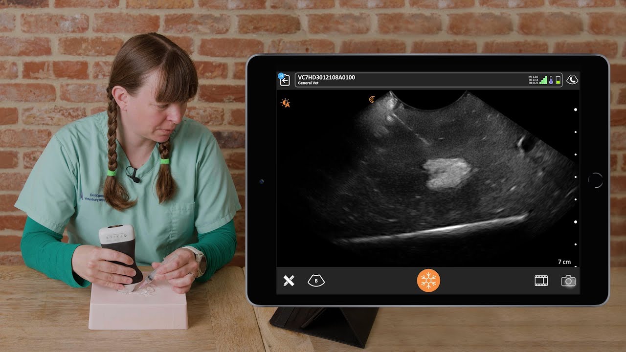
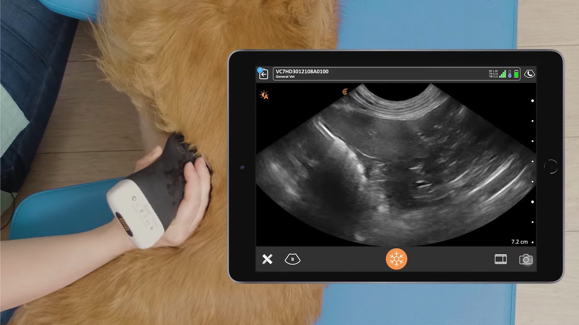
Watch this video to see the ultrasound exam performed in under a minute!
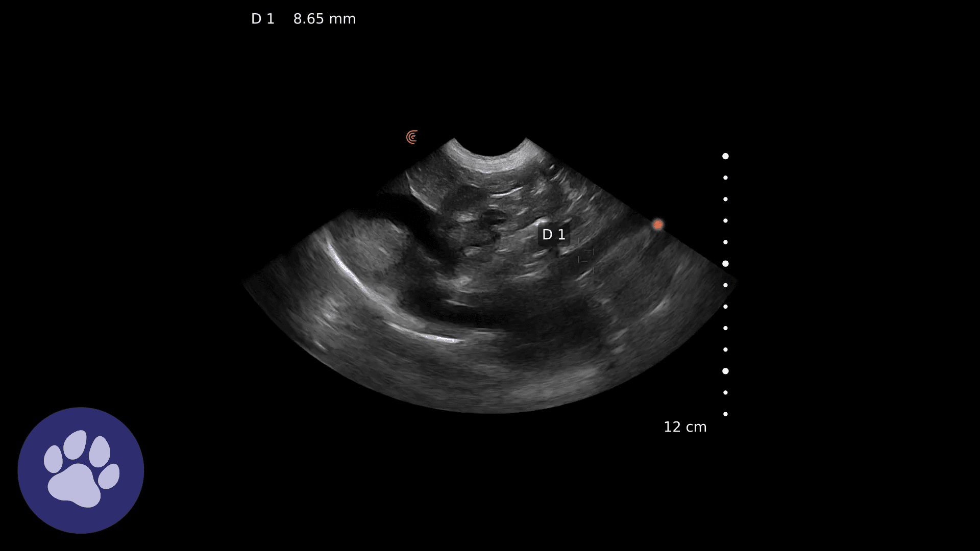
Zola’s 5-point Abdominal POCUS Findings
- Subxiphoid site – free fluid between the diaphragm and the liver and separating the liver lobes. Flat caudal vena cava supporting fluid bolus administration. Unremarkable for other findings at this location
- Umbilical site – large amount of free fluid in gravity dependent areas, otherwise unremarkable for other assessments at this location
- Urinary bladder site – fluid at the apex, otherwise unremarkable for other assessments at this location
- Right paralumbar site – fluid around the right kidney, otherwise unremarkable for other assessments at this location
- Left paralumbar site – fluid around kidney and spleen, otherwise unremarkable for other assessments at this location
An abdominal paracentesis was performed and the fluid analyzed. The results, in combination with Zola’s lab results, indicate uroabdomen. Zola needs to be stabilized, and other POCUS evaluations of the heart and lungs supported the caudal vena cava findings that IV fluid can be given (cardiac POCUS suggested hypovolemia, pleural and lung assessment (PLUS) was unremarkable), and once Zola was stable, a urethrocystogram with contrast was performed, indicating bladder rupture.
Watch the webinar, « Veterinary POCUS: Assessing Acute Abdominal Conditions Using the Rapid 5-Point Abdominal POCUS Exam« , to learn Zola’s prognosis and outcome, and how ultrasound was used to monitor post-operative urine production during his hospital stay.
Upcoming Lecture on Understanding Lung Consolidation
Award-winning lecturers and leading experts in the field of veterinary point-of-care ultrasound (POCUS), Drs. Søren Boysen and Serge Chalhoub return for another highly engaging and dynamic lecture, this time focussing on something often overlooked in veterinary medicine, lung consolidation!
In this 1-hour dynamic webinar on December 12th, participants will learn proven wireless lung ultrasound exam techniques to interpret lung consolidation. Drs. Boysen and Chalhoub will:
- Describe the difference between B-lines and lung consolidation
- Demonstrate how ultrasound can be used to scan the lung surface of small animals to search for lung pathology
- Explain how to classify different types of lung consolidation and their possible underlying etiologies
Clarius Wireless Ultrasound for Veterinary Practice
Drs. Boysen and Chaloub often use the C7 HD3 Vet in their practices. To learn more about how you can add wireless ultrasound to your practice, visit our Veterinary Specialty Page. There you’ll have access to additional webinars and classroom videos. Learn about the Advanced Veterinary Package, which provides additional workflows for a wide variety of animal examinations.
Our new Clarius HD3 Vet scanners are smaller and lighter. To find out which scanner is best for your practice, contact us today, or request a virtual ultrasound demo.

