

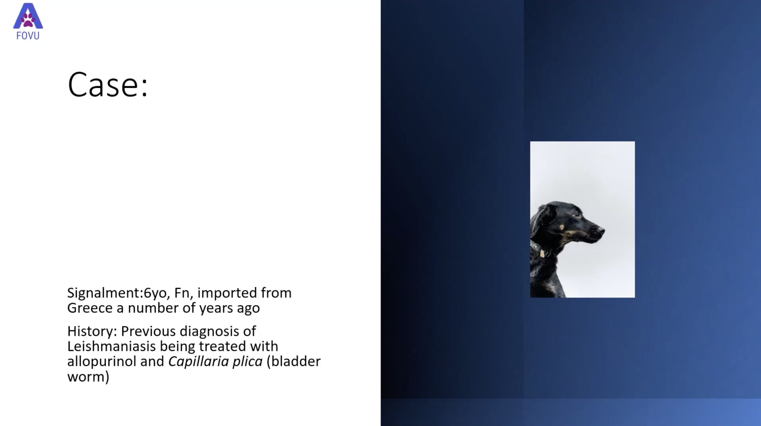
Dr. Camilla Edwards, DVM and founder of First Opinion Veterinary Ultrasound (FOVU) recently shared a detailed case study using ultrasound to identify a mass in the stomach of one of her feline patients.
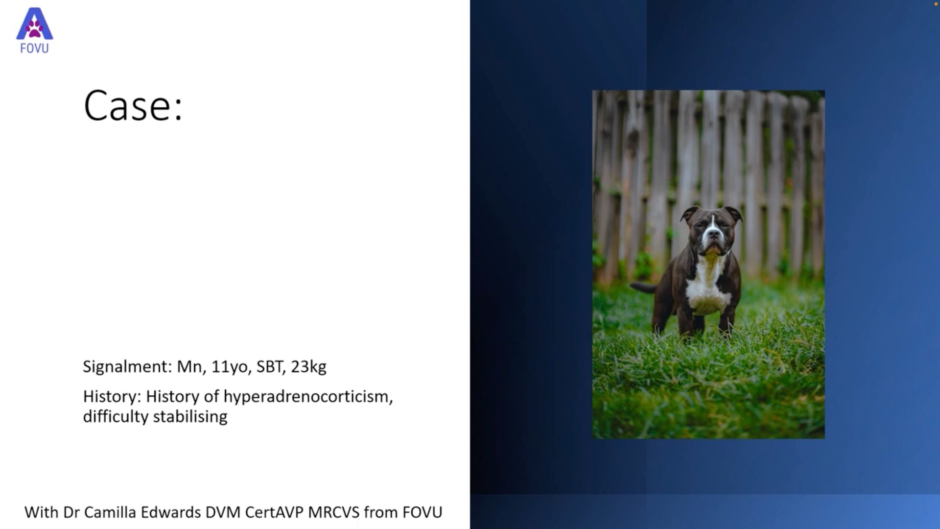
History: This 12-year-old f/n Turkish Van feline presented with a history of vomiting, which improved slightly with prednisolone. She had borderline chronic kidney disease. Naturally, Dr. Edwards used her ultrasound expertise to have a look under the fur!
Ultrasound Findings
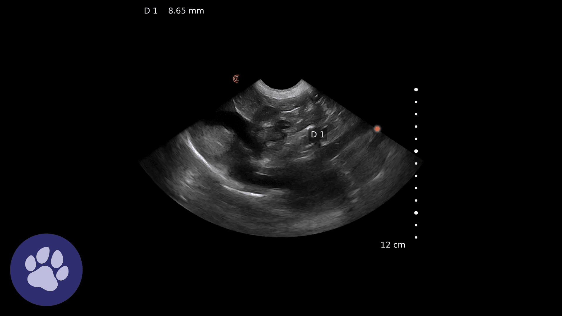
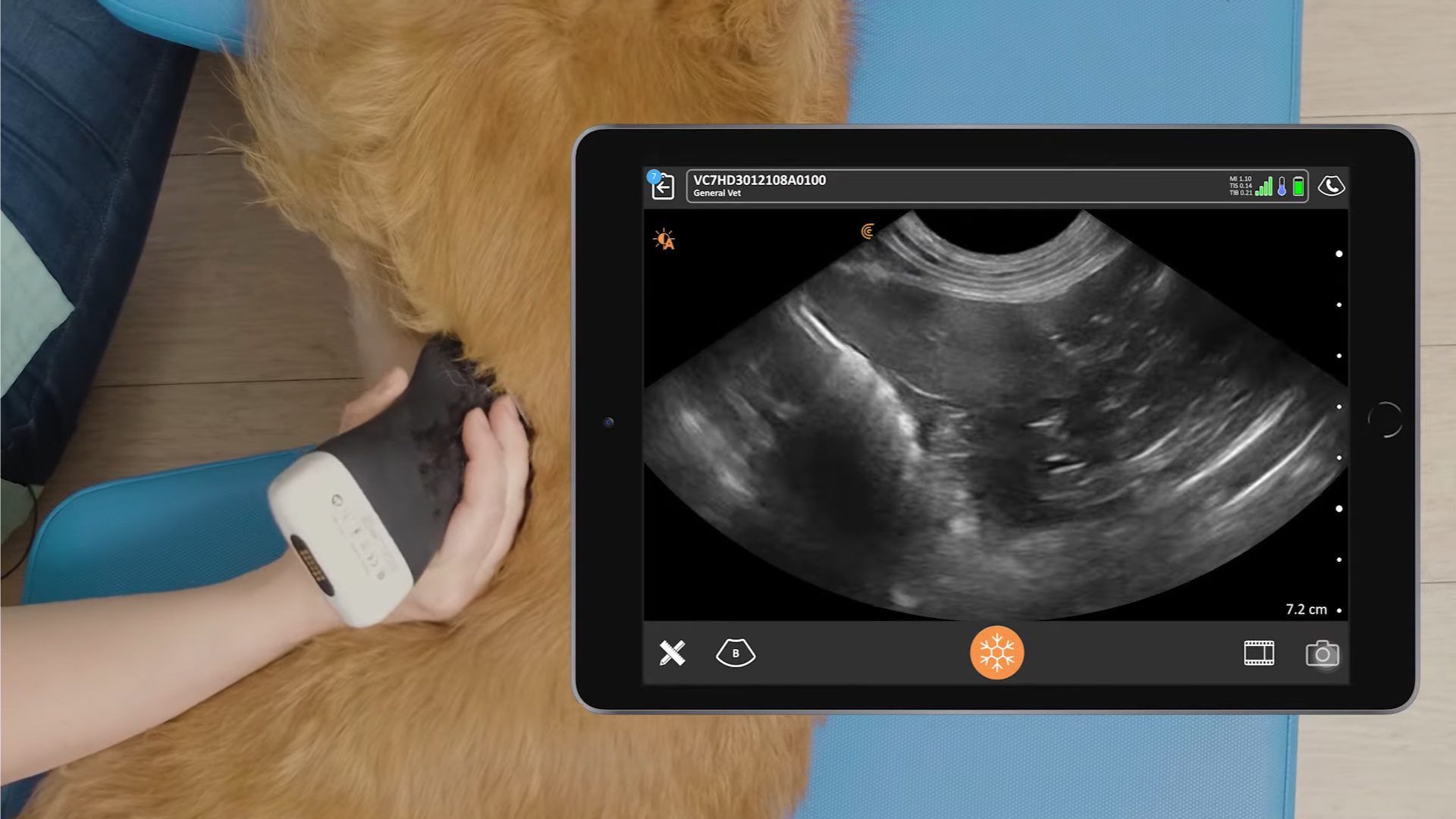
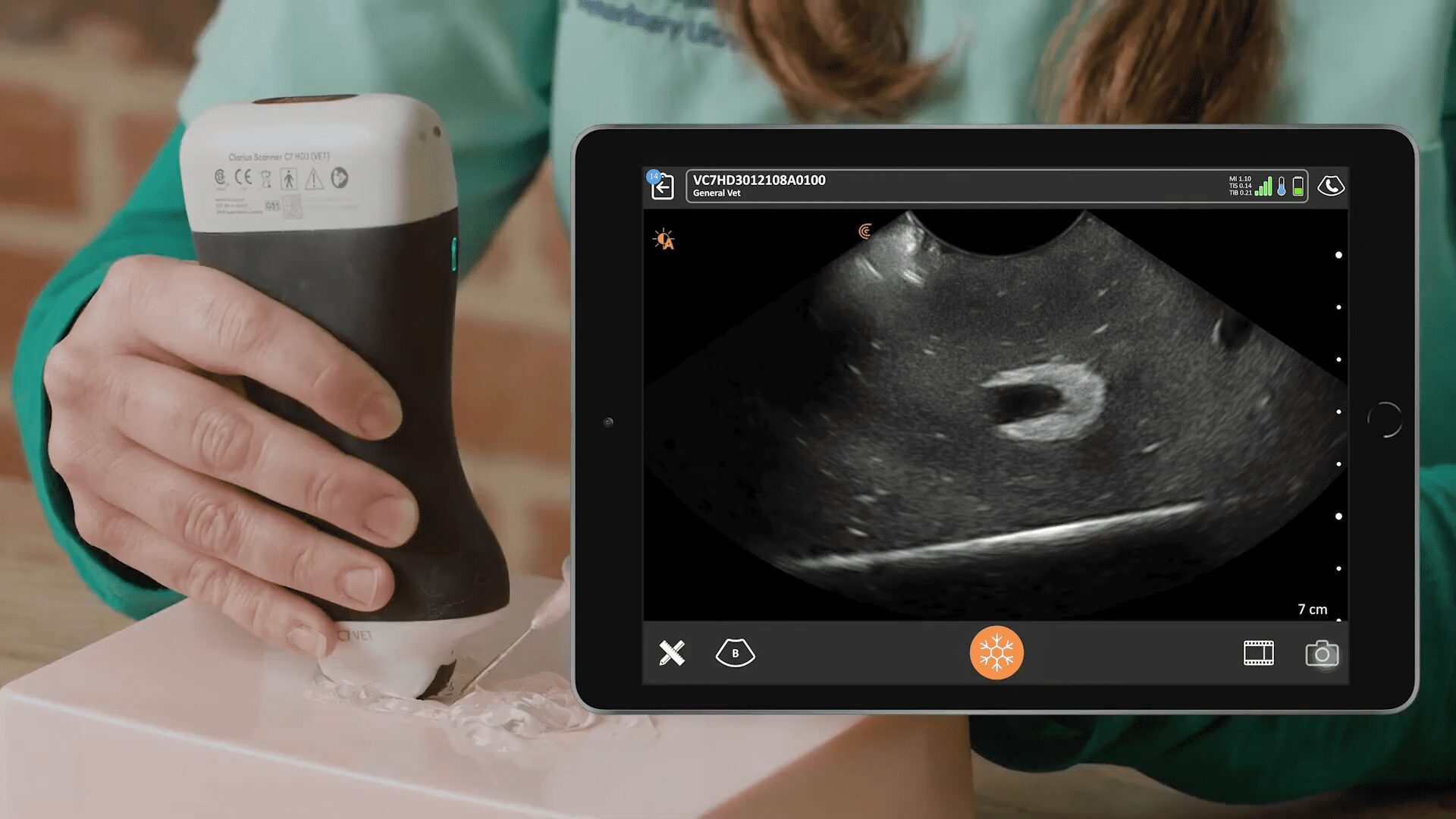
Imaging in the region of the stomach showed some areas of normal-appearing wall consisting of mucosa, submucosa, and muscularis layers. There is a large area that shows a very thick stomach wall with loss of wall layering. Caudal to the thick stomach wall, there is a large, hypoechoic and rounded lymph node that can be easily identified.
The thickness of the normal stomach wall is <= 0.25 cm. In this case, the diameter measured 2 cm. The lymph node had a normal measurement at 0.5 cm, but looked atypical.
Video: Dynamic ultrasound identifying the abnormal area of the stomach and an abnormal adjacent lymph node
Ultrasound-guided fine needle aspirations (FNA) of the stomach wall were performed in attempt to determine pathology, but the samples were inadequate, and therefore inconclusive.

The lymph node was not sampled due to its tricky location. There was risk of bleeding with passing the needle through the liver, or peritonitis if the needle passed through the intestines.
Outcome & Discussion
In cats, a focal mass in the stomach can be neoplastic or inflammatory, but there’s no way of knowing by just looking. The enlarged, rounded, and hypoechoic lymph node supports a diagnosis of inflammation or neoplasm as well – Dr. Edwards says the larger the node, the more likely the mass is neoplastic. The cat is being weaned off steroids so a biopsy can be performed for a definitive diagnosis.
Watch this Clarius Classroom video to learn how to systematically perform an ultrasound of the canine or feline stomach and rule out abnormalities of the stomach wall.
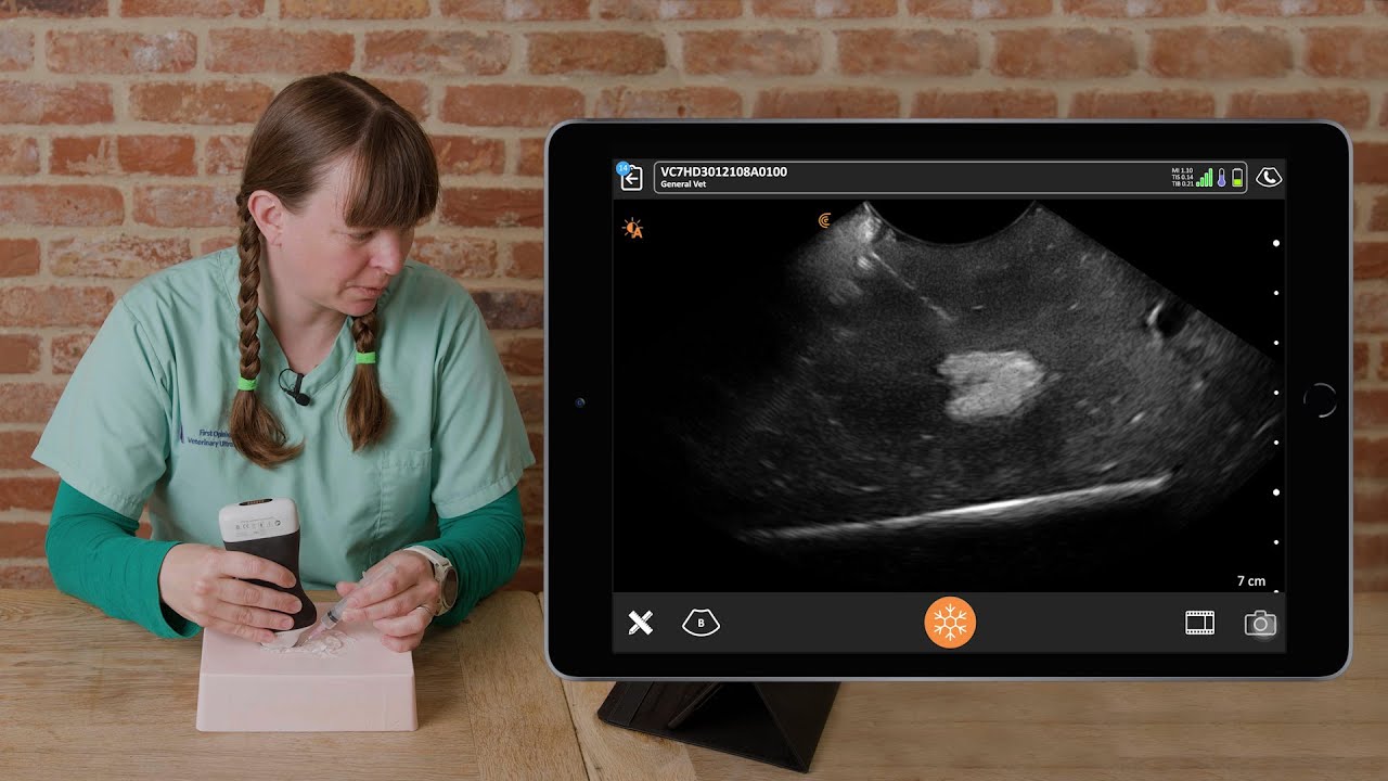
Dr. Edwards uses the Clarius C7 HD3 Vet, which is specifically designed for clinical imaging of small and medium-size animals. Our Advanced Veterinary Package offers more flexibility for users who need additional workflows for various animal exams.
To learn about how easy and affordable it is to add Clarius handheld ultrasound to your veterinary practice, visit our Clarius veterinary ultrasound page for product details. Or contact us today to discuss which scanner is right for your veterinarian practice.

