

Fine needle aspirations (FNA) of abdominal organs and masses are becoming more common in veterinary practices as they can often provide a definitive diagnosis. One of the challenges, though, is obtaining a sample that is diagnostic. The use of ultrasound to guide the needle to the best location is proving to be extremely valuable in increasing the amount of clinically useful samples.
We recently partnered with Dr. Camilla Edwards of First Opinion Veterinary Ultrasound in a webinar dedicated to fine needle aspirations with ultrasound guidance. Dr. Edwards is a peripatetic veterinary sonographer who teaches ultrasound with IMV-imaging and Celtic SMR and delivers her own courses. She uses ultrasound extensively in her practice and believes that everyone in her profession deserves to have the skills and confidence to use veterinary ultrasound to help improve the care of their patients.
Read on for highlights, or watch the free on-demand webinar Practical Small Animal Ultrasound: Guiding Safe, Accurate Fine Needle Aspirations.
Why Should We Aspirate?
Fine needle aspiration is inexpensive, fast, and easy to perform. The need for general anesthesia is rare, and because a small gauge needle is used, there are very few complications. Using ultrasound to plan the safest and shortest route to the target, while avoiding blood vessels and other organs helps to minimize risks, reduce repeat sampling, increase diagnostic yield, and avoid complications.
What Can We Aspirate?
- A Lesion or mass for cytology
- Free fluid for chemistry or cytology
- Fluid from an organ lumen for cytology or chemistry
- Treatment – abscess drainage, pleural effusion, etc.
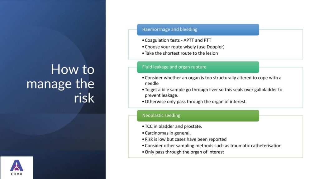
Are There Risks?
According to Dr. Edwards “The complications are pretty rare, having done lots of fine needle aspirates myself, it’s very, very rare that I’ve had complications, but we do have to be aware of them so that we can minimize the risks”. Some potential complications associated with FNA are:
- Hemorrhage and bleeding
- Fluid leakage and organ rupture
- Neoplastic seeding
How to Take an FNA
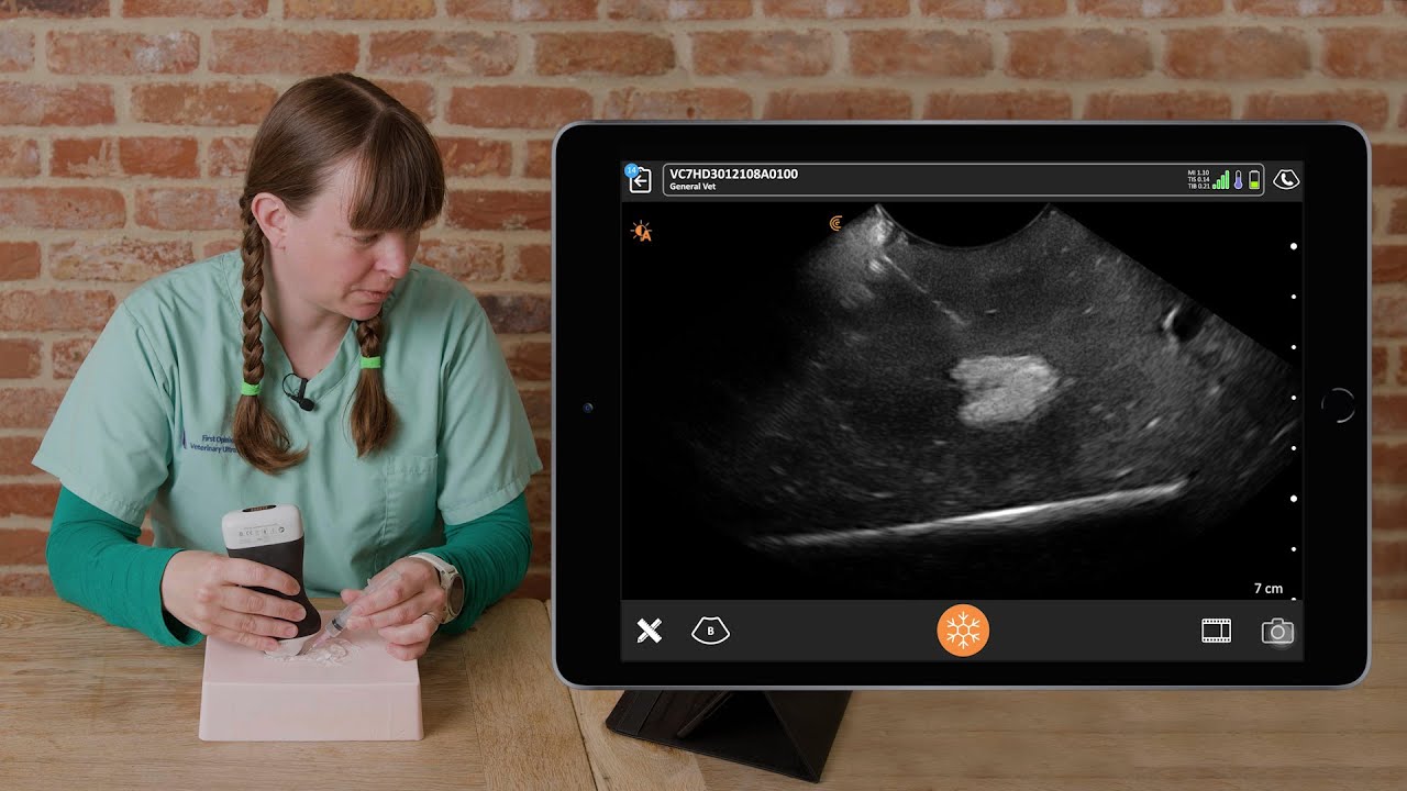
Practice makes perfect, and Dr. Edwards recommends honing your skills using something that can be imaged with ultrasound. There are simulators and ultrasound phantoms available for purchase, but substitutes like a jelly mold, meat, or tofu work very well, especially if something can be inserted into the medium to mimic a mass.
Needle size depends on the size of the patient and the depth of the structure being aspirated. Typically, 21- or 23-gauge needles are suitable. For multiple samples at the same site, use the same syringe, but a new needle for each sample.
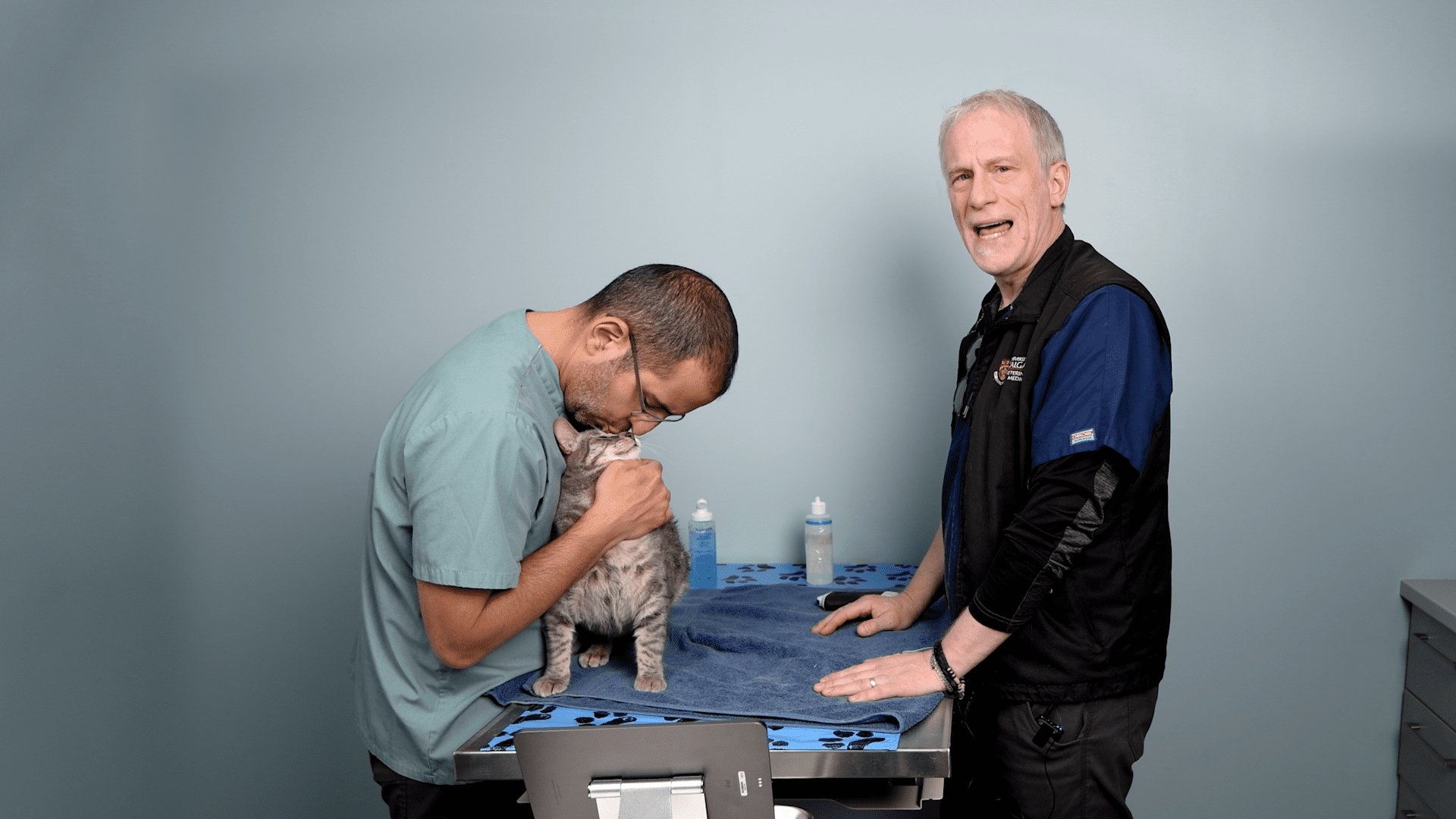
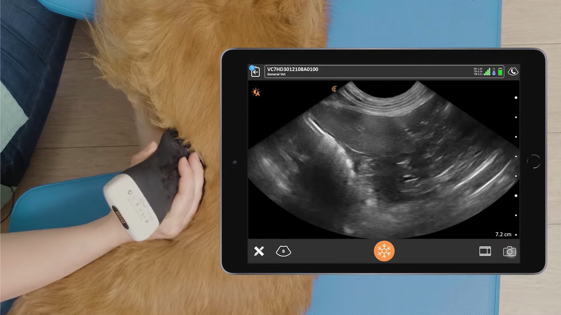
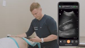
Watch this short video to see how Dr. Edwards does it.
Once you’ve guided your needle to the target area, use a “woodpecker technique” to take multiple samples – the more samples taken, the more likely a diagnosis will be achieved. Immediately after the procedure, repeat imaging with ultrasound to ensure there’s no evidence of bleeding or organ damage, and monitor accordingly.
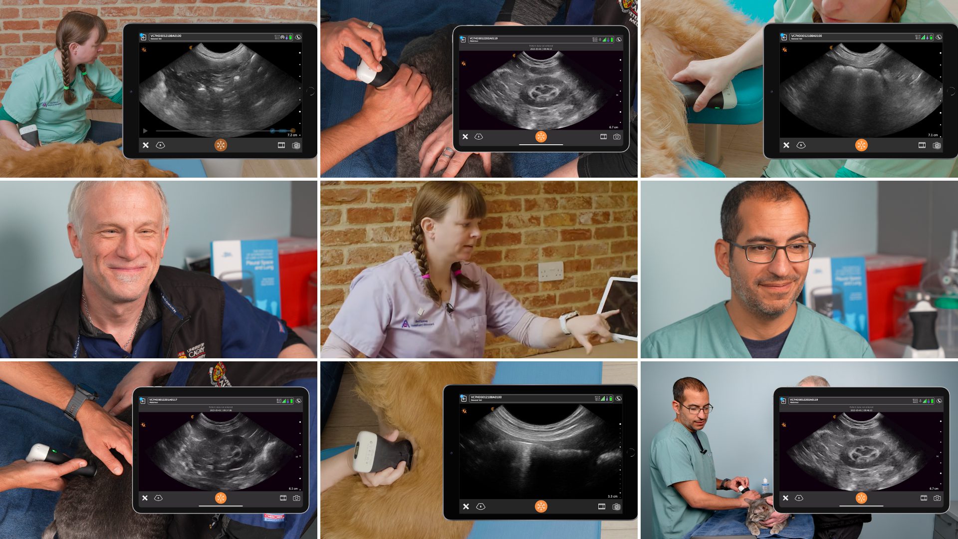
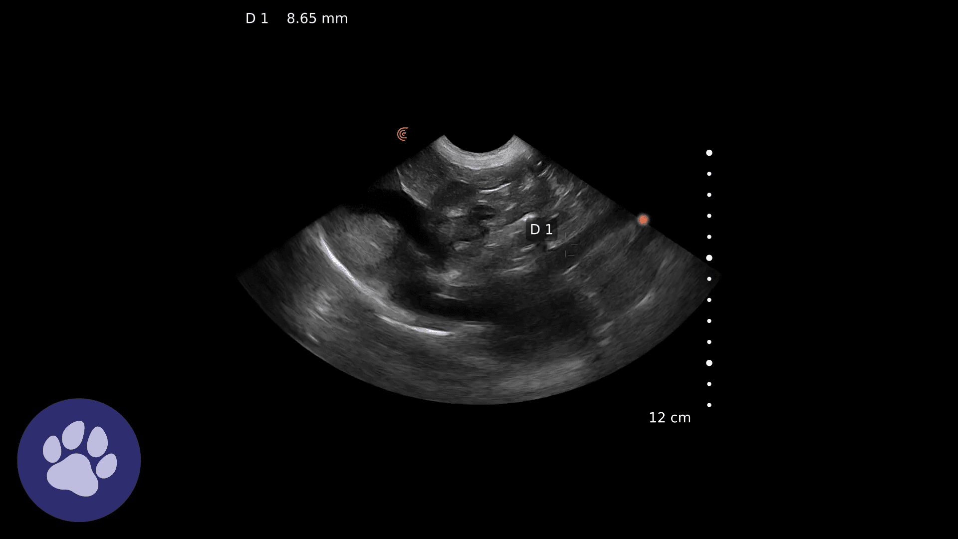
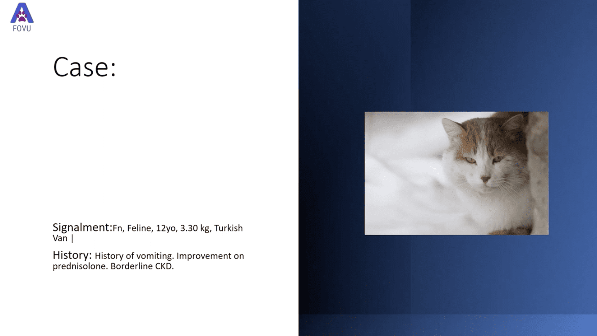
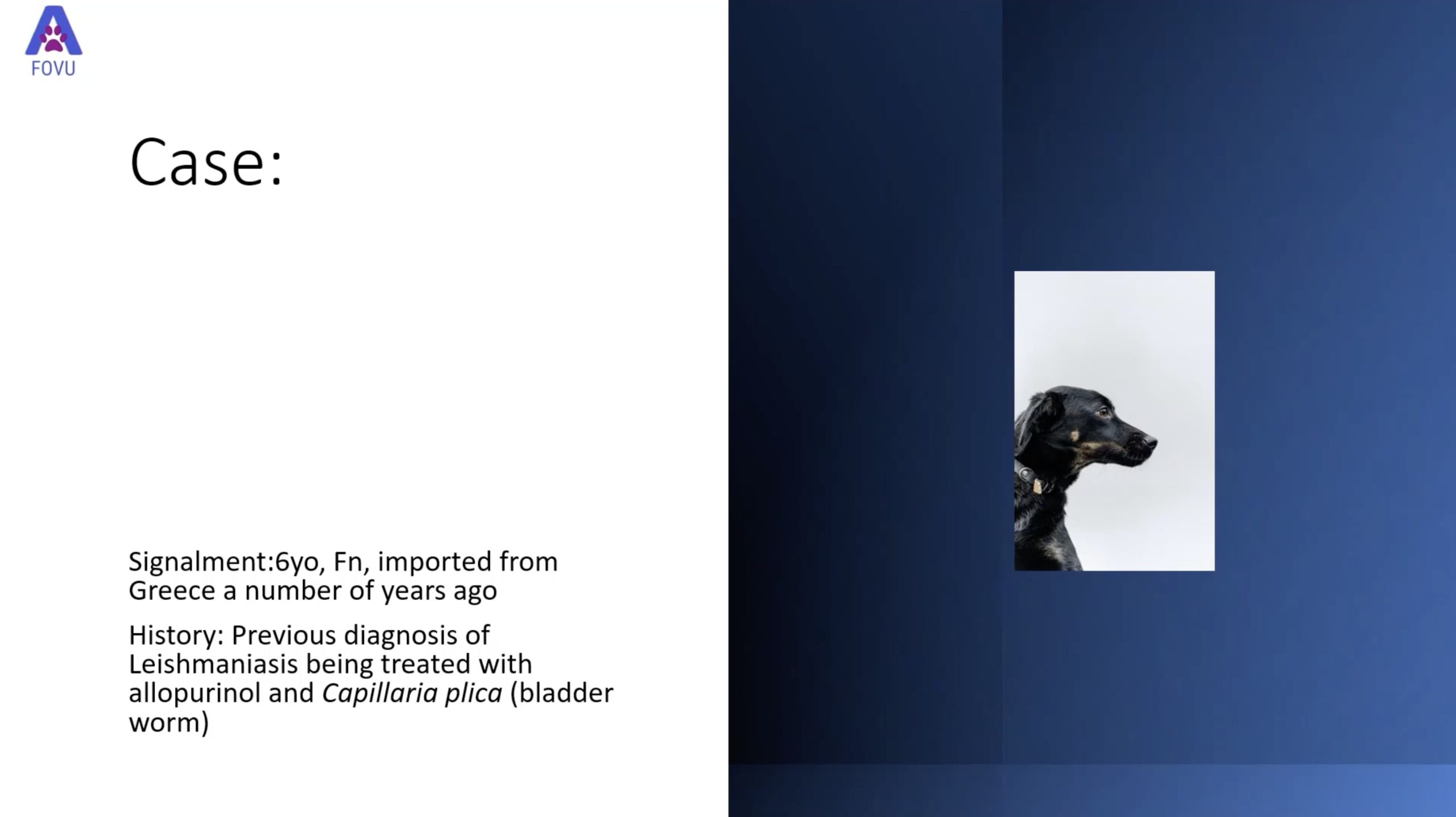
In the webinar, Dr. Edward presents 2 interesting cases complete with patient history and high-resolution ultrasound imaging of pathology prior to FNA. She describes the ultrasound findings, pre-procedure preparations, planning the trajectory of the needle, and live videos showcasing the ultrasound-guided FNA procedures.
For more information about Dr. Edwards and the educational services she provides, visit her website, or get in touch at [email protected].
Clarius Wireless Ultrasound for Veterinary Practice
Dr. Edwards uses the Clarius C7 Vet HD3 scanner in her small animal practice. To learn more about how you can add wireless ultrasound to your practice, visit our Veterinary Specialty Page. There you’ll have access to additional webinars and classroom videos. Learn about the Advanced Veterinary Package provides additional workflows for a wide variety of animal examinations.
Our new Clarius HD3 Vet scanners are smaller and lighter. To find out which scanner is best for your practice, contact us today, or request a virtual ultrasound demo.

