


When the Anatomy is Bigger than the Frame
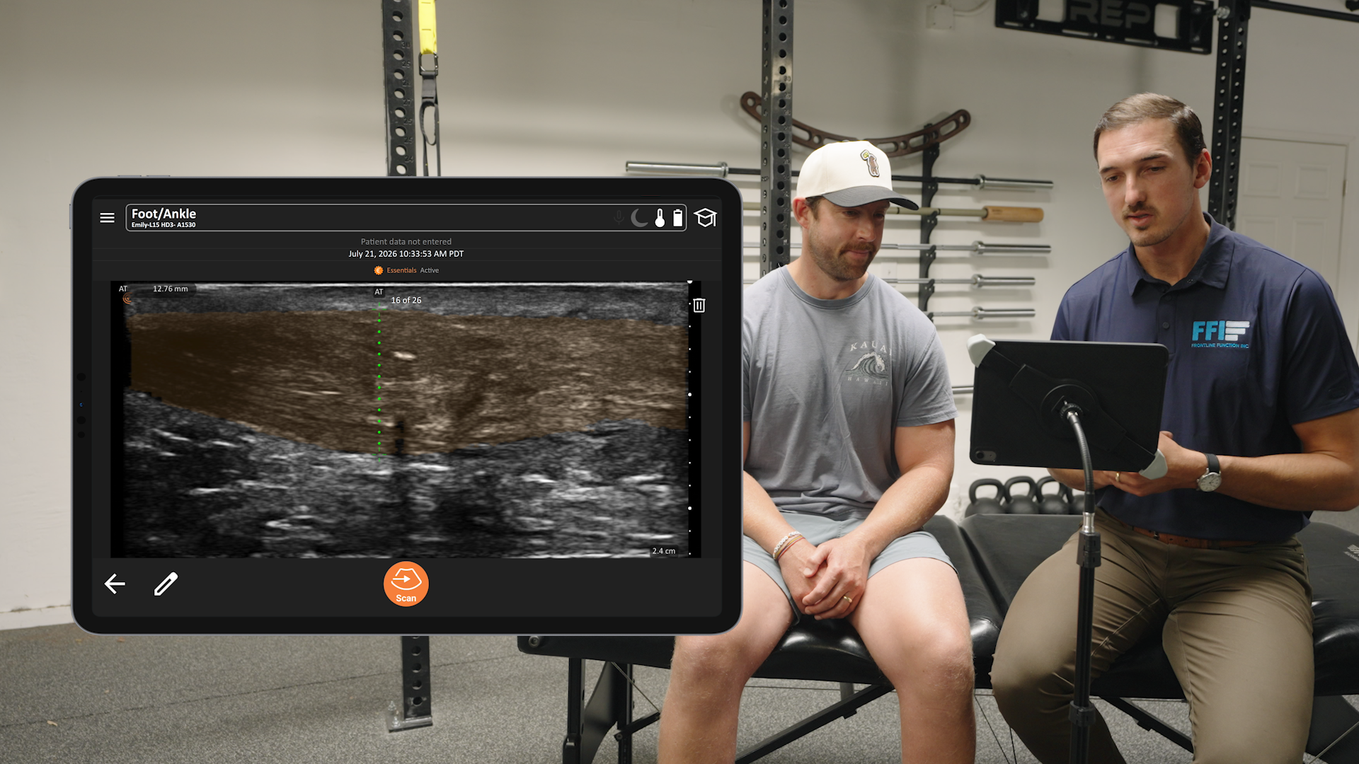
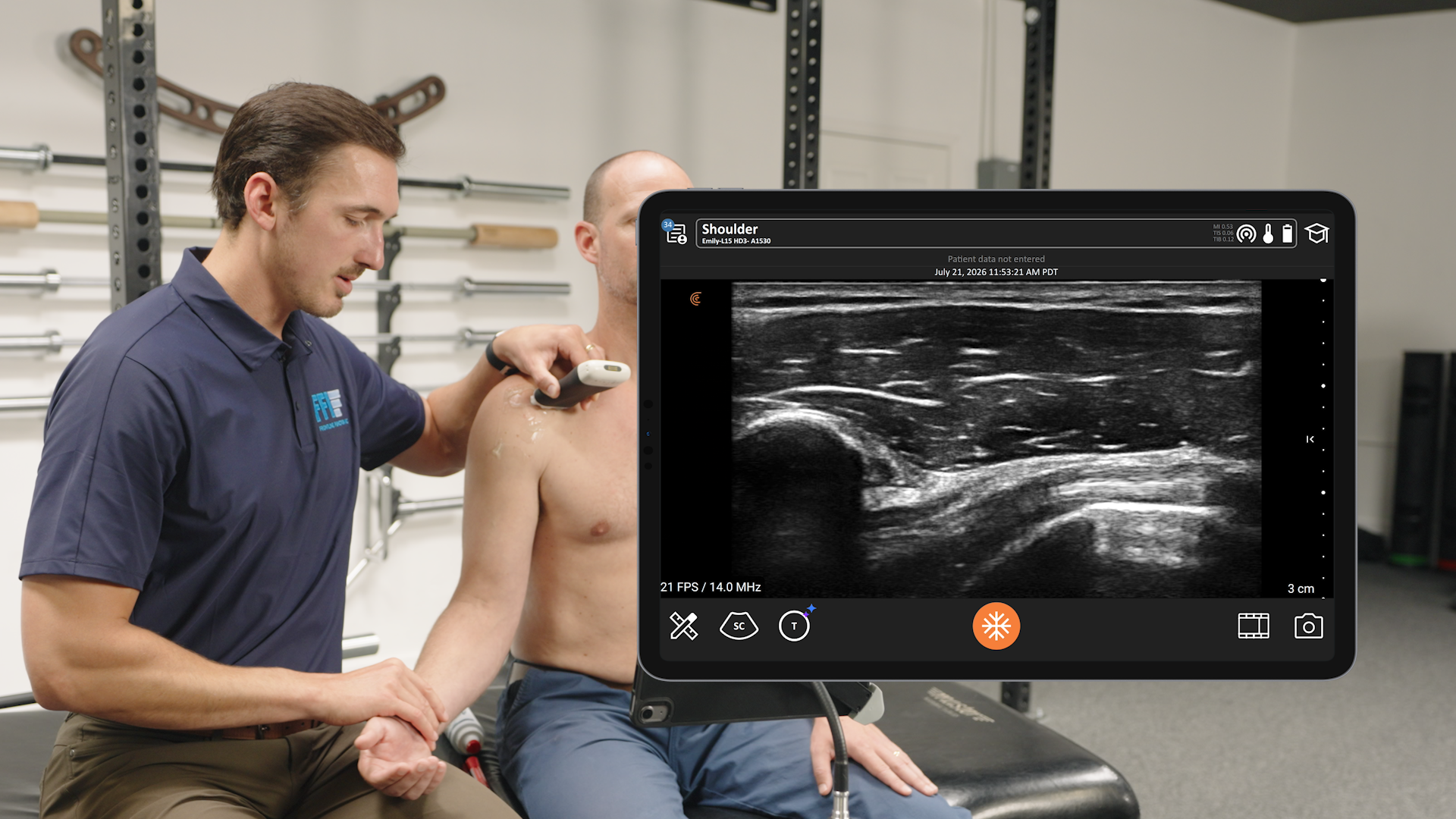
Explaining an injury to a patient is always easier when they can actually visualize it. But with standard ultrasound, long anatomical structures—like the Achilles tendon, quadriceps tendon, or hamstring muscle bellies—rarely fit into a single frame. Showing a patient disjointed 4 cm windows or scrolling through a cine loop often leaves them trying to mentally assemble the pieces.
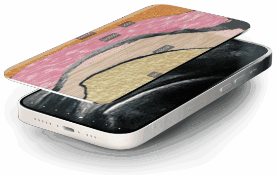
Clarius Panorama changes that. Available now with the release of Clarius App 13.1, it turns one slow sweep into a single image of the full Achilles tendon, hamstrings, quadriceps tendon, or any structure longer than your linear scanner’s footprint. You can show patients the whole structure, not just a slice of it.
Many of the structures MSK clinicians assess with musculoskeletal ultrasound, such as the Achilles tendon, hamstrings, and quadriceps tendon, are longer than a single image. Until now, the options were to record a cine of the sweep or piece the anatomy together mentally. Both are difficult to document and explain to patients.
Extended field of view imaging has been standard on cart-based ultrasound systems for years. Clarius now brings Panorama to its wireless handheld scanners, enabling users to capture structures that are larger than a standard ultrasound image.
How to Use Panorama Mode on Clarius Ultrasound
Panorama runs as an imaging mode within your existing exam, so the workflow is familiar:
- Using a linear scanner, tap the Modes menu, and tap PAN
- The screen splits, with live B-mode displayed for reference, and the panoramic image building alongside it.
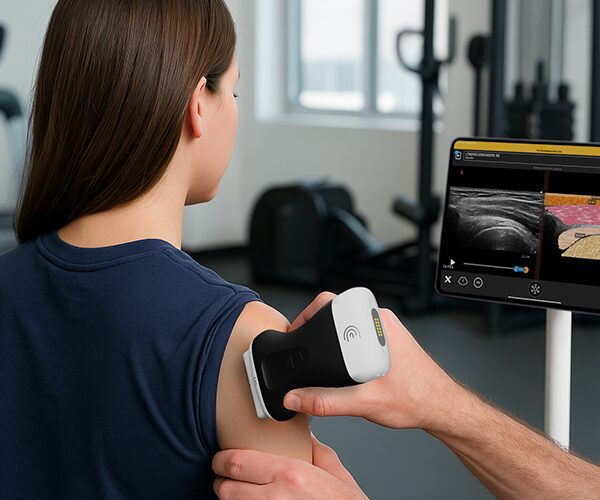
- Maintain good contact (lots of gel) and sweep slowly along the length of the anatomy. The composite image builds in real time.
- Freeze once the full structure is captured, then measure, annotate, and save as you would any other image.
To acquire a new image, unfreeze and sweep again. Each unfreeze starts a new panorama.
Benefits of Clarius Panorama in Clinical Practice
Better patient compliance and communication: When patients see their entire tendon—with the tear, tendinopathy, or repair shown in relation to surrounding healthy tissue—treatment plans and rehab restrictions make immediate sense.
Actionable documentation for referrals: Instead of attaching five separate still frames, a single panoramic image of a long tendon or muscle belly can be added directly to notes or referrals to physicians, surgeons, and physical therapists with unambiguous anatomical context.
Objective progress tracking: Capturing the complete structure across serial visits lets you and your patient track healing, scar remodeling, and structural recovery over the course of rehabilitation
Tips for Capturing a Clean Panoramic Ultrasound Image
- Sweep slowly along the length of the scanner array. A steady, controlled sweep produces the cleanest composite.
- Use sufficient gel, and place the gel along the anatomy to be scanned to ensure a smooth acquisition
- Maintain contact. Place the probe on the patient before or as you activate Panorama. Frames without skin contact are skipped automatically, so a brief lift-off leaves the image intact.
- Interpret measurements with care. Distances measured on a panoramic image may differ from those on a single 2D frame, and the app displays a reminder to this effect.
Availability
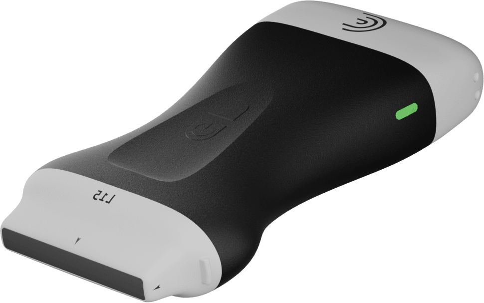
Clarius Panorama is available on the Clarius L7 HD3, L15 HD3, L20 HD3, and PAL HD3. It is included with Clarius Essentials and Clarius membership as part of Panoramic Imaging.
Get Started
The next time a structure extends beyond your standard field of view, activate PAN from the modes menu and start to build your extended image.
To see Panorama in action, watch the Clarius Classroom walkthrough below.
Not yet a member? Learn more about Clarius Essentials to start using Panorama.
What Defines the Best Ultrasound System for MSK Practice?
If you’re evaluating a new handheld ultrasound for MSK, consider looking at four clinical pillars:
- High-Resolution Linear Imaging: Clear visualization of tendons, ligaments, muscle, and nerves, from superficial structures like the patellar tendon and plantar fascia to deeper muscle bellies in the thigh and calf.
- Extended Field of View: Panoramic imaging that captures the full length of a tendon or muscle in a single image for documentation, referrals, and side-to-side comparison.
- Ultrasound-Guided Procedures: Real-time imaging to guide joint, bursa, and tendon sheath injections at the point of care.
- Seamless In-Clinic Workflow: Wireless portability that moves between treatment rooms, the sideline, and home visits, with images that go straight into patient notes and referrals.
Book a private virtual demo to see if Clarius ultrasound is right for your practice.