By providing my email, I consent to receive Clarius webinar invitations, case studies, whitepapers, and more. I can unsubscribe anytime. Privacy Policy.
FREE WEBINAR
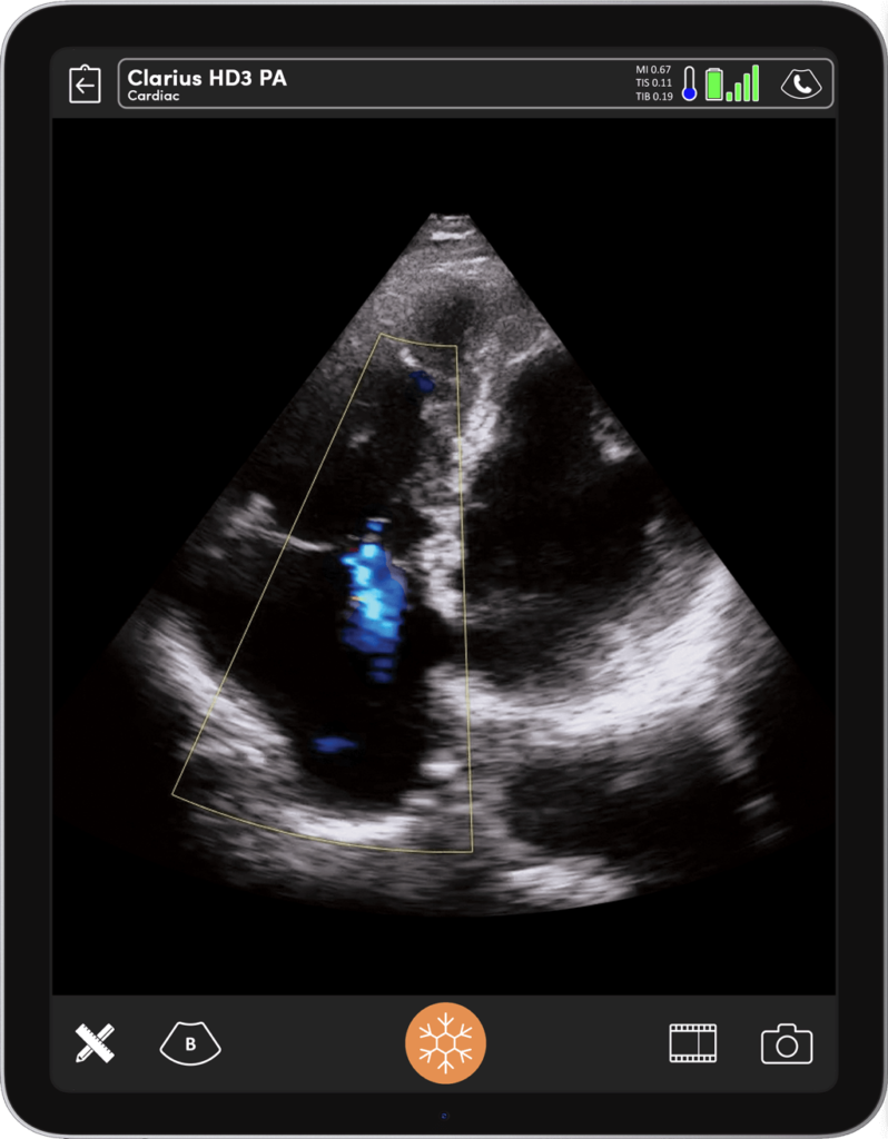
Cardiac POCUS Part 2: Techniques for Assessing Right Ventricular Function
Watch On-Demand
Watch Now
Emergency physician and renowned ultrasound educator Dr. Tom Cook will teach how to accurately assess for signs of right ventricular strain and dysfunction in your patients with chest pain, dyspnea, or unstable vital signs.
In this 1-hour webinar, you will learn to quickly and accurately use POCUS to evaluate the role of the right ventricle in your patient’s pathologic presentations. Dr. Cook will teach how to:
- Visually estimate and quantify the degree of tricuspid regurgitation
- Estimate the pulmonary artery systolic pressure using the tricuspid regurgitation jet
- Estimate the severity of right ventricle dysfunction using the tricuspid annular plane systolic excursion (TAPSE)
- Determine if a patient is showing signs of sub-massive or massive pulmonary embolism
- Identify the different signs of acute versus chronic right heart strain
Pulmonary embolism and other causes of right heart strain lurk on the differential diagnosis for most patients with chest pain or dyspnea. Relying on standard blood testing and radiography protocols can create delays to diagnosis, leaving patients without what can sometimes be life- and function-saving treatment. A quick point-of-care ultrasound exam can make all the difference.
POCUS can rapidly identify a dysfunctional right heart in patients with hypotension immediately narrows the differential diagnosis and can guide clinicians to life-saving treatments. Similarly, diagnosing right heart strain in a patient with suspected pulmonary embolism can immediately triage them as a high risk case and expedite both further diagnostic strategies and treatment.
Dr. Cook will give you the tools you need to quickly, accurately, and reliably determine whether the right ventricle is showing signs of dysfunction or increased pressure. He will teach how to integrate this knowledge into a global assessment and determine the next best steps to care for your patient.
In this dynamic presentation, you’ll see real cases demonstrating healthy and pathological images to help you develop the skills to generate and interpret ultrasound images of patients both already sick or becoming so. Come learn practical knowledge you can use on your next day in the ER, hospital ward, or intensive care unit.
Dr. Cook will be joined by emergency physician Oron Frenkel and sonographer Shelley Guenther for live scanning to help you build practical skills that you can use with your next patient. Don’t miss this webinar – register today!
Emergency Physician
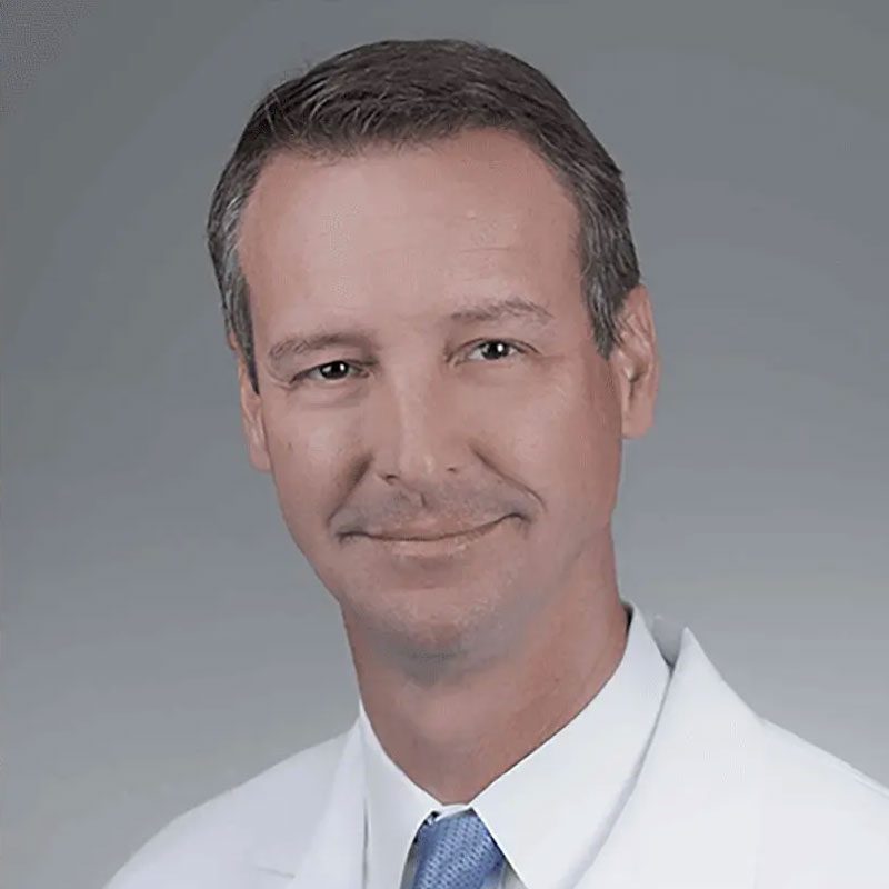
Dr. Tom Cook
Dr. Cook came to Palmetto Health after leaving the Army Medical Corp in 1996. Born in El Paso, Texas, he was raised in Northern Virginia and received his bachelor's degree from the College of William and Mary and attended the University of Virginia for Medical School. He trained in emergency medicine residency at Fort Hood, Texas before taking on a rugged three-year tour at Tripler Army Medical Center in Honolulu, Hawaii. After leaving the army in 1996 he joined the emergency department staff at Palmetto Health where he initially developed the emergency ultrasound curriculum and then founded the nationally recognized 3rd Rock Ultrasound and the Emergency Ultrasound Course. In 2001 he became program director. He lives with his family on Lake Murray in Lexington.
Emergency Physician
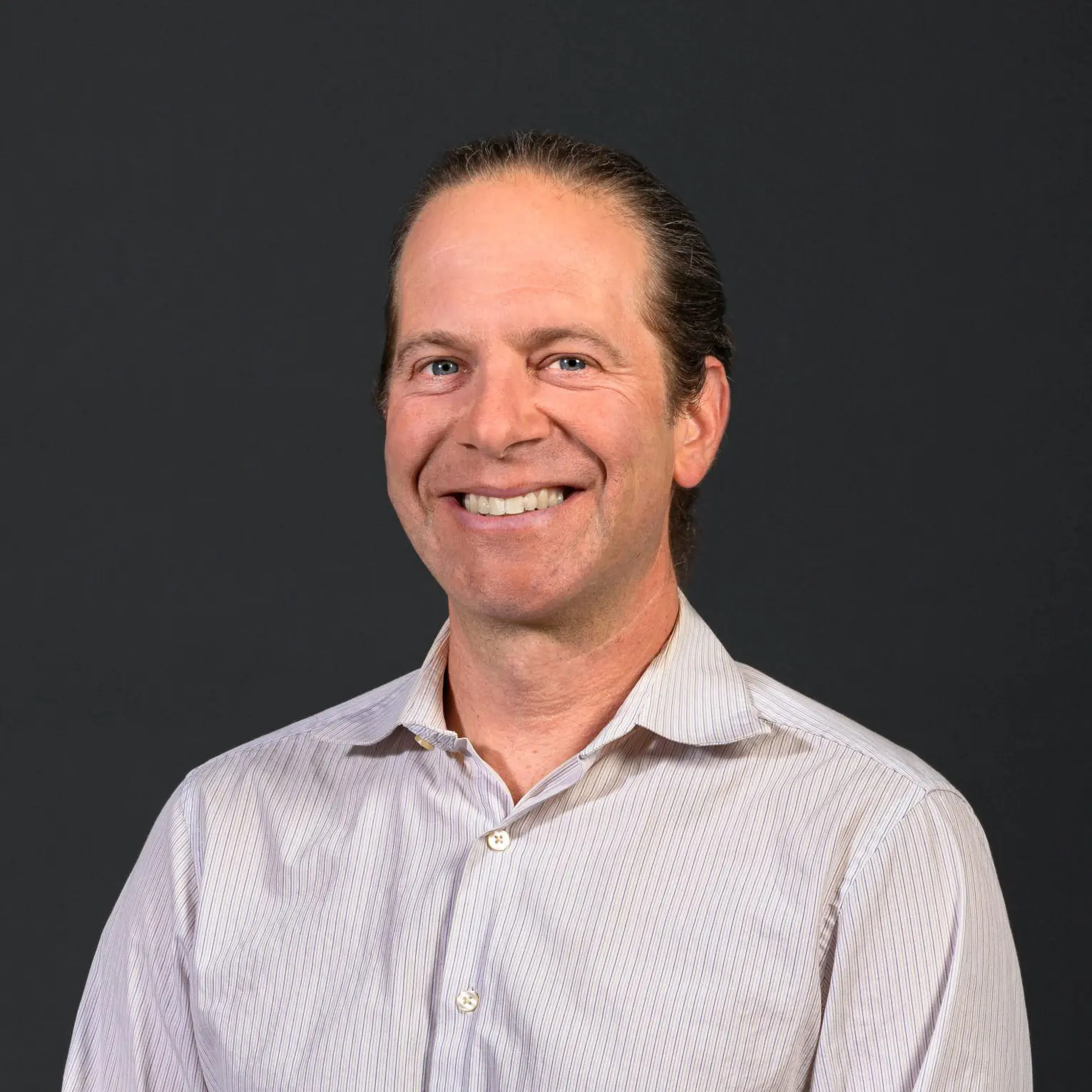
Oron Frenkel, M.D., M.S.
Dr. Oron Frenkel completed his MS and MD simultaneously at the University of California Joint Medical Program in Berkeley and San Francisco, completing his residency in Emergency Medicine followed by a fellowship in Point-of-Care Ultrasound at Alameda County Medical Center in Oakland, California. He moved to British Columbia with the goal of increasing use of point-of-care ultrasound across the province, especially among rural practitioners. An avid educator, Dr. Frenkel is constantly evaluating the best teaching methods for disseminating this technology, how to measure competency in its practice, and its effects on outcomes for individual patients. Dr. Frenkel serves as Chairman of the Clarius Medical Advisory Board.
Medical Sonographer
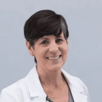
Shelley Guenther, CRGS, CRCS
Shelley Guenther worked as a Nuclear Medicine Technologist for 2 years before entering into the ultrasound program at the Royal Alexandra Hospital in Edmonton. After graduating with specialties in general ultrasound as well as echocardiography, she worked as a clinical expert in the commercial world of ultrasound for over 25 years. As Clinical Manager at Clarius, Shelley Guenther is dedicated to providing the highest quality educational content for clinicians looking to add wireless ultrasound to their practice, including practical webinars and Clarius Classroom video tutorials.