

By providing my email, I consent to receive Clarius webinar invitations, case studies, whitepapers, and more. I can unsubscribe anytime. Privacy Policy.
FREE WEBINAR
Practical Small Animal Ultrasound: Guiding Diagnosis and Management of Liver and Gallbladder Disease
Watch On-Demand
Watch Now
RACE Approved for 1 CE/CPD Credit
Ultrasound educator Dr. Camilla Edwards, DVM, CertAVP, MRCVS is back to share POCUS techniques to identify liver neoplasia and gallbladder disease, specifically mucoceles and sludge, using wireless ultrasound.
During this 1-hour webinar, Dr. Edwards will present feline and canine patient case studies, illustrating her ultrasound techniques and skills for obtaining detailed ultrasound images liver and gallbladder. She’ll teach:
- How to perform a thorough ultrasound exam of the liver
- The ultrasound appearance of normal liver and how to identify neoplasia
- The importance of ultrasound-guided sampling for a definitive diagnosis and prognosis
- Ultrasound tips and patient positioning to optimize visualization of the GB
- How to distinguish between sludge vs mucocele in the gallbladder, and the clinical significance of both
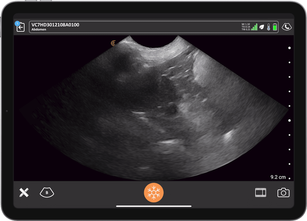
Ultrasound is a commonly used, non-invasive imaging tool for examining abdominal organs. In this webinar, Dr. Camilla Edwards will explore the role of wireless ultrasound in the investigation of liver and gallbladder abnormalities and share her systematic hands-on scanning techniques for getting the most diagnostic images possible.
Liver neoplasia and vacuolar hepatopathy in dogs often pose diagnostic challenges due to overlapping clinical and imaging features. The discovery of liver masses in dogs typically requires ultrasound-guided fine-needle aspiration to confirm the diagnosis, but there are ultrasound characteristics that can help guide next steps in your patient management.
The ultrasound discovery of a neoplastic hepatic mass in cats, however, is more likely to be metastatic spread from an abdominal source, as hepatocellular carcinoma in cats is very rare.
When ultrasound uncovers an abnormally distended gallbladder in a patient with elevated liver values, it’s important to be able to distinguish between benign sludge and potentially life-threatening mucocele. Patient positioning and imaging findings can play a role in accurately differentiating between the two conditions.
This webinar will equip veterinary practitioners with the knowledge and tools necessary to confidently diagnose and manage the above-mentioned diseases, enable clinicians to make informed decisions regarding patient management and surgical intervention when necessary.
Don’t miss this educational webinar – register today to brush up on your practical veterinary ultrasound skills!

Peripatetic Veterinary Ultrasonographer | Educator | First Opinion Veterinary Ultrasound
Dr. Camilla Edwards, DVM, CertAVP, MRCVS
Dr. Camilla Edwards is passionate about first opinion level small animal veterinary ultrasound. She travels with her dog Pippi (a Nova Scotia Duck Tolling Retriever) within 50 miles of Cambridge as a peripatetic veterinary ultrasonographer, Camilla teaches ultrasound through FOVU and has built a thriving Facebook community for First Opinion Small Animal Vets. Through her website, www.fovu.co.uk, she reviews ultrasound machines with general practice small animal vets in mind. Camilla qualified as a vet in 2006 and has worked all over East Anglia, UK. Camilla is experienced in emergency and critical care, having gained her CertAVP in 2018.
Emergency Physician
Oron Frenkel, M.D., M.S.
Dr. Oron Frenkel completed his MS and MD simultaneously at the University of California Joint Medical Program in Berkeley and San Francisco, completing his residency in Emergency Medicine followed by a fellowship in Point-of-Care Ultrasound at Alameda County Medical Center in Oakland, California. He moved to British Columbia with the goal of increasing use of point-of-care ultrasound across the province, especially among rural practitioners. An avid educator, Dr. Frenkel is constantly evaluating the best teaching methods for disseminating this technology, how to measure competency in its practice, and its effects on outcomes for individual patients. Dr. Frenkel serves as Chairman of the Clarius Medical Advisory Board.
Medical Sonographer
Shelley Guenther, CRGS, CRCS
Shelley Guenther worked as a Nuclear Medicine Technologist for 2 years before entering into the ultrasound program at the Royal Alexandra Hospital in Edmonton. After graduating with specialties in general ultrasound as well as echocardiography, she worked as a clinical expert in the commercial world of ultrasound for over 25 years. As Clinical Manager at Clarius, Shelley Guenther is dedicated to providing the highest quality educational content for clinicians looking to add wireless ultrasound to their practice, including practical webinars and Clarius Classroom video tutorials.