By providing my email, I consent to receive Clarius webinar invitations, case studies, whitepapers, and more. I can unsubscribe anytime. Privacy Policy.
FREE WEBINAR
Pragmatic MSK Ultrasound: 8-Step Protocol for Assessing the Distal Biceps Tendon
Watch On-Demand
Watch Now
Renowned SonoSkills founder and educator Mr. Marc Schmitz teaches his 8-step ultrasound protocol for confidently assessing the distal biceps tendon and diagnosing pathologies.
In this 1-hour webinar, learn MSK ultrasound techniques to improve your scanning skills and gain a deeper understanding of the distal biceps tendon. With a focus on anatomy, biomechanics, and pathophysiology, expert sonographer Mr. Schmitz teaches:
- Normal anatomy including origins, insertions, and surrounding structures
- Common pathologies, injuries, and pathophysiologic mechanisms in the region
- The ultrasound appearance of the distal biceps tendon and connected structures
- Eight approaches to examine the distal biceps with ultrasound
- Scanner and patient positioning for optimal diagnostic imaging
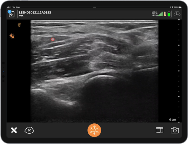
With distal biceps tendon injuries, early diagnosis of tendon tear or rupture with high-resolution ultrasound is key to early treatment and better outcomes. SonoSkills educator Mr. Schmitz demonstrates techniques using high-definition wireless ultrasound to diagnose tendon pathologies without relying on other medical imaging modalities.
Scanning the distal bicep tendon and insertion onto the radial tuberosity can be challenging due to its small size, deep location, and oblique course. Additionally, the surrounding structures such as the radial tuberosity, brachialis muscle, and bicipital aponeurosis can create artifacts and obscure visualization. For these reasons, accurate assessment has historically required a combination of imaging modalities including MRI and CT. Mr. Schmitz demonstrates eight approaches for a thorough, accurate, and confident point-of-care ultrasound assessment to limit dependance on other imaging modalities.
Mr. Schmitz demonstrates the ventral approach, providing a look at the bicep tendon and the lacertus fibrosus. With proper patient positioning, the tendon is then be examined in transverse, where it can be seen between the brachial artery and cephalic vein, and in long axis, where fluid in or around the tendon can be easily identified.
Examination of the bicep tendon from the dorsal approach is a favorite for Mr. Schmitz. He’ll show how to perform the exam with the patient’s arm in a “cobra” position, which provides access to the bicep tendon insertion onto the radial tuberosity. He localizes the tendon and then performs both static and dynamic assessments to look for bursitis and impingement, and in long axis, visualize the broad tendon insertion.
Mr. Schmitz demonstrates the lateral approach, which is great for evaluating the distal tendon, not including the insertion. Dynamic pro- and supination are helpful in evaluating tendon fibers and can confirm if the insertion is intact. In this position, Mr. Schmitz explains how to evaluate tendon thickness and identify abnormalities such as fluid and calcifications.
Lastly, he explores the medial approach, which requires the forearm to be slightly flexed, with the scanner positioned in line with and over the distal humerus. From here, the brachial artery is an important landmark, and Mr. Schmitz uses it as an acoustic window to enhance visualization of the bicep tendon, including the insertion.
Mr. Schmitz is joined by your host Shelley Guenther. Together, they show you how to perform a thorough diagnostic examination of the distal biceps using high-resolution wireless ultrasound. You’ll learn a systematic protocol you can use for your next biceps tendon exam for a confident diagnosis. Don’t miss this webinar – watch now!

Founder & CEO of SonoSkills
Marc Schmitz, MSc
During his MSc. at the Faculty of Medicine and Pharmacy at the Vrije Universiteit Brussel, Marc’s interest in functional human anatomy grew tremendously while dissecting and studying cadavers. It was here that his interest in observing functional human anatomy in “real-time” peaked, which led to the start of embracing musculoskeletal ultrasound. After completing numerous courses and attending conferences on the topic, he started developing his own course and SonoSkills was formed in 2010. Since then SonoSkills has taught its courses in 30 countries. Marc’s team consists out of 22 expert trainers. He teaches, next to managing SonoSkills, the foundational course and some intermediate level courses. Marc has a special interest in the shoulder joint.

Medical Sonographer
Shelley Guenther, CRGS, CRCS
Shelley Guenther worked as a Nuclear Medicine Technologist for 2 years before entering into the ultrasound program at the Royal Alexandra Hospital in Edmonton. After graduating with specialties in general ultrasound as well as echocardiography, she worked as a clinical expert in the commercial world of ultrasound for over 25 years. As Clinical Manager at Clarius, Shelley Guenther is dedicated to providing the highest quality educational content for clinicians looking to add wireless ultrasound to their practice, including practical webinars and Clarius Classroom video tutorials.