


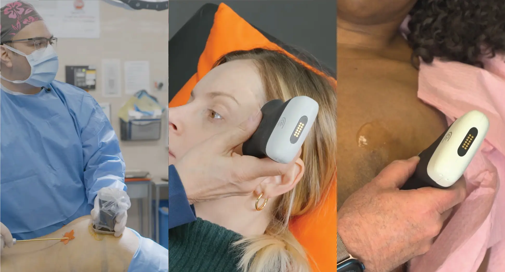
Recent advancements in breast cancer surgery have highlighted the critical role of ultrasound technology in enhancing precision and improving patient outcomes. A review of published studies reveals compelling evidence supporting the use of ultrasound throughout the surgical journey – from preoperative planning to postoperative care.
Dr. Muneer Ahmed, a leading, innovative, and enthusiastic breast cancer surgeon practicing in the United Kingdom, is a passionate ultrasound advocate and shares his best practices for improving outcomes for breast cancer patients, in a one-hour webinar: Enhancing Precision in Oncoplastic Breast Surgery: The Role of Ultrasound in Pre-, Peri- and Postoperative Assessment. Watch the webinar recording at your convenience to learn how easy and affordable it is to add handheld ultrasound to your practice to improve oncologic effectiveness and cosmetic results. Scroll down for some highlights of the presentation.
Preoperative Applications for Handheld Ultrasound
Surgical Planning
Ultrasound proves invaluable in surgical planning, allowing surgeons to:
- Precisely locate lesions
- Determine optimal incision sites
- Plan appropriate oncoplastic techniques
By scanning patients in the clinic, surgeons can visualize the exact location of tumors, facilitating more informed decisions about surgical approaches.
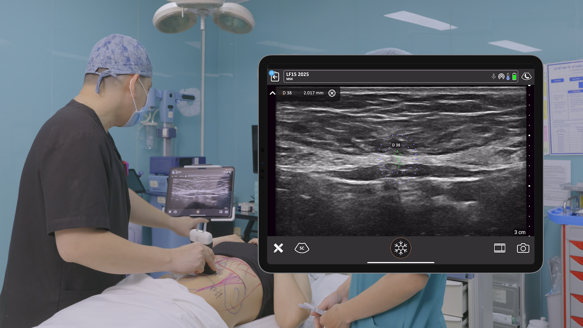
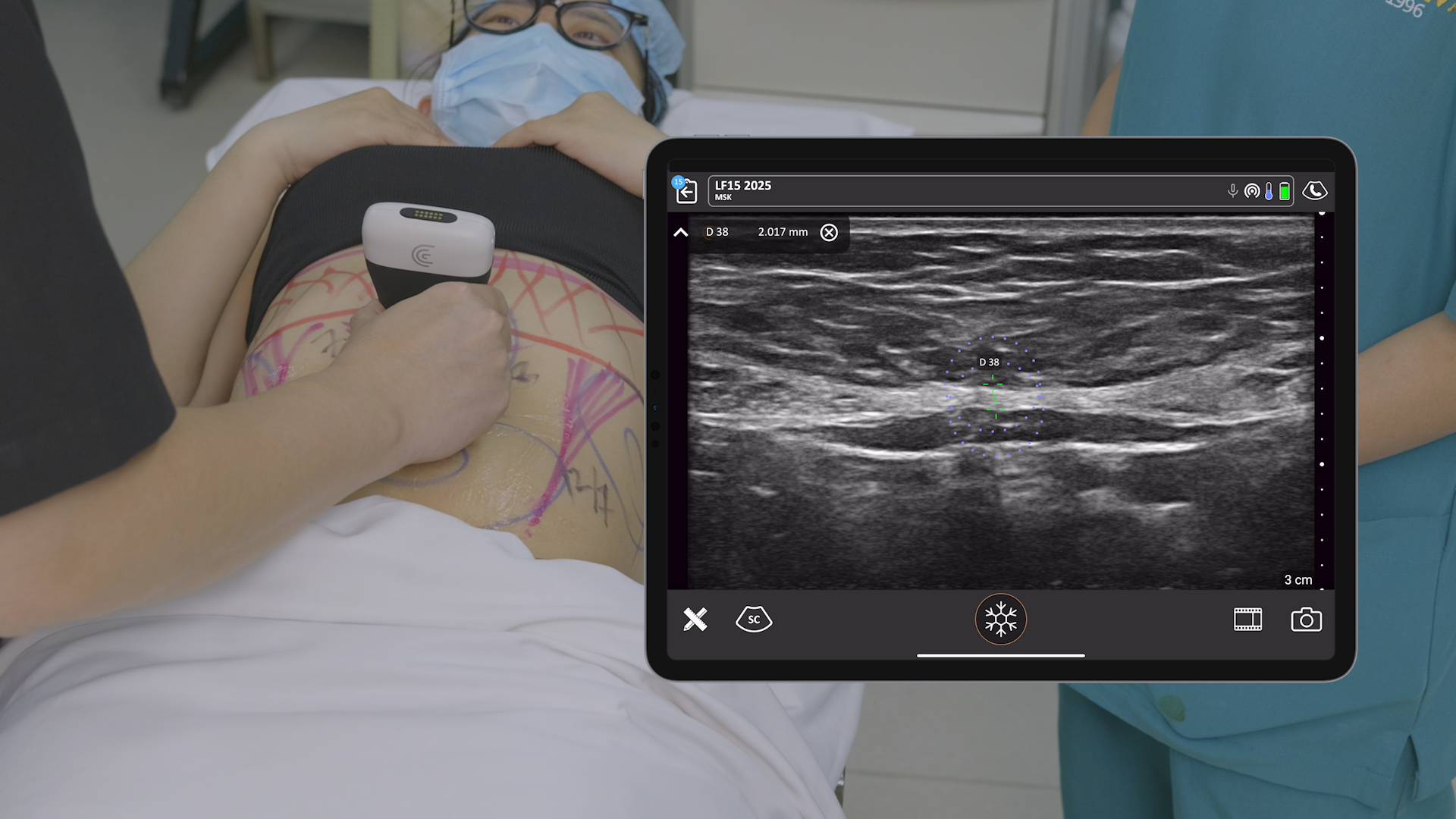
Intraoperative Uses
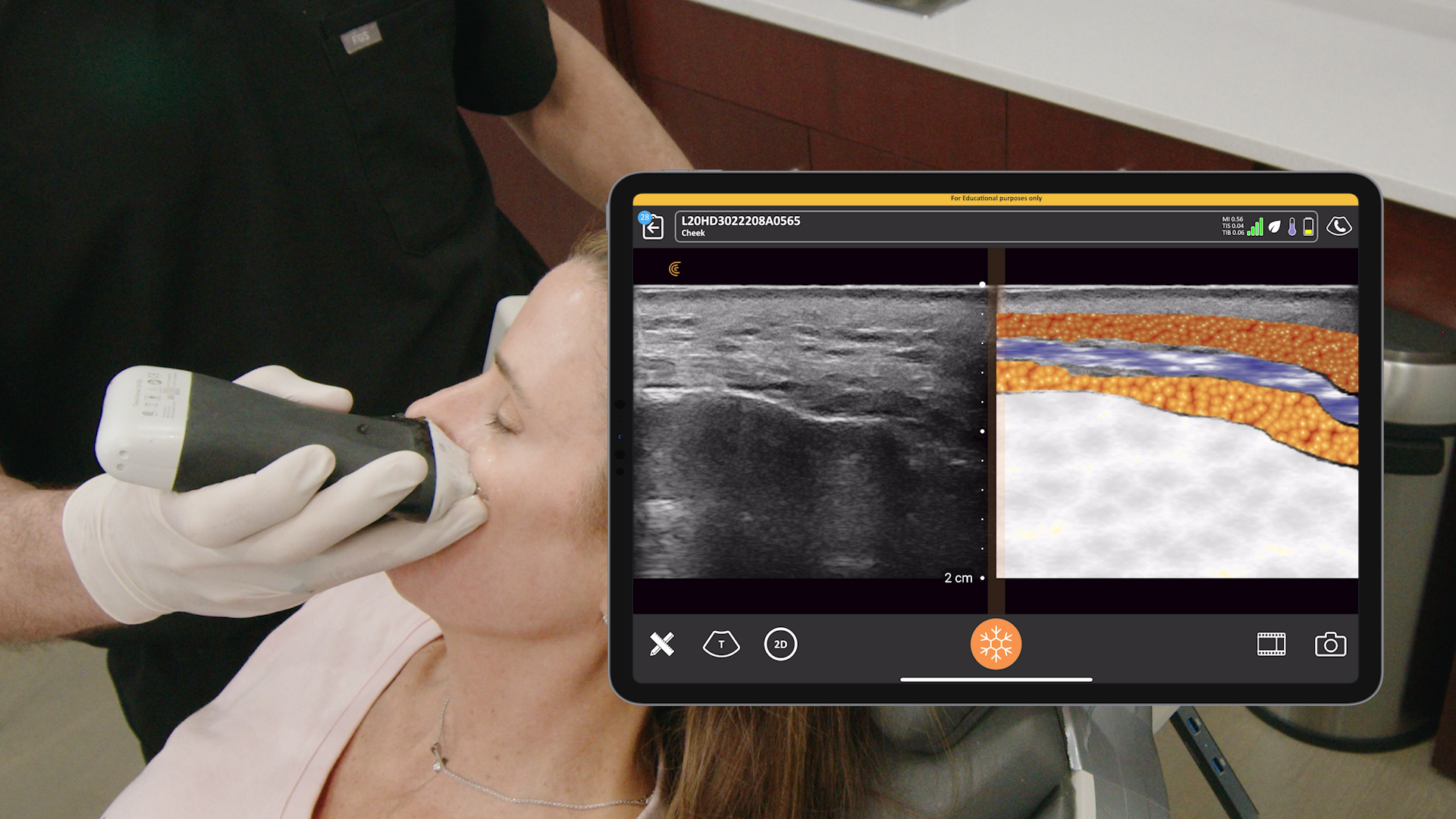
Real-Time Visualization
Intraoperative ultrasound offers surgeons real-time visualization of tumors during excision. This approach has demonstrated several advantages:
- Eliminates the need for fine wire localization
- Provides greater logistical flexibility
- Allows for precise tumor removal with adequate margins
Margin Assessment
Surgeons can perform ex-vivo margin assessment immediately after tumor excision, potentially reducing re-excision rates.
Postoperative Care
Ultrasound continues to play a crucial role postoperatively:
- Assessing and managing seromas or abscesses
- Evaluating skin flap thickness for implant-based reconstruction
- Monitoring response to neoadjuvant chemotherapy
Learning Curve and Accuracy
Encouragingly, the learning curve for surgeons to become proficient in intraoperative ultrasound is relatively short. Studies suggest that surgeons with prior ultrasound experience may only need about 5 supervised cases to achieve competency. Research indicates that surgeon-performed ultrasound in outpatient settings shows high concordance with radiologist interpretations, with discordance rates below 4%.
The integration of ultrasound technology throughout the breast cancer surgical journey offers numerous benefits. From enhancing surgical precision to improving postoperative care, ultrasound is proving to be an indispensable tool for modern breast surgeons. As more surgeons adopt this technology, we can expect to see continued improvements in patient outcomes and satisfaction.
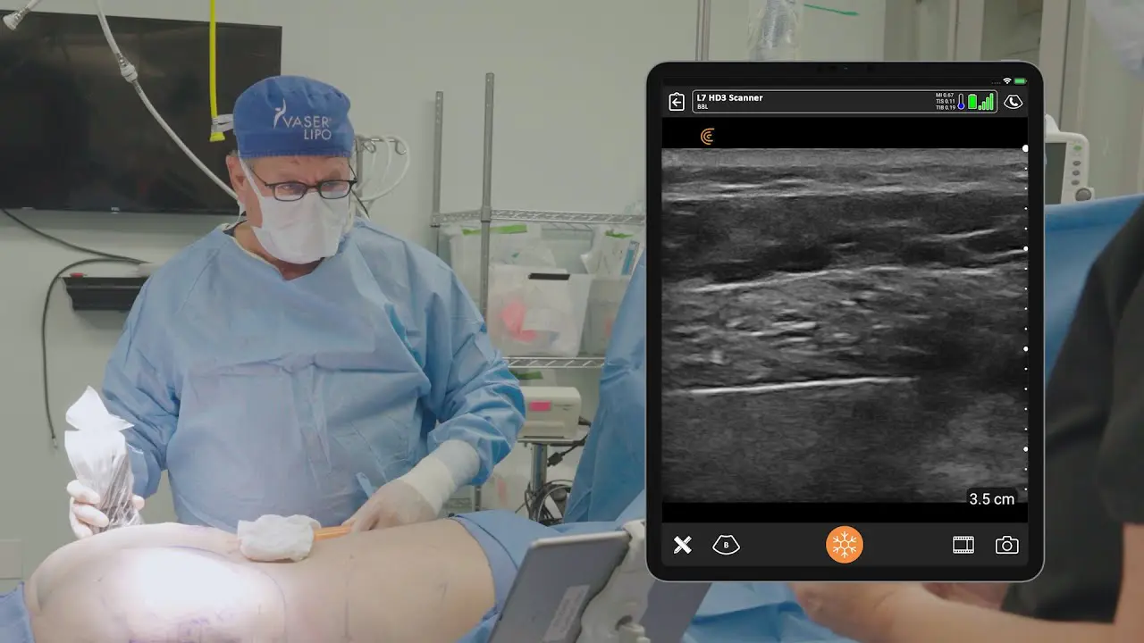
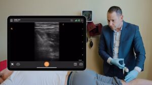
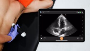
Video Demonstration: Intraoperative Breast Cancer Clip Localization with Ultrasound
Watch this 3-minute video to see Dr. Ahmed use high-resolution ultrasound in the operating room to locate multiple marker clips in a patient with breast tumours to reduce the excision of margins intra-operatively.
About Dr. Muneer Ahmed
Muneer Ahmed is a leading, innovative, and enthusiastic breast cancer surgeon. He believes in enhancing patient experiences through the application of minimally invasive techniques for bespoke breast cancer management. He trained at the most advanced and comprehensive breast surgical oncology and reconstructive centres in the UK, Europe, and Japan.
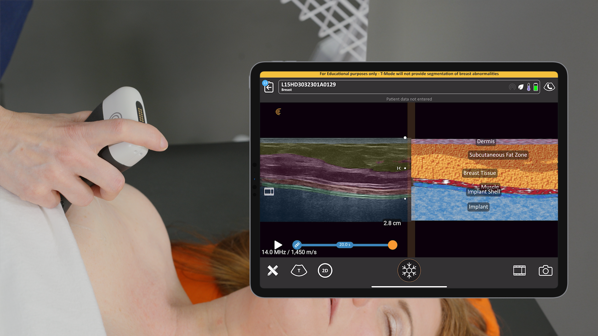
Clarius Ultrasound for Breast Surgery
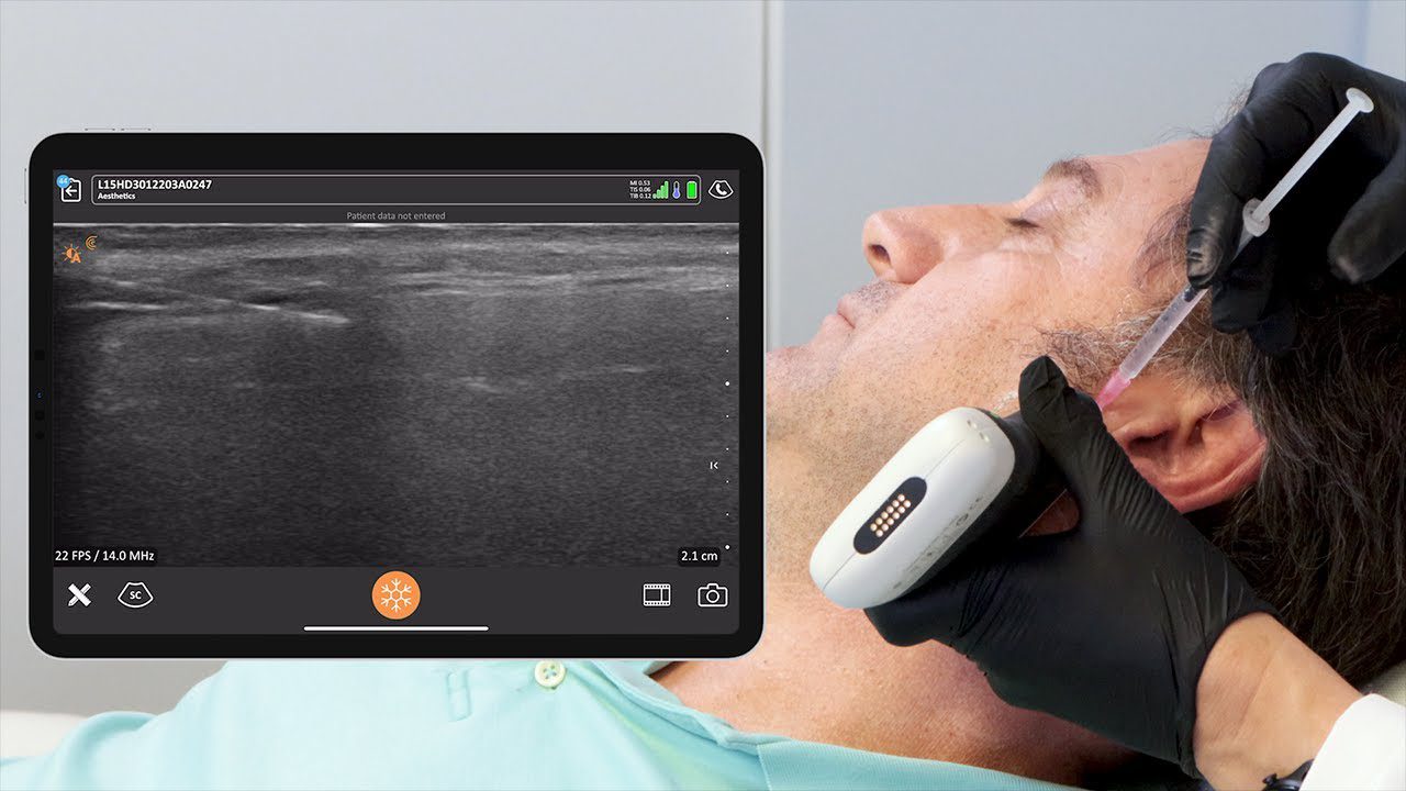
Dr. Ahmed uses the Clarius L15 HD3 high-frequency linear scanner at his practice with the Advanced Breast Package, which offers additional customizations for breast examinations and procedures. The software package is included when a membership is purchased with the Clarius L7 HD3 or Clarius L15 HD3 linear scanners.
To learn which Clarius scanner is right for your practice, book a demo today with a Clarius expert.

