A safe, painless, and inexpensive exam, ophthalmic ultrasound has become an important tool to diagnose and monitor a wide range of eye conditions.
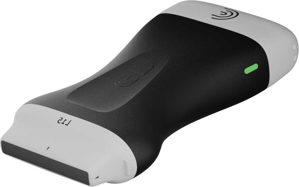
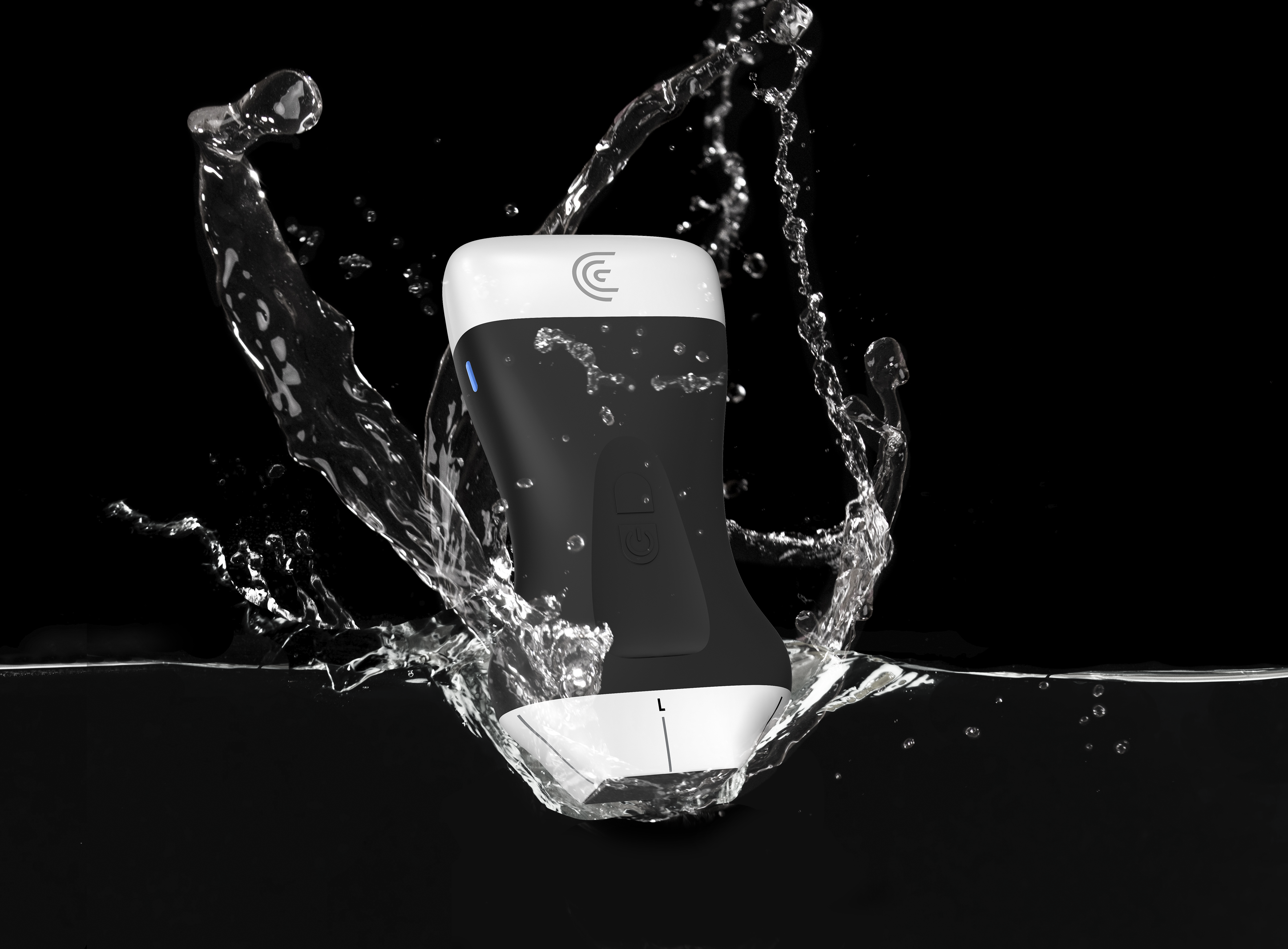
Since the introduction of the Clarius L20 HD3, the world’s first ultra-high frequency wireless handheld scanner, it has become a popular choice for ophthalmologists, enabling real-time, detailed imaging of the structures and movements of the eye.
With the release of its latest software update, Clarius is pleased to announce the availability of A-Mode, or Amplitude mode, which enables measurements of the axial length of the eye, aiding in the diagnosis and monitoring of conditions such as myopia, hyperopia, and cataracts. It is also valuable for assessing retinal and choroidal pathologies.
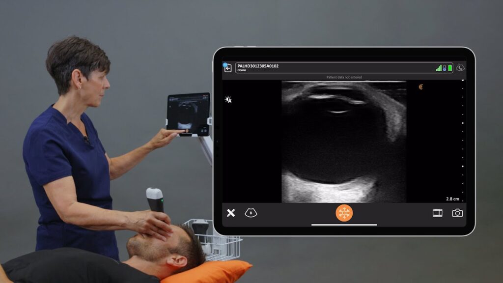
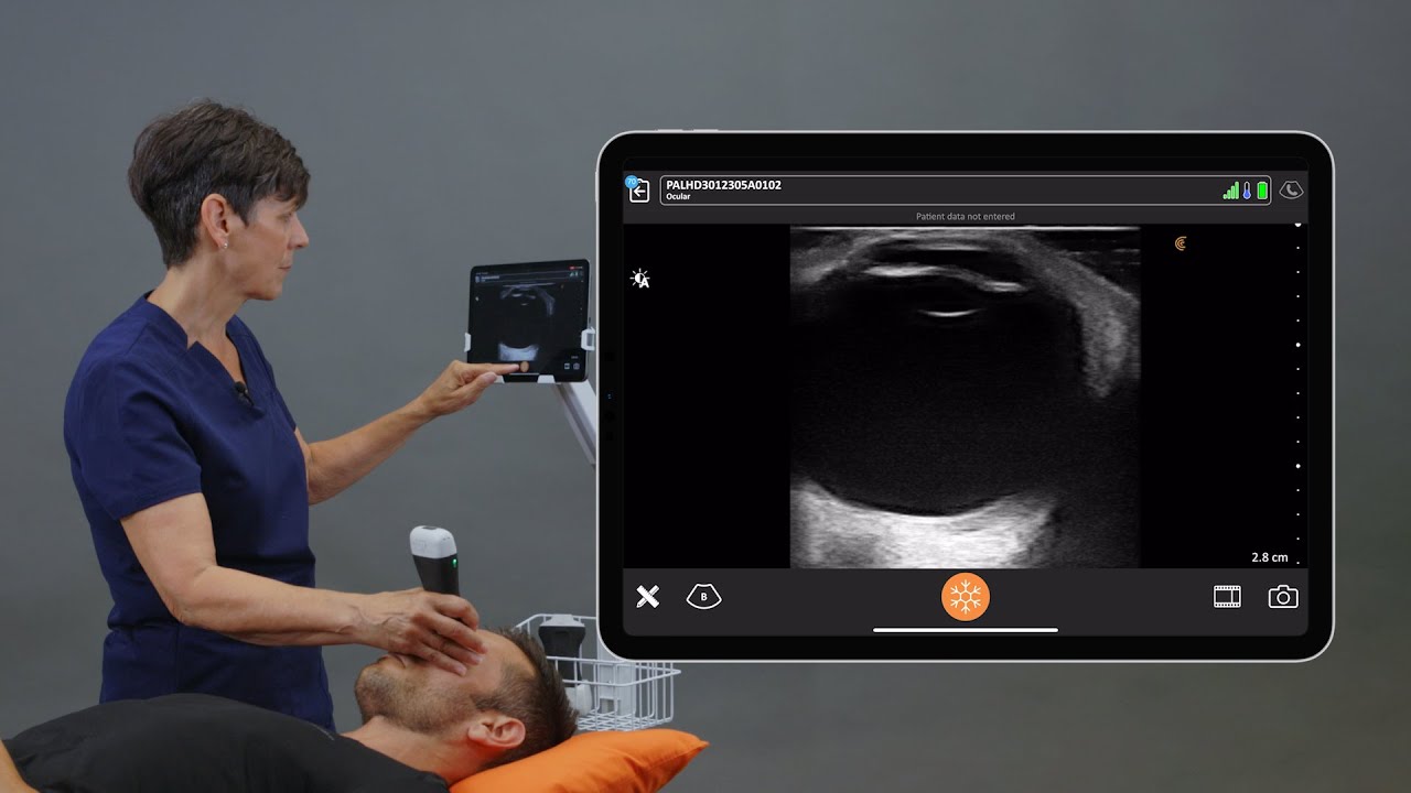
Watch this 90-second video to see a demonstration of A-Mode by Shelley Guenther, CRGS, CRCS. She uses the Ophthalmology Preset to ensure optimal image clarity with minimal adjustments.
Clarius L20 HD3 Ranks Highest for Ophthalmic Imaging Among Five POCUS Devices
In 2023, the Clarius L20 HD3 received a first-place ranking for orbital imaging in an independent research study conducted at the Keck School of Medicine of University of Southern California by Kristen E Park, Preeya Mehta, Charlene Tran, Alomi O Parikh, Qifa Zhou, and Sandy Zhang-Nunes.1
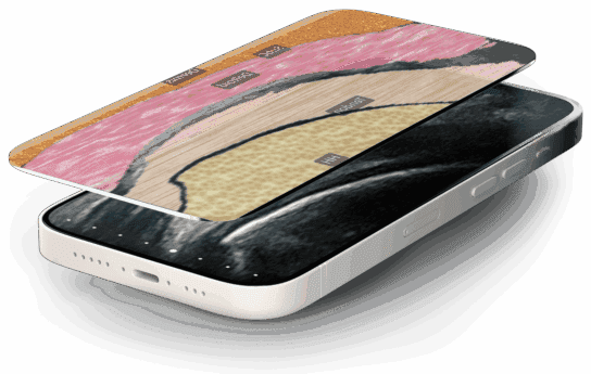
With exceptional superficial imaging, the Clarius L20 HD3 is the only specialty-designed handheld ultrasound with ultra-high frequency to 20 MHz, delivering superior visualization when assessing opacity that obscures the posterior eye. The L20’s high-frequency imaging provides unmatched visibility into delicate structures, empowering ophthalmologists to confidently evaluate and treat conditions like dense cataracts and vitreous hemorrhages.
[On-Demand Webinar] Ocular POCUS: Diagnosing Traumatic Eye Injuries and Avoiding Vision Loss
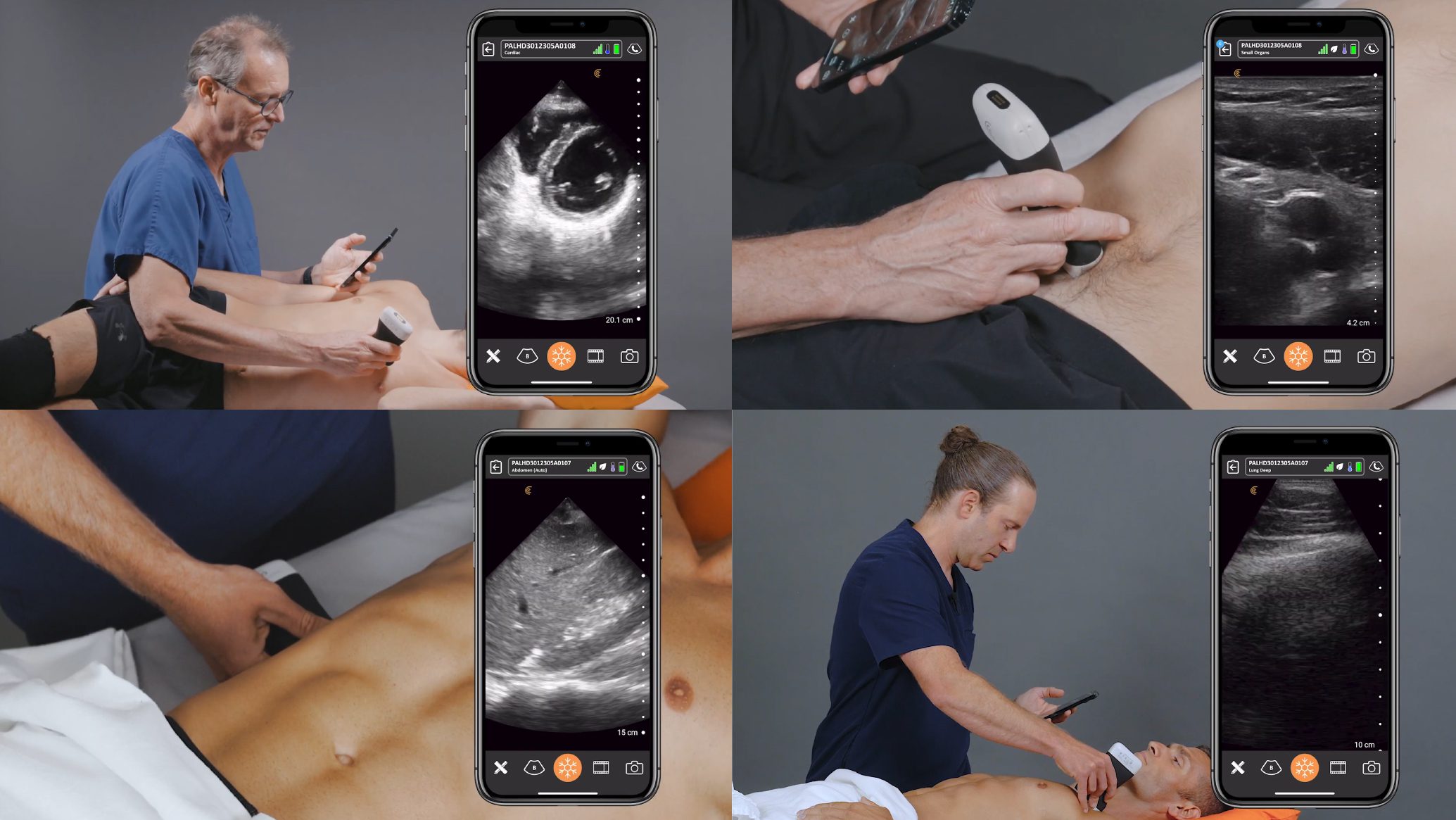
Ocular emergencies account for 3% of all ED visits in the United States. Tune in to this 1-hour webinar for a deep dive by emergency physician Dr. Tom Cook into the use of ultrasound in the initial evaluation of ophthalmic applications and the integration of ultrasound findings into clinical decision-making. You’ll learn:
- Techniques for scanning the eye
- The ultrasound appearance of retinal detachment, vitreous bleeding, lens dislocation, papilledema, and ocular trauma
- How POCUS can enhance decision-making, reduce time to treatment, and improve outcomes
Watch this 2-minute video to see how to scan and identify the eye with high-resolution ultrasound. The Clarius ocular preset has built-in thermal limits to ensure optimal patient safety. Book a personal, virtual demonstration to learn more.
- Park KE, Mehta P, Tran C, et al. Comparison of five point-of-care ultrasound devices for ophthalmology and facial aesthetics. Ultrasound. 2024 Feb;32(1):28-35. doi:10.1177/1742271X231166895
