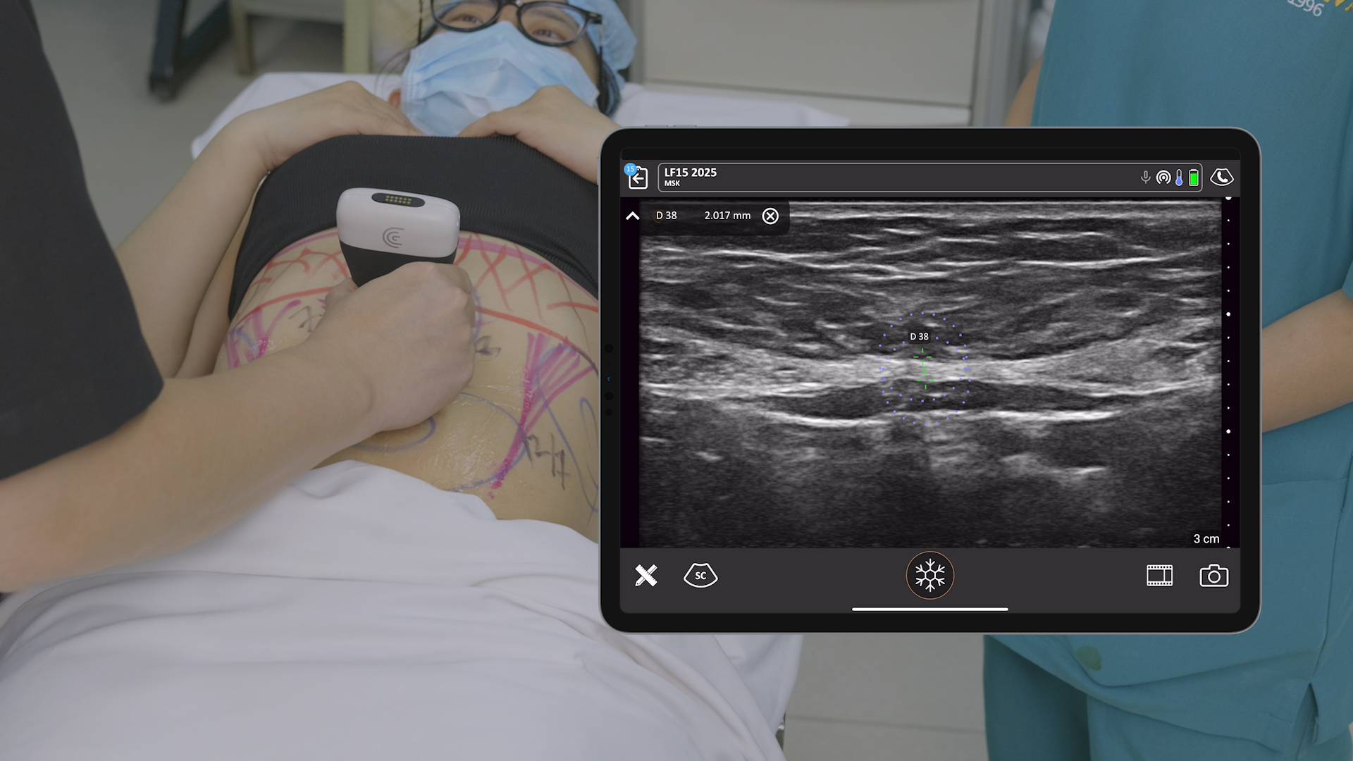
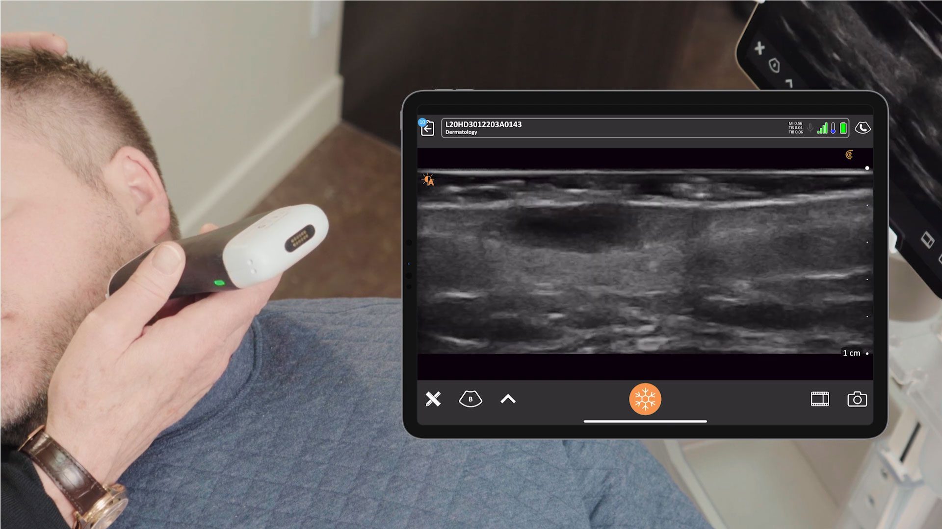
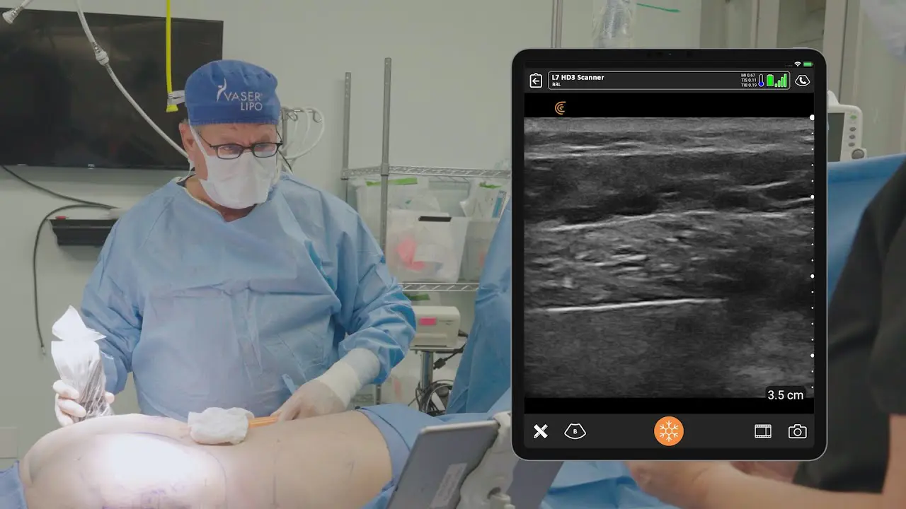
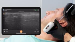
In the dynamic world of medical aesthetics, staying at the forefront of innovation is considered essential for delivering the best results to your patients. For many aesthetic clinicians, including Dr. Stefania Roberts, a renowned cosmetic physician, phlebologist, and ultrasound expert, handheld ultrasound is the newest innovation that is helping to ensure the safety and accuracy of Botulinum (Botox) and filler injections.
Dr. Roberts joined us to present a practical webinar to demonstrate ultrasound techniques for identifying anatomy that she uses day-to-day at her aesthetic practice in Victoria, Australia. Watch the one-hour webinar that is now available on demand: Ultrasound-Powered Aesthetics: Precise Masseter and DAO Botulinum Injections and Perioral Filler Placement. Scroll down for highlights of the presentation and brief video demonstrations using the Clarius L20 HD3.
Ultrasound Guidance in Masseter and DAO Botulinum Injections
Dr. Roberts emphasizes the pivotal role of ultrasound in ensuring the safety and accuracy of botulinum toxin injections. By visualizing the three distinct heads of the masseter muscle – deep, intermediate, and superficial – clinicians can precisely distribute the toxin, thereby preventing the unwanted “dullness” or bulging that can arise from uneven dosing.
Similarly, ultrasound guidance proves invaluable in depressor anguli oris (DAO) injections. By accurately identifying the DAO and its neighboring muscles, clinicians can minimize the risk of inadvertently injecting the depressor labii inferioris (DLI), which can lead to an undesirable skewed smile.
VIDEO: Botulinum Toxin Injection into Masseter
Watch this video to see Dr. Roberts identify the 3 bellies of the masseter muscle and guide her injections into each of the bellies to avoid paradoxical doubling-up of the masseter.
Ultrasound Guidance in Perioral Filler Placement
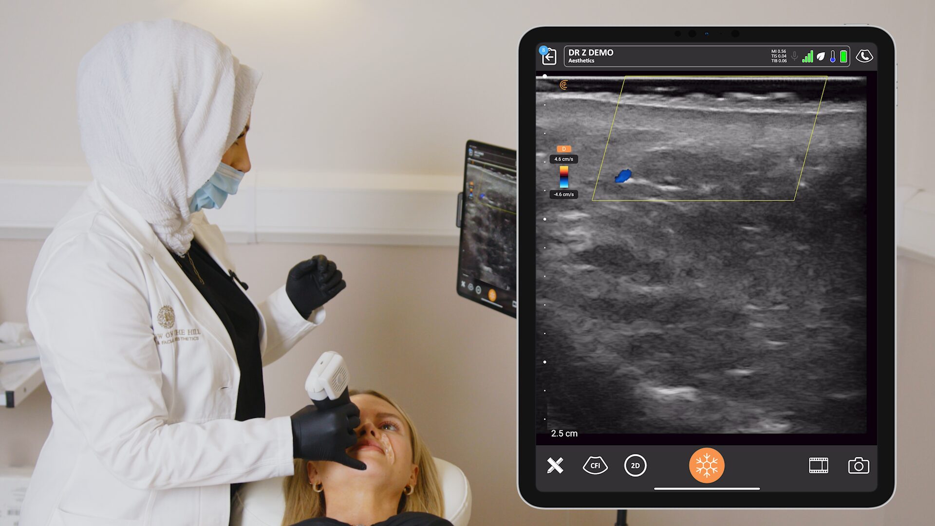
The perioral region, with its intricate vascular network, demands meticulous attention during filler injections. Dr. Roberts highlights the superior and inferior labial arteries as critical considerations.
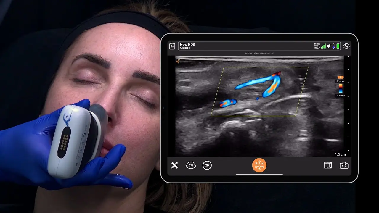
Ultrasound imaging enables clinicians to visualize these vessels, ensuring that filler injections are placed in the safe subcutaneous plane, minimizing the risk of vascular occlusion, a potentially serious complication.
VIDEO: Treating Perioral Rhytides with Filler Injections
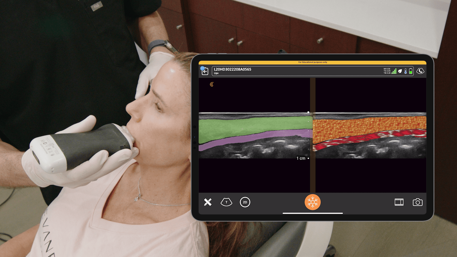
It’s important to avoid the inferior and superior labial arteries when injecting the lips with filler. In this video, Dr. Roberts demonstrates how to identify the location of these vessels using the Clarius L20 HD3 prior to injecting to avoid complications.
Clinical Benefits of Ultrasound Demonstrated by Dr. Roberts
- Ultrasound enhances the precision and safety of both botulinum toxin and filler injections.
- Visualizing the masseter muscle heads aids in the even distribution of botulinum toxin, preventing unwanted side effects.
- Identifying the DAO and DLI reduces the risk of skewed smiles associated with DAO injections.
- In perioral filler placement, ultrasound guidance helps avoid vascular occlusion by ensuring accurate filler placement.
Avoid vascular complications and confirm filler placement by looking under the skin with high-definition ultrasound
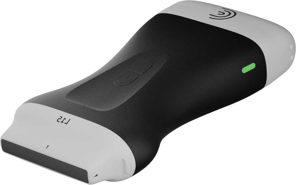
Medical aesthetic practitioners, including Dr. Stefania Roberts, are using the Clarius L20 HD3 – the world’s only ultra-high frequency ultrasound in a wireless scanner for clear imaging of the skin, muscles, vessels, and fascia to help ensure safe and consistent outcomes. It’s the only specialty-designed handheld ultrasound with ultra-high frequency of 20 MHz. Wireless and affordable, it delivers exceptional superficial imaging to 4 cm with an easy-to-use app for your iOS or Android device.
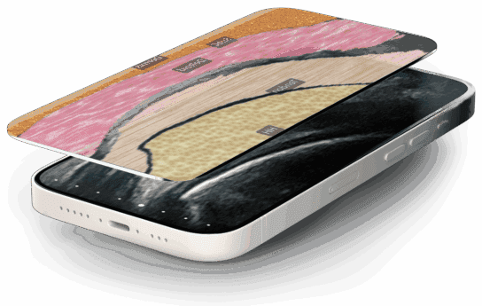
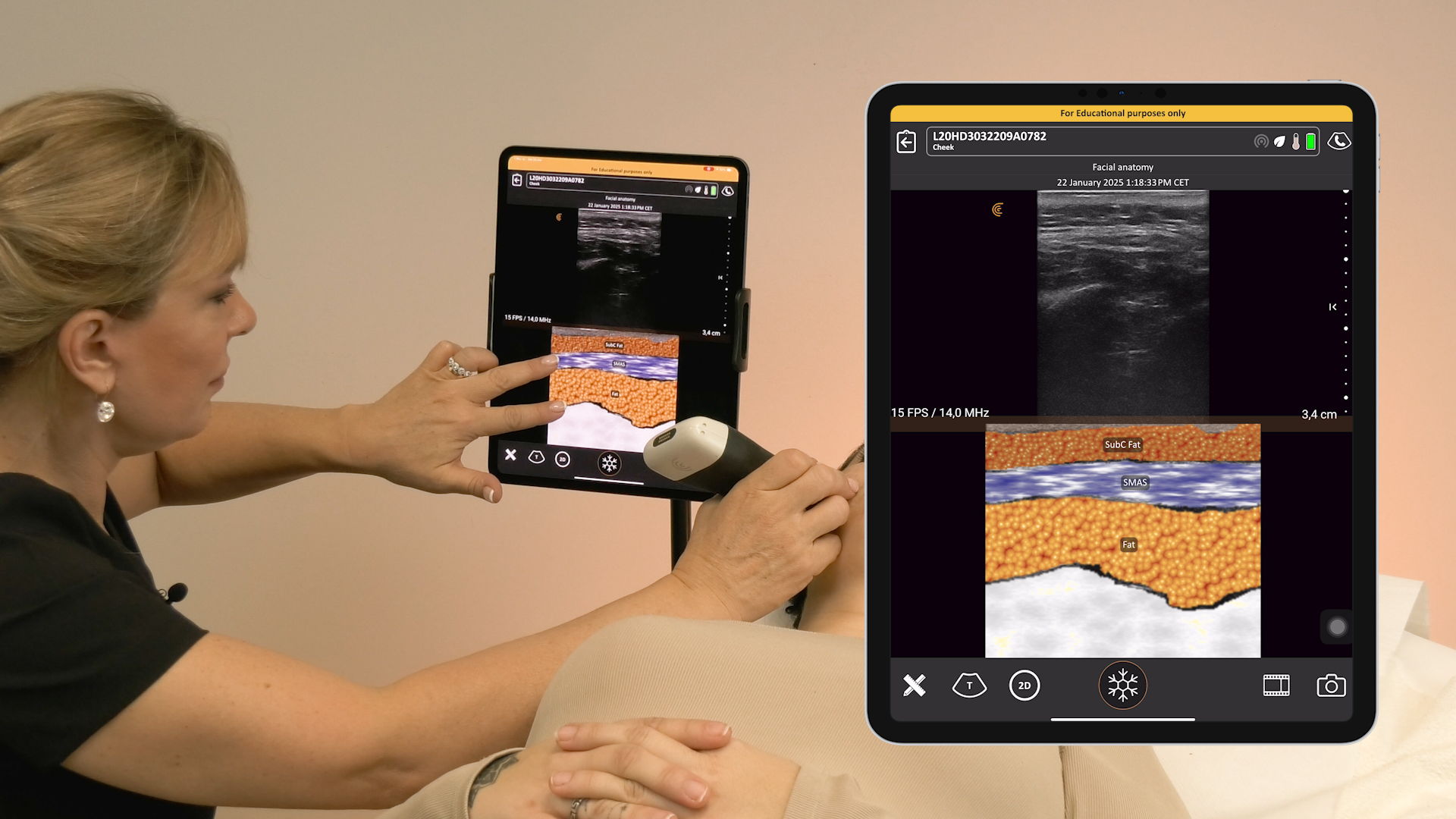
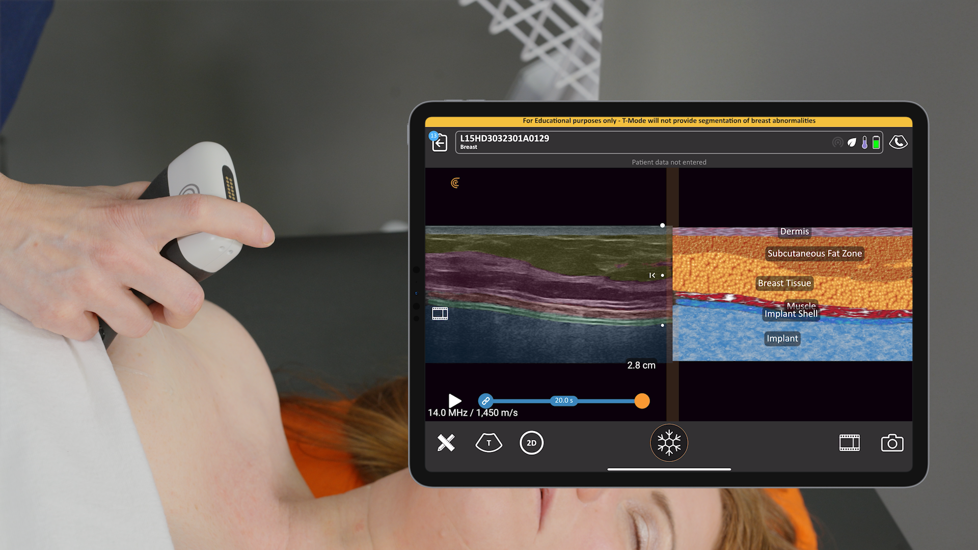
If you’re new to ultrasound, check out our new T-ModeTM AI, a groundbreaking educational technology to help new users to ultrasound advance their image interpretation skills using Clarius handheld scanners. It overlays distinctive colors, patterns, and labels to instantly identify and differentiate anatomical structures and tissue layers during aesthetic exams.
Learn more about Clarius for aesthetics or book a personal, virtual demonstration with a Clarius expert to discuss if Clarius is right for your practice.
Further Your Expertise in Aesthetic Ultrasound
Unlock the full potential of ultrasound in your aesthetic practice with tools that empower precision and safety. Pair the advanced capabilities of the Clarius L20 HD3 scanner with the invaluable insights from Ultrasound Protocol for Facial Aesthetics by Drs. Stefania Roberts and Kathryn Malherbe. This comprehensive textbook offers a deep dive into facial anatomy under ultrasound, making it an essential resource for every clinician looking to enhance their expertise. Order your copy here.
