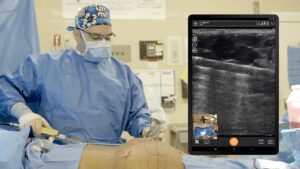
Dr. Kathryn Malherbe, CEO of MedSol AI Solutions in South Africa, passed along an ultrasound study of an umbilical hernia she detected using the Clarius L15 HD wireless scanner.
The patient presented with umbilical tenderness and a palpable lump that was most evident during lower abdominal exertion. He had undergone a laparoscopic appendectomy 7 days earlier.
Ultrasound Findings Confirm Herniated Bowel Loop
The Clarius L15 HD scanner was used to image the area of interest. Due to increased posterior acoustic shadowing related to the umbilicus, the probe was in a transverse position and at a slight angle/tilt of the probe to assess the posterior aspect of the umbilical region.
The ultrasound showed a distended, herniated bowel loop superficial to the rectus abdominus sheath, with reduced signs of vascularity during power Doppler imaging. When dynamic compression was applied with the scanner, the patient confirmed the region of tenderness corresponded to the location of herniated bowel on the ultrasound.
The neck of the herniated bowel loop measured 10.2 mm (see figure 1) and the bowel loop measured 22.3 mm x 9.8 mm (see figure 2).
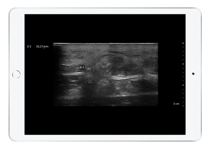
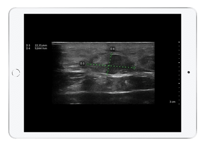
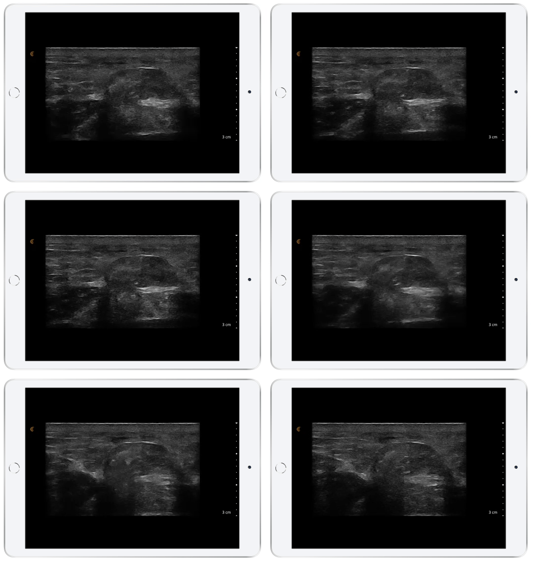
The outer bowel loop wall appeared hyperechoic on ultrasound, with loss of the typical haustra appearance. The dynamic Valsalva maneuver proved the bowel loop appeared constricted with limited motion, confirming the suspicion of a non-reducible incarcerated hernia.
Video: Dynamic Valsalva Maneuver Confirms Incarceration
Dynamic sonographic imaging of the suspected herniated bowel loop helped determine the extent of herniation as well as the presence of incarceration, which required further surgical intervention.
Treatment
The patient was referred for laparoscopy along the umbilicus, and the hernia loop was reduced through wire mesh insertion.
AI-Powered Point-of-Care Ultrasound Saves Lives
This is an example of an incisional hernia, which can occur if there is improper closure of the fascial defect at the port site following laparoscopic surgery. Some incisional hernias can develop soon after surgery, as is the case with this patient, or they can take months or years to occur.
I noticed that the patient was then referred for a cart-based ultrasound unit in hospital pre-surgery workup and the Clarius imaging surpassed that system by far!” says Dr. Malherbe.
Dr. Malherbe spends most of her time working on the development of deep machine learning and artificial intelligence software for the early detection of breast cancer in ultrasound images. MedSol software integrates with a Clarius scanner to produce quick and accurate diagnostic results, which ultimately leads to a significant reduction in mortality rates associated with breast cancer.
With high-definition images and automated settings, Clarius wireless ultrasound is the clear choice for point-of-care ultrasound. Our new HD3 scanners are 30% smaller, more affordable, with an AI-powered app that delivers optimized imaging to your Android or iOS device.
Not a Clarius user yet? We invite you to book a demo with a Clarius sonographer to see the difference high-definition imaging can make in delivering the best patient care.


