

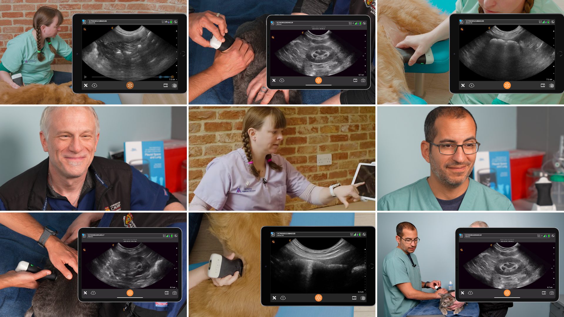
As more veterinarians join the Clarius ultrasound community, we’re producing more free educational tools to help new ultrasound users sharpen their point-of-care (POCUS) scanning skills. This week we’re featuring practical tips from Dr. Camilla Edwards, DVM, on how to integrate abdominal lymph node scanning into routine abdominal ultrasound exams of feline and canine patients.
A passionate veterinary educator, Dr. Edwards offered clear and methodical scanning techniques and pathology interpretation in a recent one-hour webinar: Watch the full webinar, « Practical Small Animal Ultrasound: POCUS Techniques for Imaging Abdominal Lymph Nodes« , to learn:
- How to locate the left and right medial iliac, proximal colic and jejunal lymph nodes using easily identifiable landmarks
- Characterize the ultrasonographic appearance of abdominal lymph nodes in dogs and cats
- Identify primary and secondary diseases of the lymph nodes
- Interpret ultrasound images with examples of inflammatory and neoplastic changes
Or read on for highlights on how to scan the medial iliac and jejunal lymph nodes in cats and dogs.
Indications for Scanning the Abdominal Lymph Nodes
Dr. Edwards scans the lymph nodes as part of her full abdominal ultrasound protocol to look for signs of cancer or inflammation. If there’s an area of concern, for example neoplasia in the hind limbs, the draining nodes can be evaluated.
Ultrasound Appearance of Normal Nodes– similar in cats and dogs
- Ovoid in shape
- Slightly hypoechoic compared to surrounding fat
- Homogeneous, but may have hyperechoic centre
- Length varies, so always measure the width
- Dogs <1 cm width
- Cats < 0.5 cm width
- Nodes are larger and more prominent in younger patients
Normal Anatomy
The Medial Iliac Lymph Nodes are parietal nodes and drain the colon, rectum, reproductive organs, urinary bladder, and urethra. They have great landmarks which makes them easy to find.
Ultrasound technique for imaging the medial iliac lymph nodes in left lateral recumbency for the right, and right lateral recumbency for the left:
- Locate the abdominal aorta in long axis at the level of the kidney
- Follow the aorta caudally to the trifurcation and fan in this area
- Look for medial iliac node in close proximity to the iliac artery
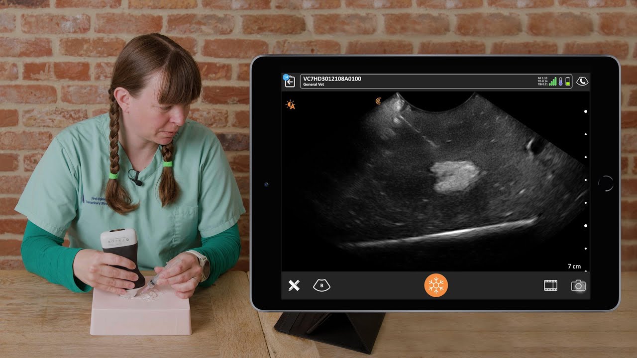
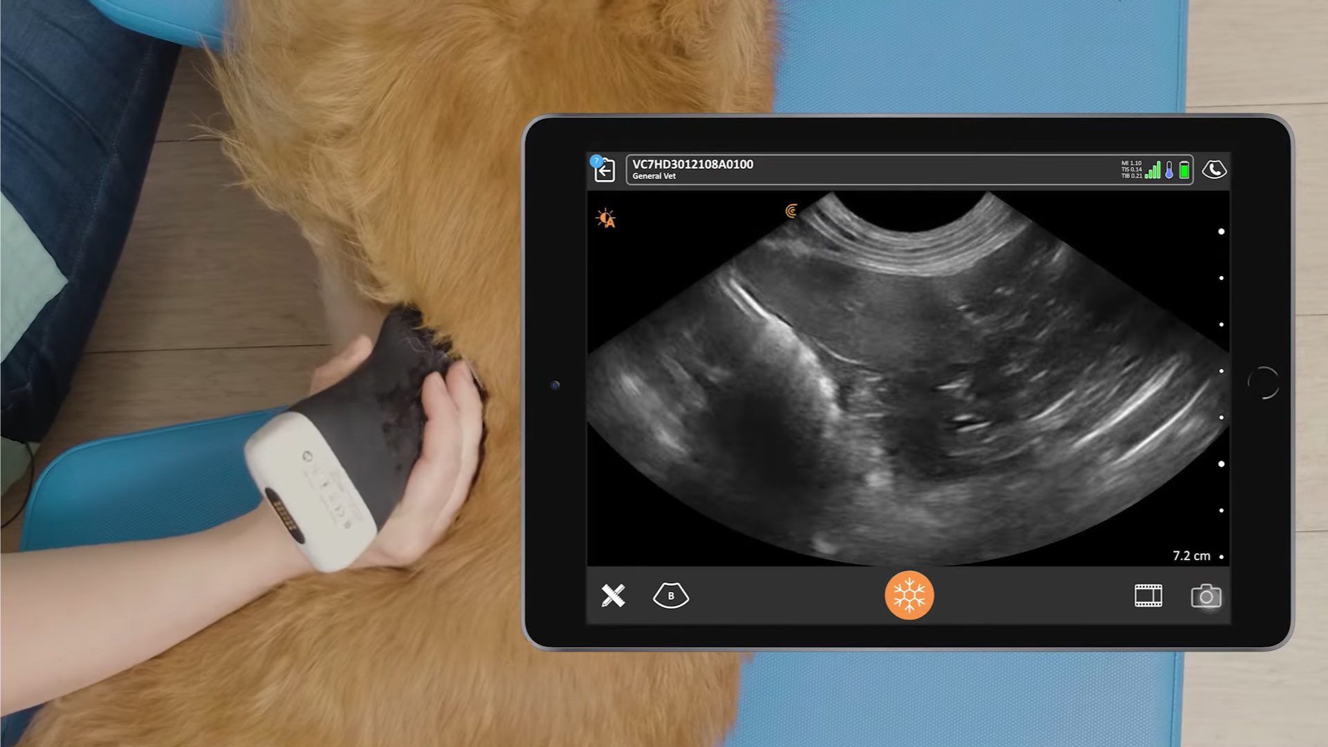
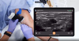
Watch this video to see how Dr. Edwards scans the left medial iliac node with the Clarius C7 HD3 Vet scanner.
The Jejunal Lymph Nodes are visceral nodes, draining the jejunum, ileum and pancreas. These are the largest nodes in the abdomen, which makes them easier to find. They are often paired and have a blood vessel running between them.
Ultrasound technique for imaging the jejunal lymph nodes, more likely to see in left lateral recumbency:
- Locate the caudal end of the right kidney
- Slide ventrally down the body, scan slowly, in a castle pattern
- Look for 2 longer hypoechoic structures
- Cranial mesenteric artery is often seen between the nodes
In this video, Dr. Edwards demonstrates how to scan the jejunal lymph nodes with the Clarius C7 HD3 Vet scanner.

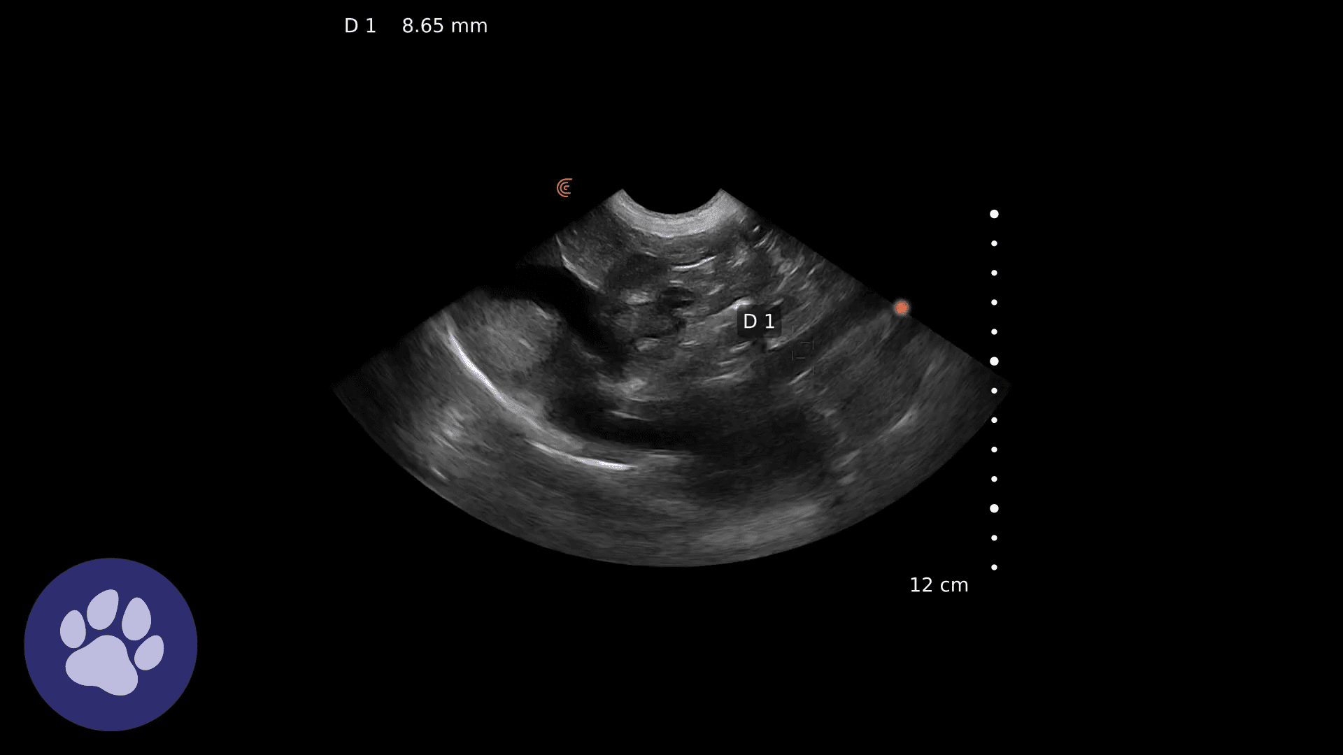
How to Identify an Abnormal Node
- Enlarged
- Rounded
- Hypoechoic/heterogeneous
Pathology can be reactive, primary neoplasia, or metastatic disease, which can be confirmed by FNA or biopsy.
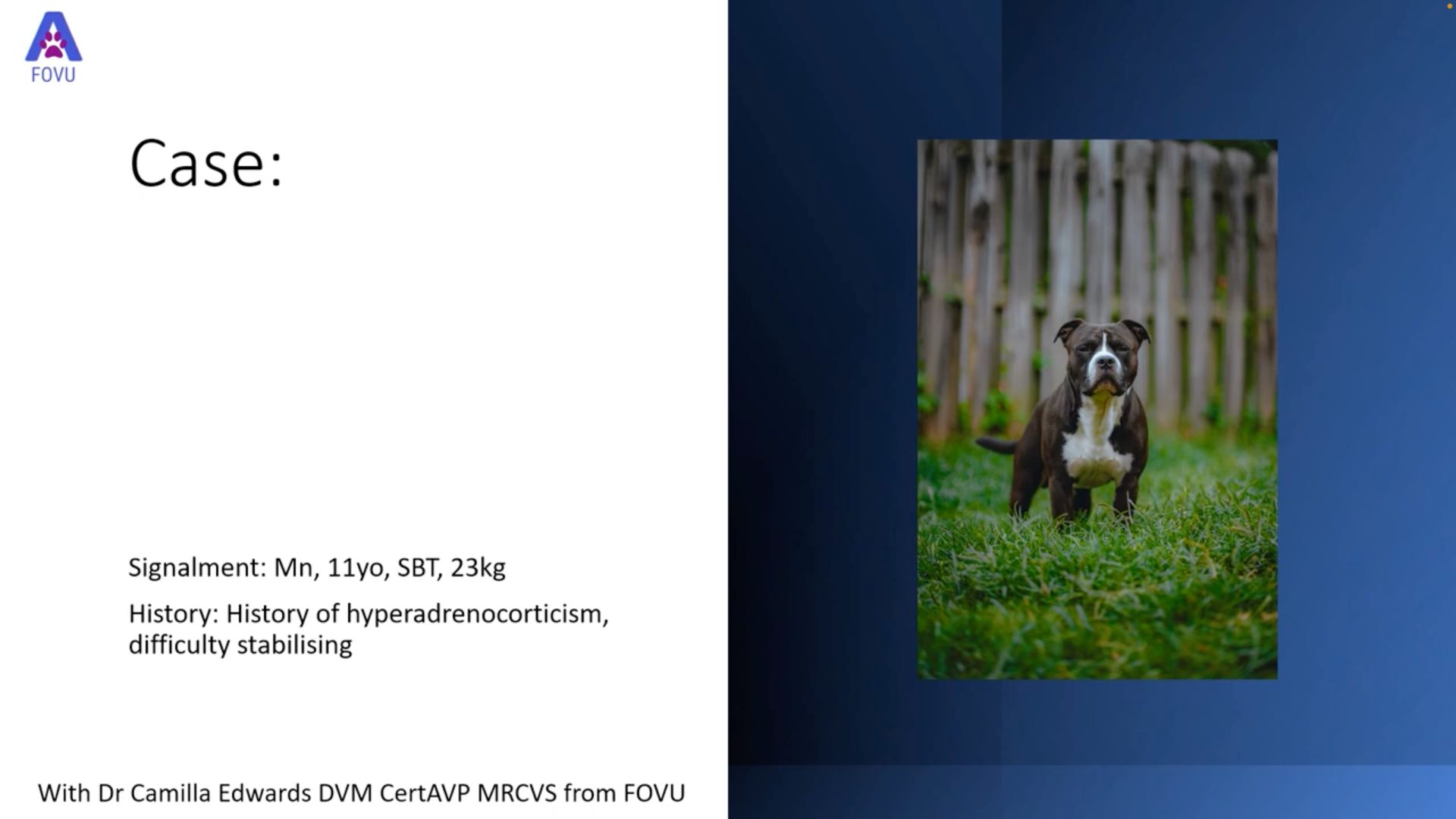
During the webinar, Dr. Edwards shares 2 case studies that demonstrate pathological abdominal lymph nodes. Watch the full webinar, « Practical Small Animal Ultrasound: POCUS Techniques for Imaging Abdominal Lymph Nodes« , for information about how using ultrasound helped determine the next steps in the treatment of these pets.
For more information about Dr. Edwards and the educational services she provides, visit her website, or get in touch at [email protected].
Clarius Wireless Ultrasound for Veterinary Practice
Dr. Edwards uses the Clarius C7 HD3 Vet scanner in her small animal practice. To learn how you can add wireless ultrasound to your practice, visit our Veterinary Specialty Page. There you’ll have access to additional webinars and classroom videos. Learn about the Advanced Veterinary Package provides additional workflows for a wide variety of animal examinations.
Our new Clarius HD3 Vet scanners are smaller and lighter. To find out which scanner is best for your practice, contact us today, or request a virtual ultrasound demo.

