By providing my email, I consent to receive Clarius webinar invitations, case studies, whitepapers, and more. I can unsubscribe anytime. Privacy Policy.
FREE WEBINAR
Cardiac POCUS: Coronary Artery Disease for the Non-Cardiologist
Watch On-Demand
Watch Now
Emergency physician and ultrasound educator Dr. Tom Cook teaches how to accurately assess left ventricular wall motion in the presence of coronary artery disease, for improved patient management.
In this 1-hour webinar, you will learn how to extend your cardiac POCUS repertoire to identify regional abnormalities in contractile function of the left ventricular walls in patients with suspected acute coronary artery syndromes (ACS) or older infarcts. You’ll learn:
- Indications for cardiac wall motion assessment
- Cardiac views that will give you the best bang for your buck
- The association of ECG findings to ultrasound findings
- Common patterns of wall motion abnormalities and corresponding coronary artery distribution
- How POCUS can enhance decision-making, reduce time to treatment, and improve outcomes
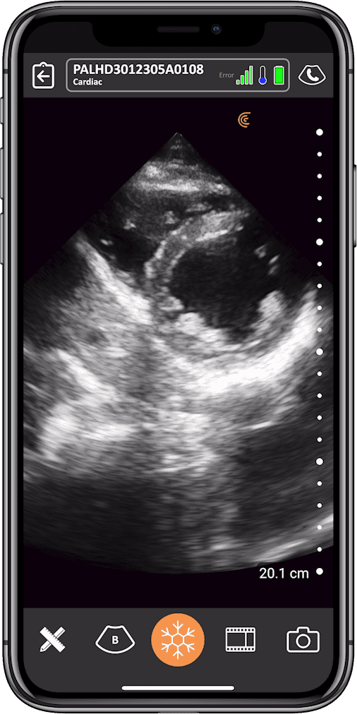
Ischemic heart disease is a common cause of LV wall motion abnormalities. Structural abnormalities can appear within seconds from the onset of myocardial ischemia, so identifying wall motion abnormalities in patients presenting to the ED with chest pain or suspected ACS could lead to earlier intervention.
Cardiac POCUS can be a valuable tool for the evaluation of cardiac function and structure in patients presenting to the ED with chest pain. Bedside echo can enhance clinical, lab and ECG findings by providing a rapid screening assessment of LV wall motion, expediting treatment, and saving myocardium.
Dr. Cook will give you the tools you need to quickly and reliably evaluate left ventricular wall segments to assess for hypo- or akinesis of one or more segments, which may be indicative of myocardial infarction. He will teach how to use this information, along with ECG and lab values, to determine if your patient may benefit from early invasive intervention like angiography and stenting.
As always, Dr. Cook will review real cases with healthy and pathological images and video clips to help you develop the skills to generate and interpret ultrasound images on your patients. Join this free webinar to gain practical knowledge you can use on your next day in the ER, hospital ward, or intensive care unit.
This webinar is a must for healthcare professionals seeking to improve their bedside cardiac ultrasound skills.
Emergency Physician
Dr. Tom Cook
Dr. Cook came to Palmetto Health after leaving the Army Medical Corp in 1996. Born in El Paso, Texas, he was raised in Northern Virginia and received his bachelor's degree from the College of William and Mary and attended the University of Virginia for Medical School. He trained in emergency medicine residency at Fort Hood, Texas before taking on a rugged three-year tour at Tripler Army Medical Center in Honolulu, Hawaii. After leaving the army in 1996 he joined the emergency department staff at Palmetto Health where he initially developed the emergency ultrasound curriculum and then founded the nationally recognized 3rd Rock Ultrasound and the Emergency Ultrasound Course. In 2001 he became program director. He lives with his family on Lake Murray in Lexington.
Medical Sonographer
Shelley Guenther, CRGS, CRCS
Shelley Guenther worked as a Nuclear Medicine Technologist for 2 years before entering into the ultrasound program at the Royal Alexandra Hospital in Edmonton. After graduating with specialties in general ultrasound as well as echocardiography, she worked as a clinical expert in the commercial world of ultrasound for over 25 years. As Clinical Manager at Clarius, Shelley Guenther is dedicated to providing the highest quality educational content for clinicians looking to add wireless ultrasound to their practice, including practical webinars and Clarius Classroom video tutorials.