


Time is of the essence when patients present to the ED with trauma and point-of-care ultrasound can play a key role in the initial evaluation of various trauma-specific applications and can guide decision-making in minutes,” says Dr. Tom Cook, a renowned emergency physician and educator, who shared his expertise on using point-of-care ultrasound (POCUS) in trauma care during a recent webinar.
More than 4,000 clinicians registered for the session: Emergency POCUS: Mastering Trauma Care with Handheld Ultrasound. The well-reviewed session is now available to watch on demand. Read on for a few highlights and video demonstrations.
The Evolution of POCUS in Emergency Medicine
Host Dr. Oron Frenkel, emphasized the increasing integration of POCUS into clinical practice, driven by technological advancements and its proven utility in rapid diagnosis and clinical decision-making. POCUS has become an indispensable tool throughout the patient journey, from pre-hospital settings to the emergency department, operating room, and intensive care unit.
FAST Exam: A Crucial Tool in Trauma Assessment
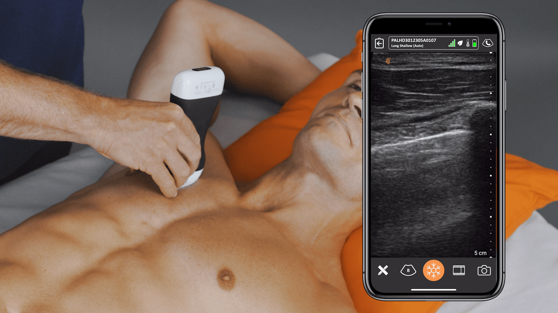
Dr. Cook reviewed the Focused Assessment with Sonography for Trauma (FAST) exam, a standard protocol for evaluating trauma patients. “All of these different views shouldn’t take more than 30 seconds to do, so your total exam’s just a few minutes,” he explained. The FAST exam allows for rapid assessment of the peritoneal and thoracic cavities, looking for critical conditions such as:
- Intraperitoneal fluid
- Pneumothorax
- Hemothorax
- Pericardial effusion
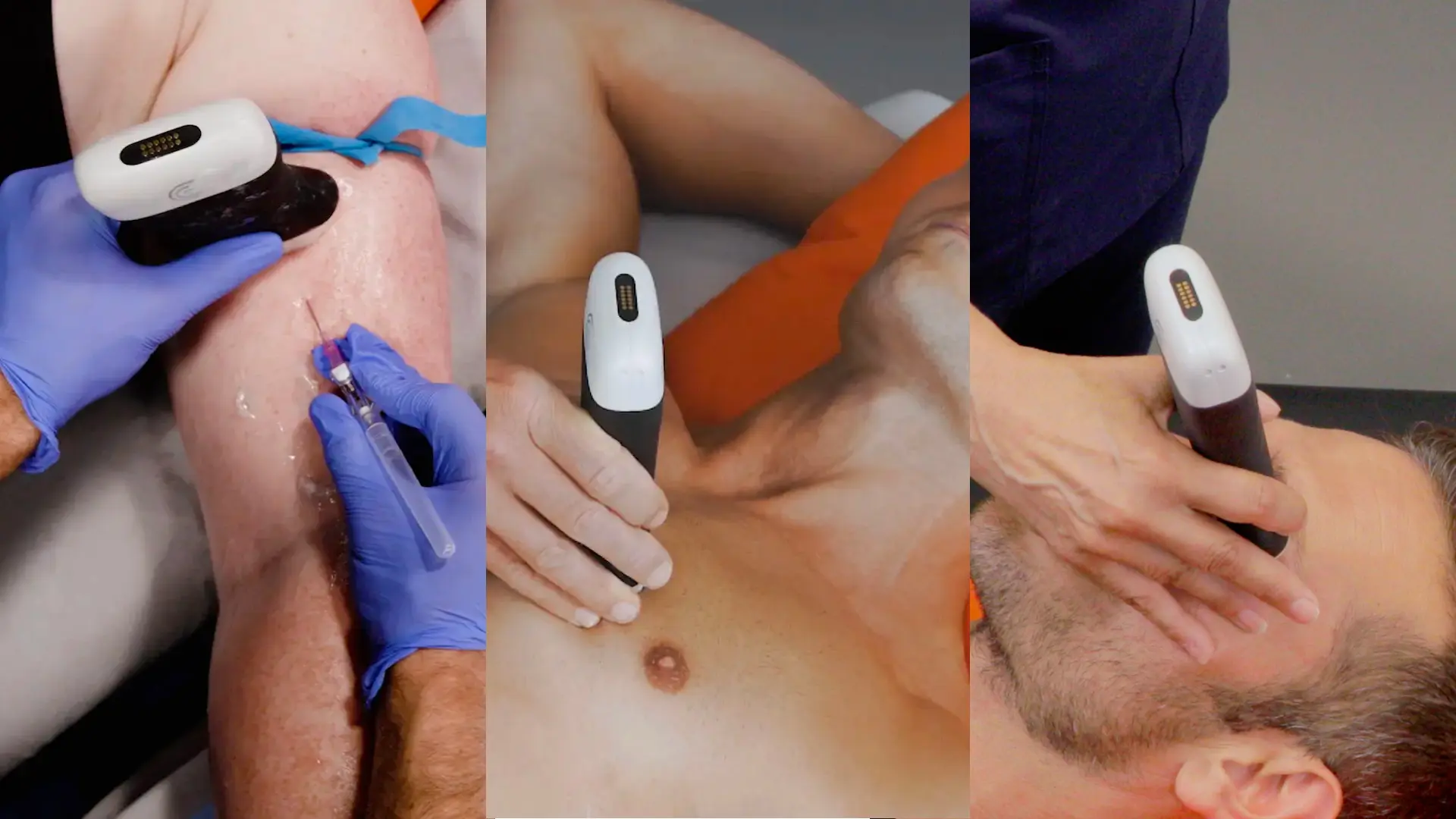
Watch Dr. Cook demonstrate the eFAST exam in the 5-minute video with the Clarius PAL HD3.
Key Takeaways for Emergency Physicians
- Right Upper Quadrant: The most sensitive area for detecting intraperitoneal fluid. “If you had to learn one thing to do about trauma learn this view. This is the money view. This is the one you get positive most of the time” said Dr. Cook.
- Left Upper Quadrant: While specific, it’s not very sensitive; a negative result doesn’t rule out significant injury.
- Chest X-rays: Not reliable for diagnosing pneumothorax or hemothorax. In Dr. Cook’s words, “Chest x-rays, in a word, three words, they suck. They’re about a coin flip. Trust your ultrasound in this situation. The chest film is not so great.”
- Repeat Scans: Crucial for monitoring patients, especially those with unstable hemodynamics. Dr. Cook emphasized, “Repeat the scan. Repeat the scan. Repeat the scan”
- Clinical Correlation: Integrate ultrasound findings with the patient’s clinical status to guide management decisions.
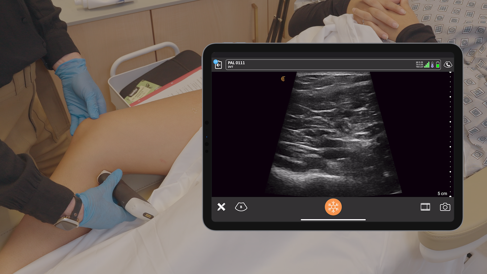
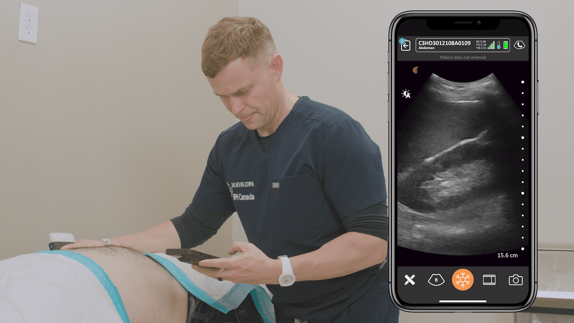
Dr. Cook’s RUSH Exam Protocol with Clarius Ultrasound
Watch this 4-minute video to see Dr. Cook perform a RUSH (Rapid Ultrasound in Shock and Hypotension) exam using the Auto Select option on the Clarius PAL HD3 for quick assessments of the heart, IVC, abdomen, and lungs.
Pearls and Pitfalls
Dr. Cook shared valuable pearls and pitfalls to help clinicians avoid common mistakes:
- IVC Assessment: Use ultrasound to assess intravascular volume status.
- Fluid Appearance: Differentiate between free fluid and other structures like the gallbladder or stomach contents. On this, Dr. Cook cautioned, “When you’re ultrasounding in the heat of the battle, you want to find something positive so you can feel good about yourself. Don’t get fooled into thinking that is free fluid.”
- Subcutaneous Air: Recognize how it can obscure the pleural line.
- Ascites vs. Blood: In cases of suspected ascites, consider aspiration to differentiate from blood.
- Fracture Assessment: Ultrasound can be a valuable tool for identifying fractures, especially in the femur.
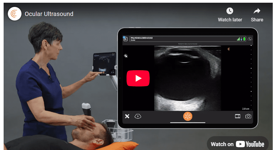
- Ocular Ultrasound: Don’t forget to include the eyes in your examination; ultrasound can quickly identify a ruptured globe. Sonographer Shelley Guenther demonstrates an ocular ultrasound exam with the Clarius PAL in this 2-minute video.
Q&A Highlights
The webinar concluded with a Q&A session, where Dr. Cook addressed several important questions:
- Retroperitoneal Trauma: Ultrasound is not the ideal tool for assessing retroperitoneal injuries.
- Differentiating Fluids: Aspiration may be necessary to differentiate between urine, blood, and other fluids.
- Tension Pneumothorax: Clinically, tension pneumothorax presents with hypotension and obstructive shock.
- Training Requirements: Proficiency in trauma ultrasound depends on the frequency of use and individual learning curves; Dr. Cook suggests that 20-25 scans may be sufficient in a high-volume trauma center.
Clarius Ultrasound: A Game-Changer in Emergency POCUS
The webinar also showcased the Clarius PAL HD3, a new wireless high-definition scanner featuring both phased array and linear transducers.
Key advantages of Clarius ultrasound for emergency POCUS include:
- Versatility: The dual array design eliminates the need to switch probes, streamlining workflow.
- Portability: Wireless and compact, Clarius scanners are easy to carry and use in crowded resuscitation bays.
- Hygiene: Waterproof and submersible for easy disinfection.
- Connectivity: Seamless integration with iOS or Android devices and easy image sharing.
Experience the Freedom of Ultra-portable Wireless Ultrasound for Triage and Treatment
Dr. Cook is confident that anyone can learn to use ultrasound: “If I can do it, almost anybody can. It’s just a matter of perseverance and repetition and you can use this quite easily.” While he uses the Clarius PAL HD3 during the webinar, there are other Clarius wireless scanners that are suitable for emergency POCUS. Learn more on our emergency medicine page.
If you’d like to consult with a Clarius expert, please contact us to book a personal, virtual demonstration. For more short tutorials on trauma ultrasound, visit Clarius Classroom.


