


Non-surgical rhinoplasty has surged in popularity, offering a minimally invasive alternative to traditional surgical procedures. However, for many injectors, the intricate vascular anatomy of the nose presents significant risks, with soft tissue filler augmentation in the nose carrying the highest risk of blindness compared to any other facial region.
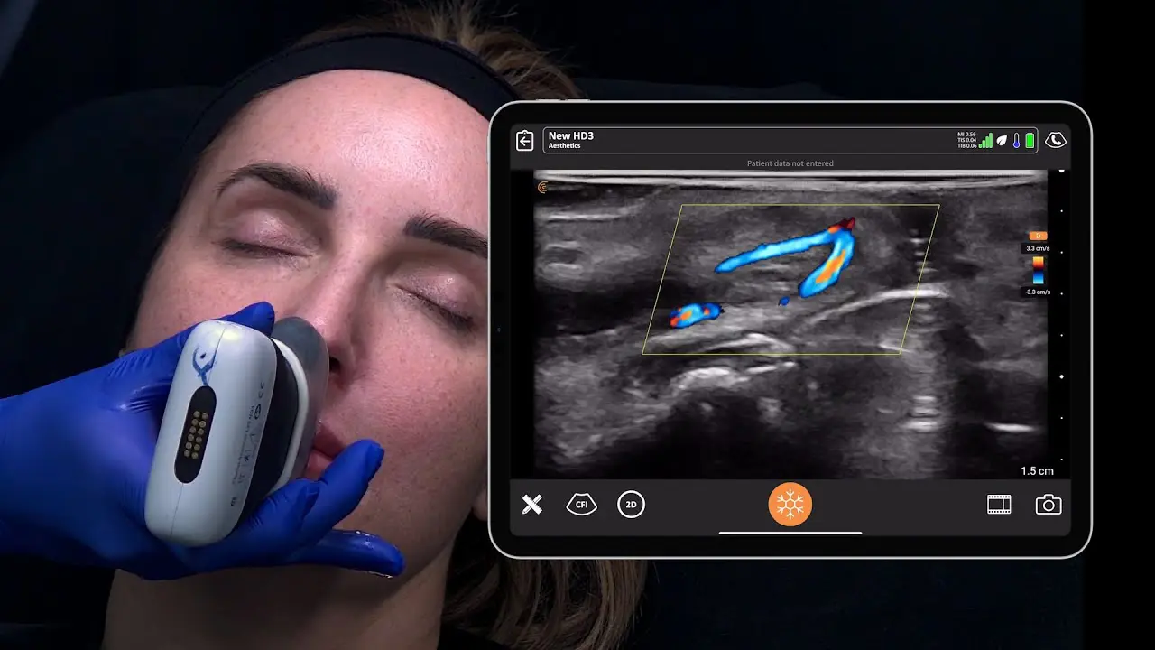
Since mastering the art of ultrasound-guided aesthetic procedures, the nose is no longer a “no-go danger zone” for Dr. Zainab Al-Mukhtar. She recently shared her expertise and experience with using ultrasound to enhance the safety and precision of non-surgical rhinoplasty during a recent webinar. Dr. Al-Mukhtar highlighted how ultrasound has revolutionized her approach to these treatments, providing a real-time view of the nose’s anatomy and helping to prevent vascular injuries.
Watch the free one-hour on-demand webinar for details – Ultrasound for Nonsurgical Rhinoplasty: Avoiding Vascular Complications and Improving Results. Scroll for quick highlights and a video demonstration.
Why the Nose is a “Danger Zone” and How Ultrasound Helps
The nose carries the highest risk of blindness among all facial regions for soft tissue filler augmentation, says Dr. Zainab. This heightened risk is due to the intricate and highly variable vascular anatomy, including an anastomosis between the external and internal carotid systems. As Dr. Zainab Al-Mukhtar emphasizes, “there is no safe zone in the nose”. This makes traditional injection techniques, even with aspiration, potentially risky.
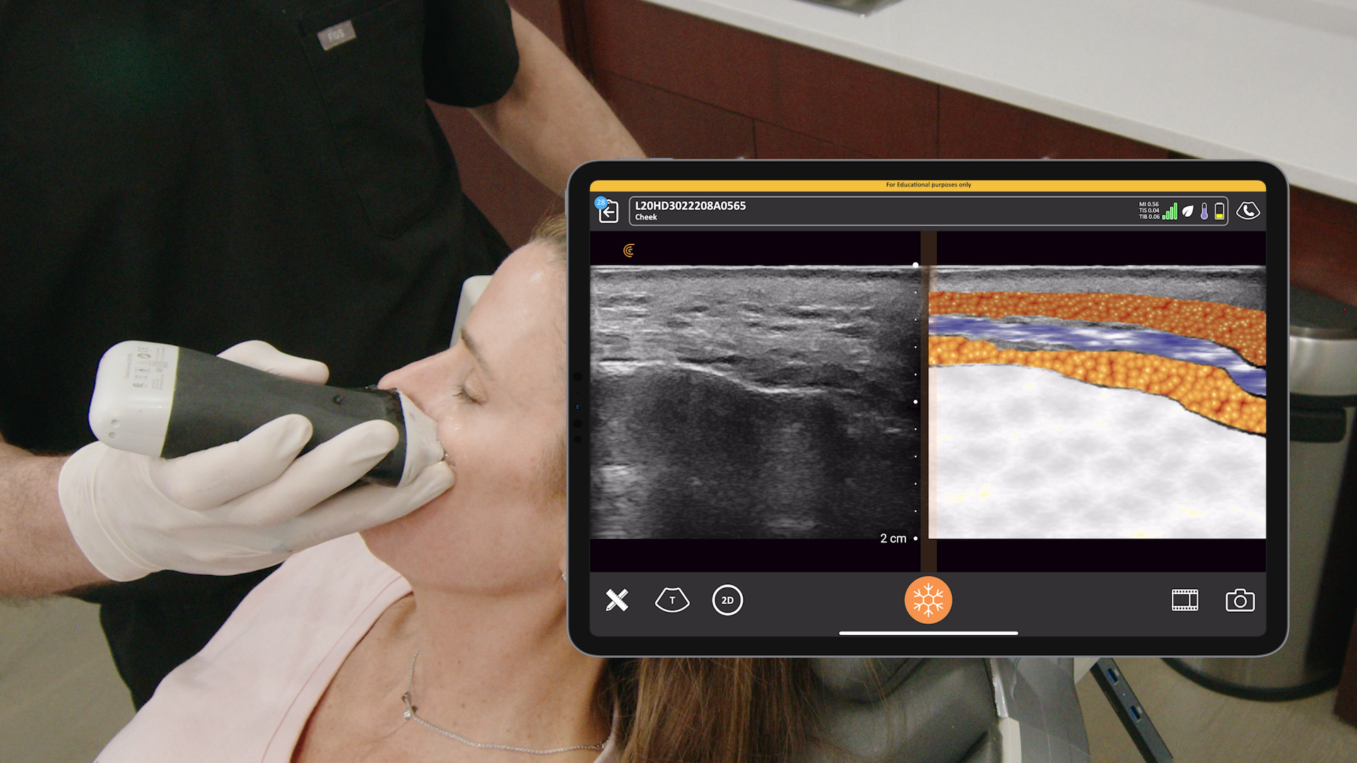
Ultrasound provides a dynamic, real-time view beneath the skin, offering invaluable information for better treatment planning and precise execution.
Key Benefits of Ultrasound in Non-Surgical Rhinoplasty
- Enhanced Safety: Ultrasound enables practitioners to visualize tissue layers, locate blood vessels, and identify anatomical variations, thereby minimizing the risk of vascular complications.
- Improved Precision: By guiding injections, ultrasound helps to optimize filler placement, leading to more predictable and aesthetically pleasing outcomes.
- Effective Complication Management: Ultrasound aids in the diagnosis and management of complications, such as vascular occlusions, allowing for targeted treatment and minimizing the use of hyaluronidase.
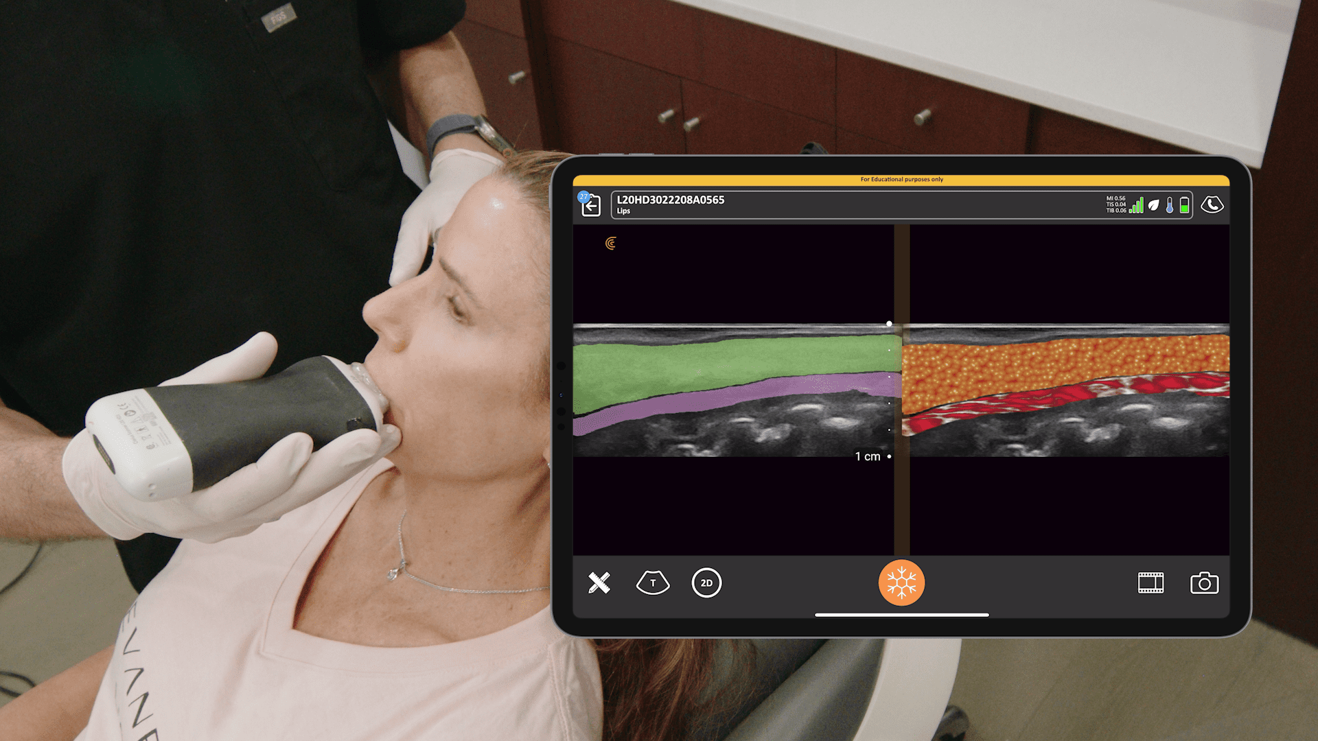
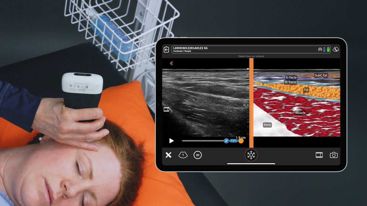
Anatomical Considerations of Nasal Anatomy Prior to Filler Injections
Dr. Al-Mukhtar emphasized the importance of understanding nasal anatomy, including the location of key vessels and the variability of tissue layers. She highlighted that there are no universal safe zones in the nose due to the high variability in vascular anatomy.
Clinical Highlights from the Webinar
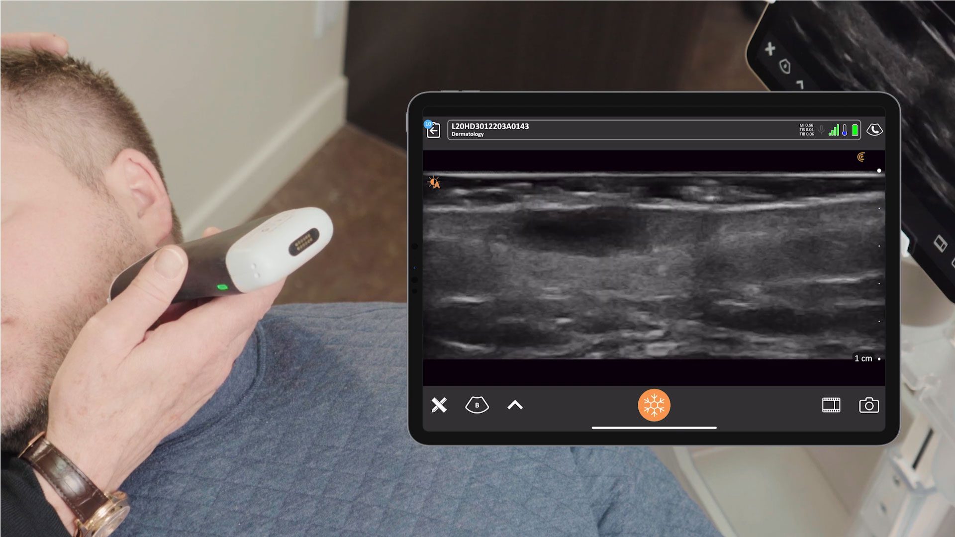
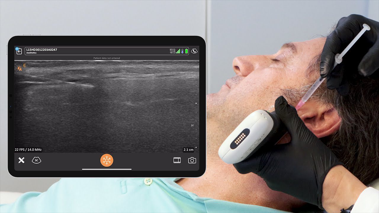
- Pre-treatment assessment: Dr. Al-Mukhtar stressed the importance of pre-treatment assessment using ultrasound to map vessels and plan injections safely.
- Injection technique: Dr. Al-Mukhtar advises injecting small amounts at a time. She also emphasized the importance of continuous skin observation during the procedure to ensure patient safety.
- Post-treatment care: Post-treatment scans can be used to confirm filler placement and assess for any potential complications.
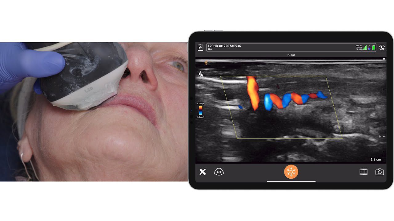
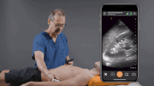
Watch this detailed video demonstration showing Dr. Zainab performing vascular and anatomical mapping with ultrasound before proceeding with filler injections to re-shape her patient’s nose.
The Value of Ultrasound Beyond Safety
While safety is a primary concern, ultrasound offers additional benefits in aesthetic practice:
- Optimized treatment outcomes: Precise depth measurements and understanding of tissue space allow for more refined and predictable results.
- Understanding filler behavior: Observing how filler evolves and interacts with blood vessels over time provides valuable insights.
Research tool: Ultrasound facilitates studies of anatomy and filler behavior, contributing to the growing body of knowledge in aesthetic medicine.
Q&A Highlights: Insights from Dr. Zainab Al-Mukhtar
The webinar concluded with a valuable Q&A session, addressing common concerns raised by practitioners attending the session:
Is aspiration enough for safety?
Dr. Al-Mukhtar states that “Aspiration is not enough”. While she still aspirates as “good practice,” she warns of “false negatives”. She notes that vascular occlusions can still occur even when practitioners have aspirated, injected slowly, and maintained a steady hand. Therefore, using ultrasound provides an additional, crucial layer of precaution.
Have you ever had persistent redness hyper vascular in the skin of the nasal tip after injection and do you dissolve in this case?
This is a common occurrence, often related to tissue thickness and volume, leading to compression of superficial vessels. Dr. Al-Mukhtar advises waiting about four weeks for swelling to subside, as it usually resolves. If it persists, she may dissolve a small, superficial amount of filler using a hyper-diluted hyaluronidase and a thin needle, without reversing the entire procedure. Patients should always be warned this can happen, and consistent follow-up is essential.
Is it possible for the filler to occlude vascular flow due to compression even without being intravascular?
“Yes,” confirms Dr. Al-Mukhtar. Vessels can collapse or spasm from irritation by nearby filler, or due to mechanical compression, even if the filler is not directly inside the vessel. This underscores the importance of staying as far away from vessels as possible, even if they appear relatively deep.
What filler brand do you prefer for nasal filling?
“I like to use Restylane Lyft. Generally, a filler that is not too hydrophilic is preferred to avoid overexpansion and maintain a sharp, defined result rather than a “spongy” or “bulbous” nose.”
For more insights from Dr. Al Mukhtar, watch the highly rated webinar that one webinar described as “an eye opener to be extra safe.”
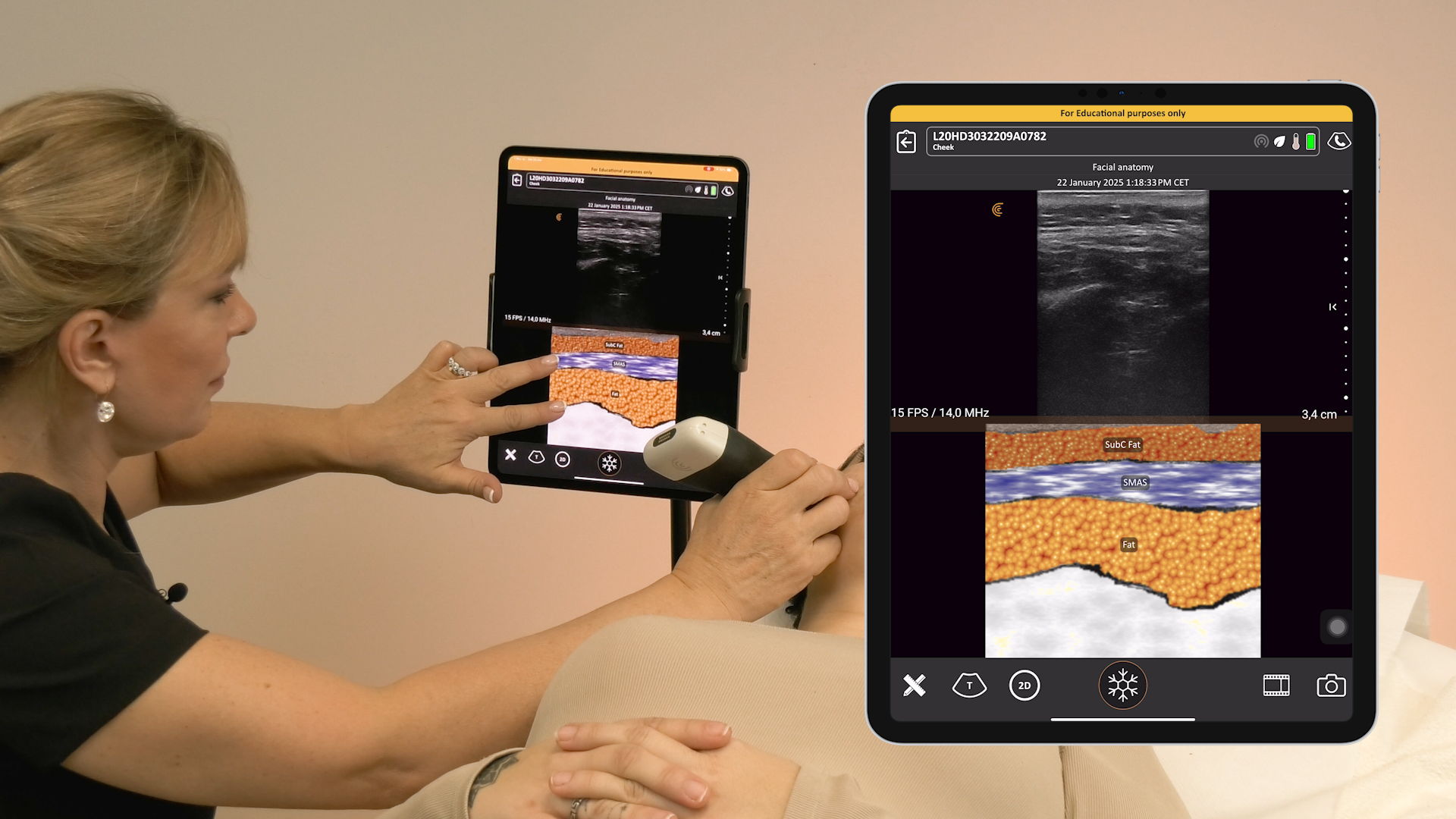
Improve Outcomes with Ultra-High-Definition Ultrasound for Facial Aesthetics
By providing detailed visualization of facial anatomy and guiding filler placement, ultrasound empowers practitioners to achieve optimal aesthetic outcomes while minimizing the risk of complications. Many aesthetic practitioners rely on Clarius wireless handheld ultrasound to clearly visualize facial and superficial anatomy in real time to safely guide procedures like cosmetic fillers and confidently treat complications. With exceptional superficial imaging, the new advanced aesthetic protocol, and new T-Mode, the Clarius L20 HD3 is the popular choice for facial aesthetics.
Learn more about Clarius AI-powered ultrasound on our aesthetics specialty page. Or contact us for a personalized virtual demonstration.
