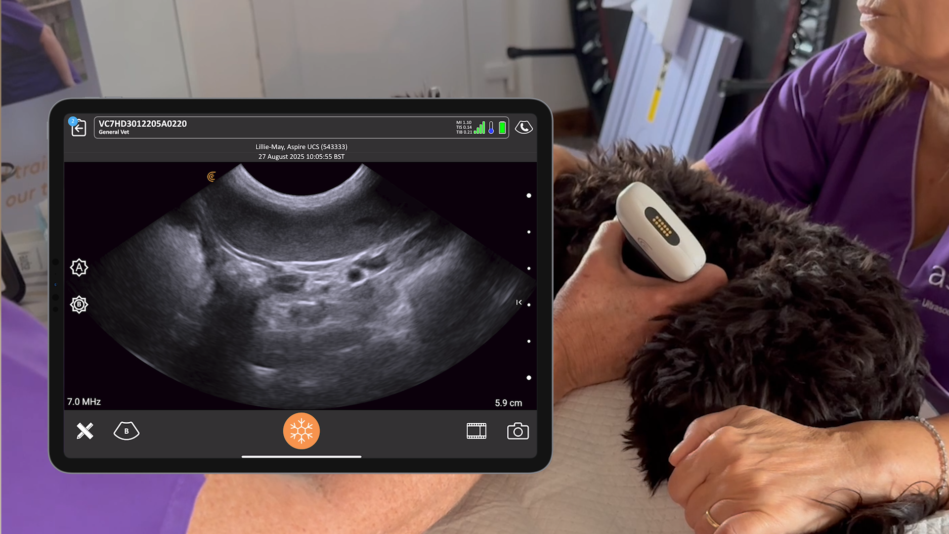
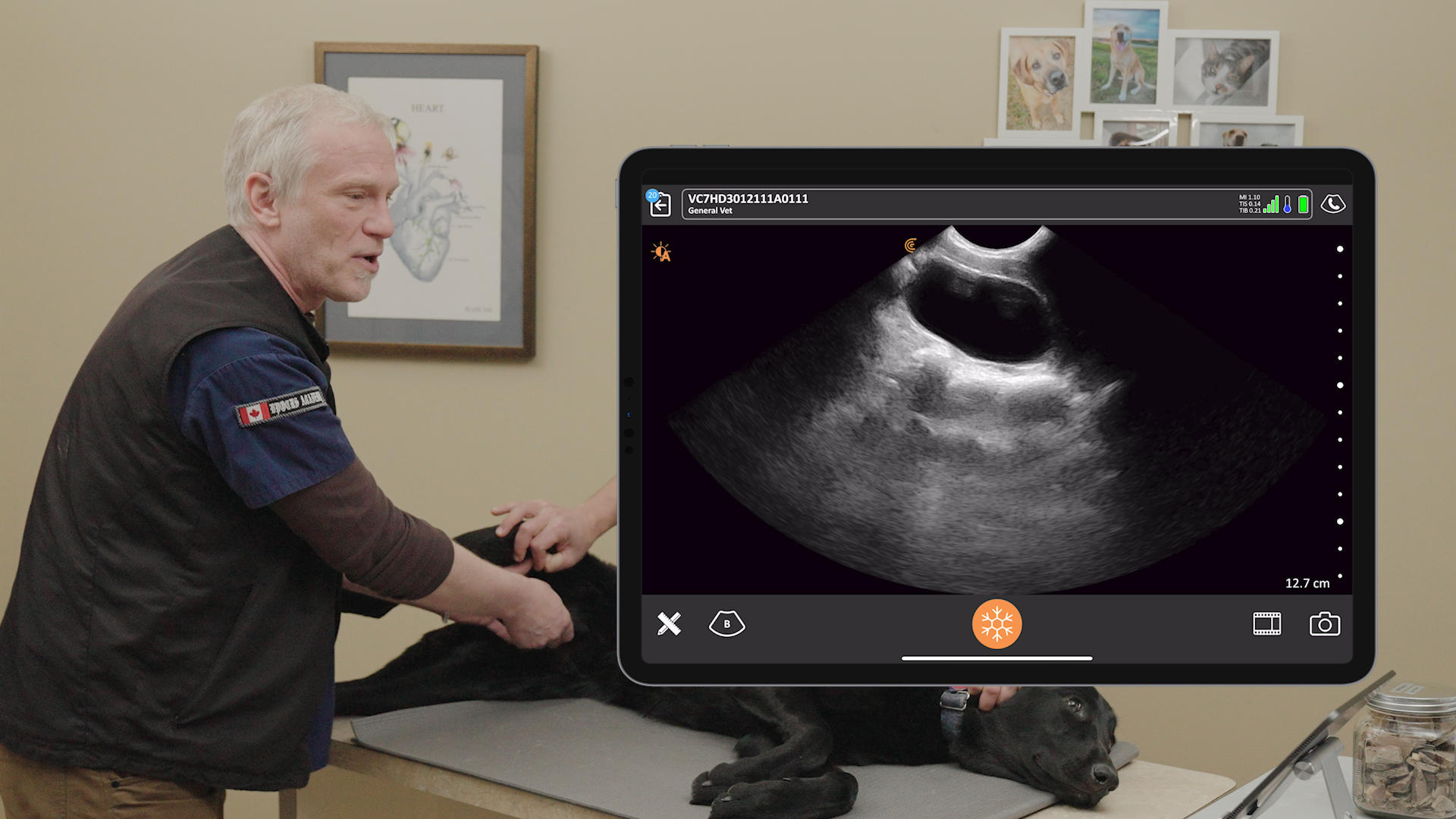
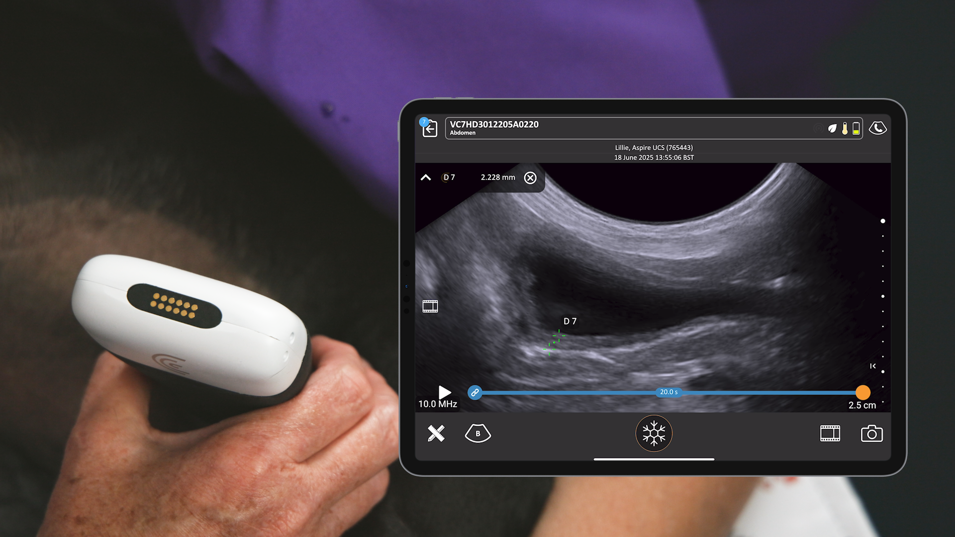
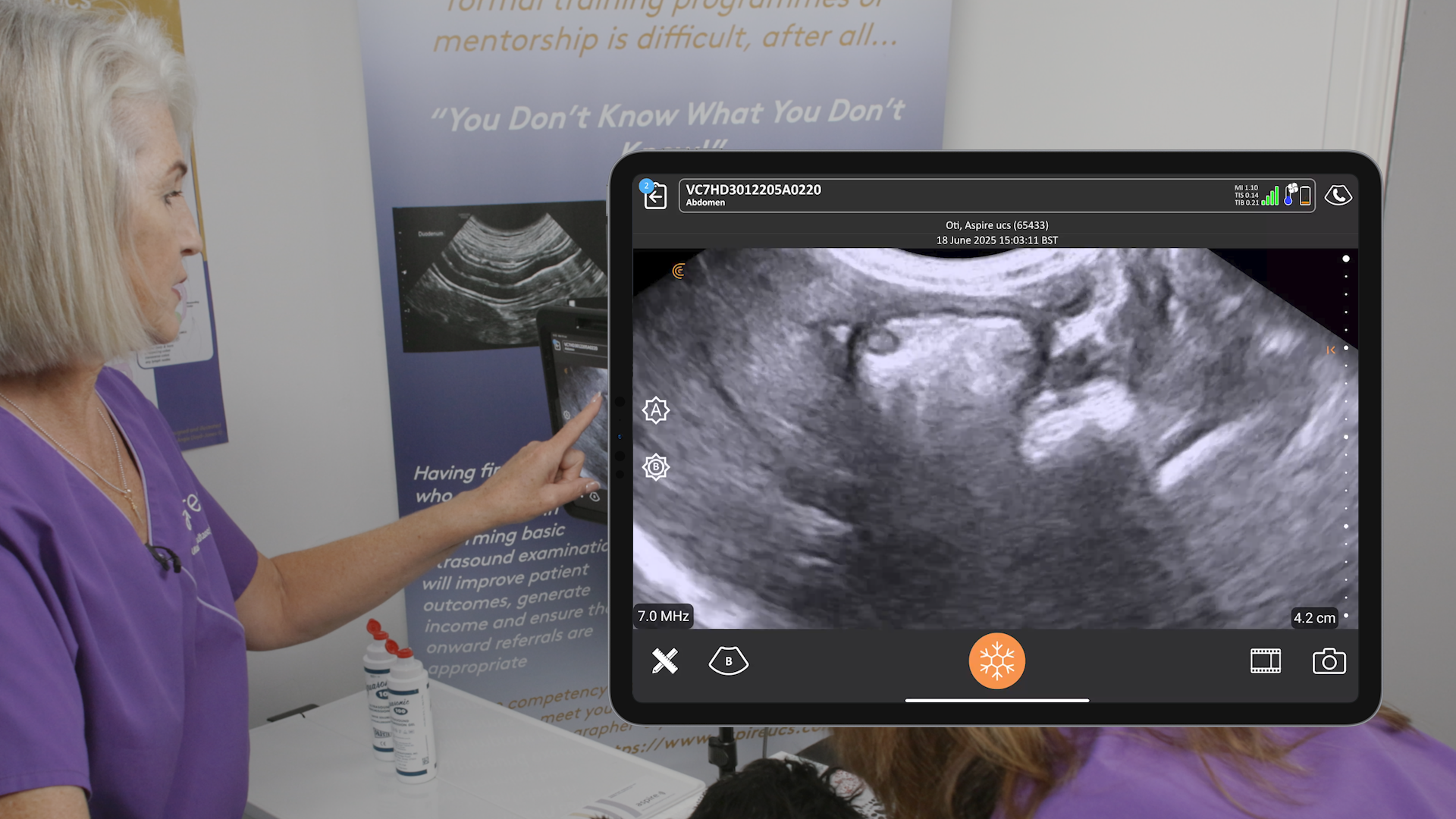
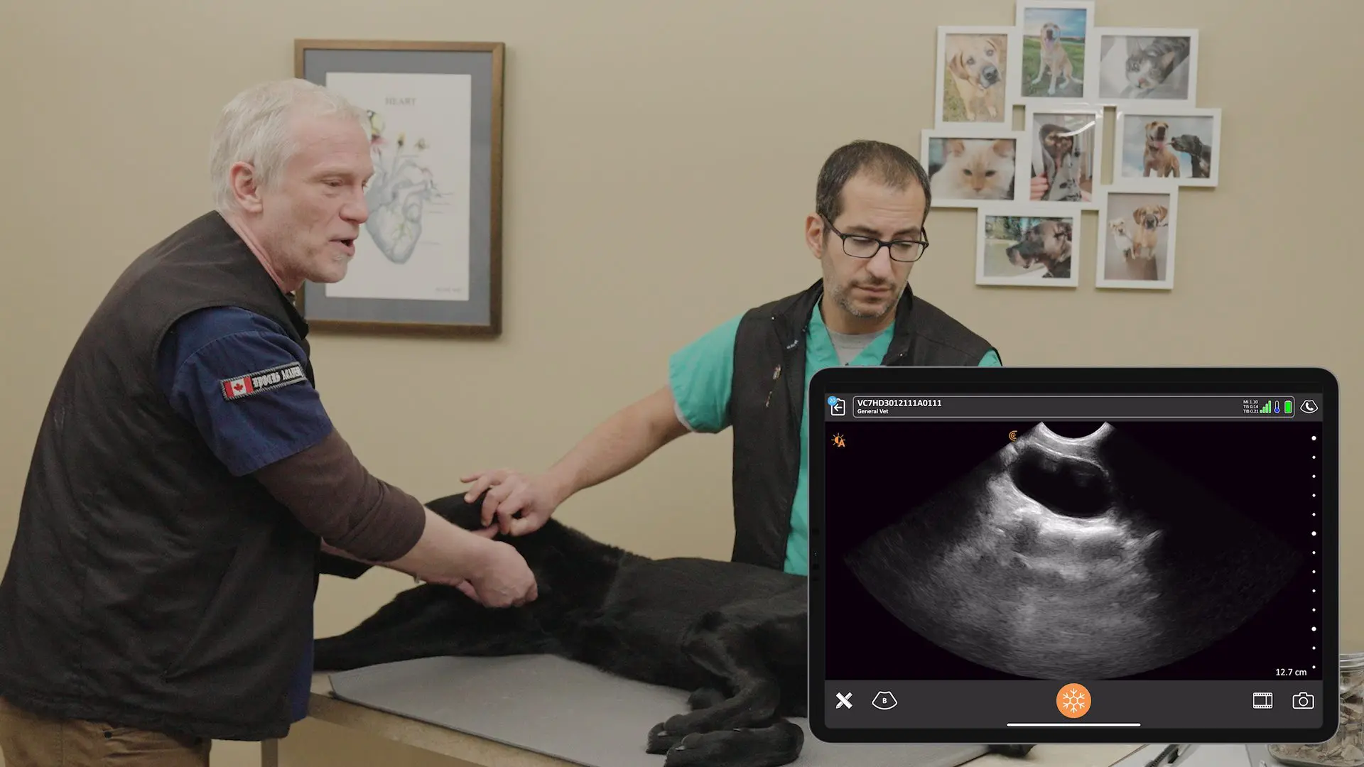
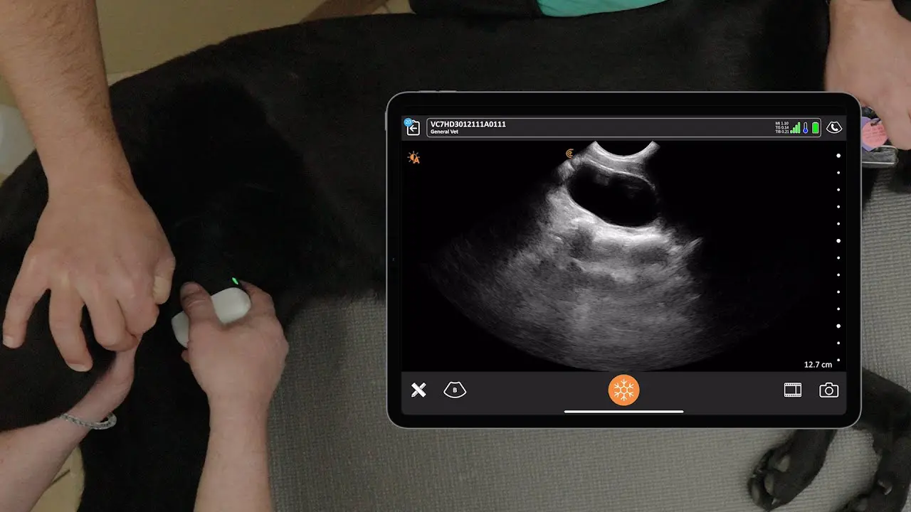
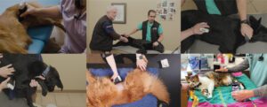
When companion animals present with palpable abdominal masses, these masses can be a source of concern for both the pet owner and the veterinarian. Ultrasound is a valuable tool for evaluating abdominal masses and guiding their diagnosis and management. It is a safe, non-invasive exam/procedure that can provide real-time images of the abdominal organs and masses, helping veterinarians to characterize masses, guide tissue sampling, and assess for local invasion or metastatic spread.
Dr. Camilla Edwards, an experienced veterinarian based in Cambridge, England, is well-versed in using ultrasound to characterize masses and safely obtain tissue samples. She shares her expert techniques in a one-hour webinar available on demand: Practical Small Animal Ultrasound Guiding Diagnosis and Management of Palpable Abdominal Masses.
Read on for highlights from her presentation and the Q&A session with the audience.
Benefits of Ultrasound for Abdominal Masses
- Non-invasive: Ultrasound does not require incisions or injections, making it a safe and well-tolerated procedure for most animals.
- Real-time imaging: Ultrasound provides real-time images of the abdominal organs and masses, allowing for immediate assessment of their size, shape, and location.
- Characterization of masses: Ultrasound can help differentiate between cystic and solid masses and provide information about a mass’s vascularity.
- Guided sampling: Ultrasound can guide the collection of tissue samples from abdominal masses, which can then be sent for cytological or histological evaluation.
How to Perform an Ultrasound-Guided FNA
Fine needle aspiration (FNA) is a minimally invasive procedure used to collect tissue samples from masses. Ultrasound guidance can improve the accuracy and safety of FNA. Following are the steps Dr. Edwards outlines for performing an ultrasound-guided FNA:
- Prepare the patient: The patient should be sedated or anesthetized, and the area to be aspirated should be clipped and cleaned.
- Identify the mass: Use the ultrasound probe to identify the mass, its depth, and surrounding structures.
- Insert the needle into the mass under ultrasound guidance.
- Collect the sample: Use the “woodpecker” technique or gentle aspiration to collect a tissue sample.
- Prepare the slides: Smear the collected sample onto glass slides and allow them to air dry.
- Submit the slides to a cytologist or pathologist for evaluation.
Practicing FNAs
There are a number of ways to practice ultrasound-guided FNA. You can purchase a commercially available phantom, or you can make your own phantom using materials such as chicken breast, olives, or tofu.
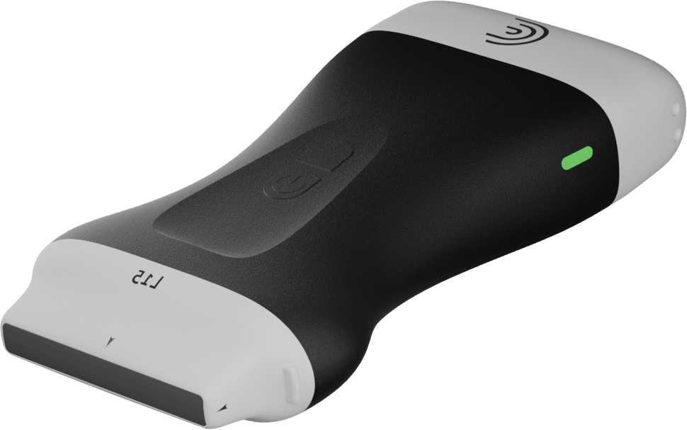
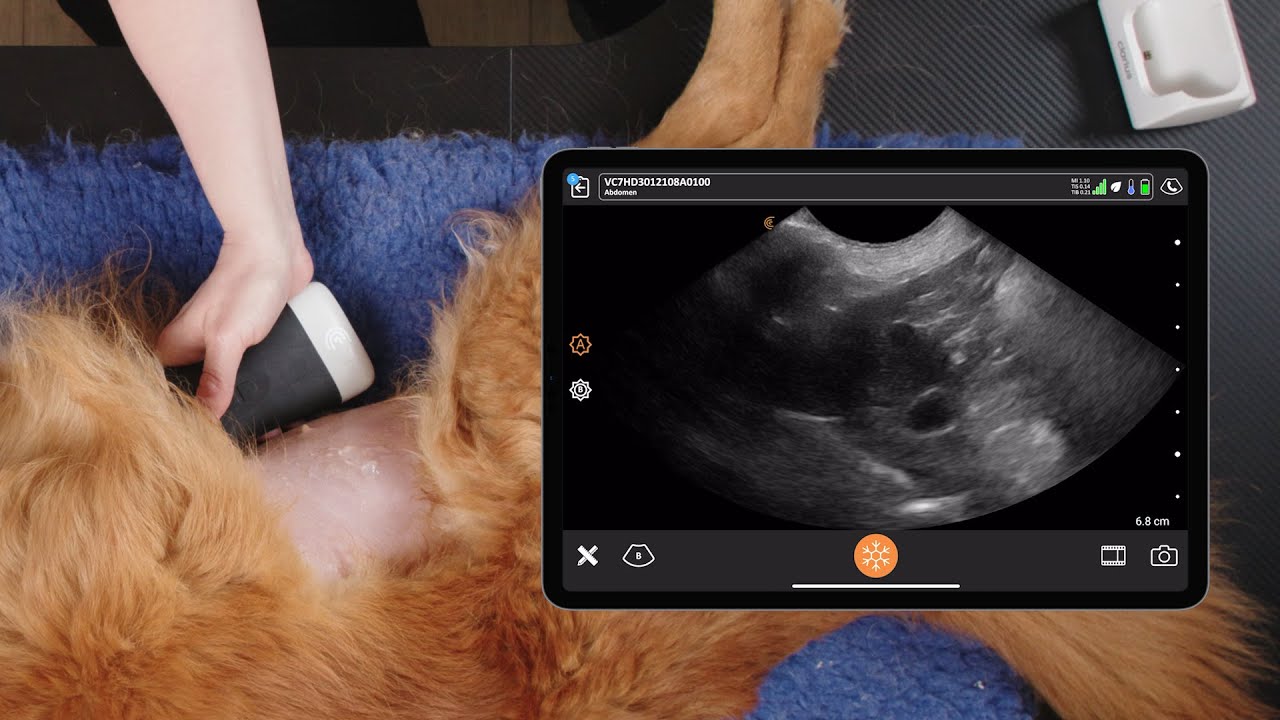
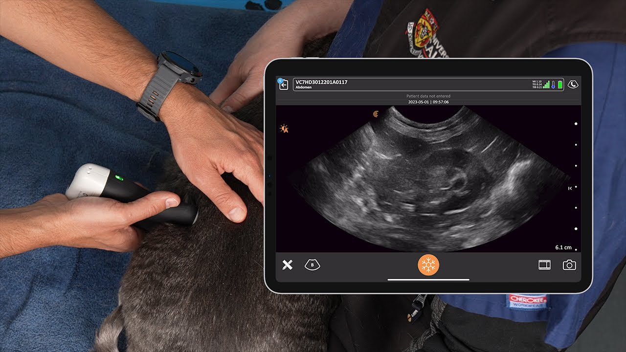
Watch this 90-second video to see Dr. Edwards demonstrate how to use tofu to practice ultrasound-guided FNA. She uses the Clarius C7 Vet HD3, which is designed for imaging small to medium-sized animals.
Visit Clarius Classroom to watch more veterinary ultrasound tutorials by ultrasound experts.
Tips for Successful ultrasound evaluation of abdominal masses
- Perform a systematic examination: Scan the entire abdomen in two planes to rule out other abnormalities and to identify any potential metastatic spread.
- Characterize the mass: Describe the size, shape, location, echogenicity, and echotexture of the mass.
- Look for local invasion: Assess whether the mass has invaded any surrounding organs or blood vessels.
- Use ultrasound-guided sampling: Obtain tissue samples for cytological or histological evaluation to confirm the diagnosis.
Dr. Camilla Answers Questions from the Audience During the Webinar
What are the bleeding risks of FNA?
The bleeding risks of FNA are minimal. The needles used for FNA are very small, and the procedure is usually performed under ultrasound guidance.
Are there any masses that should not be aspirated?
Yes, there are a few masses that should not be aspirated. These include masses in the urinary tract and masses that are particularly vascular.
Should aspiration be used during FNA?
Aspiration is not always necessary during FNA. In many cases, the “woodpecker” technique can be used to collect a sufficient sample. If aspiration is used, it should be done gently to avoid aspirating from surrounding tissues.
What is the smallest animal that can be aspirated?
Ultrasound-guided FNA can be performed on animals of all sizes, including kittens, puppies, Guinea pigs, and rabbits.
What is the least favorite organ to aspirate?
The liver can be a challenging organ to aspirate due to its location under the ribs.
Is sedation always necessary for FNA?
Sedation is not always necessary for FNA. If the animal is compliant, an opioid may be sufficient to relax them enough for the procedure.
Clarius Wireless Ultrasound for Veterinary Practice
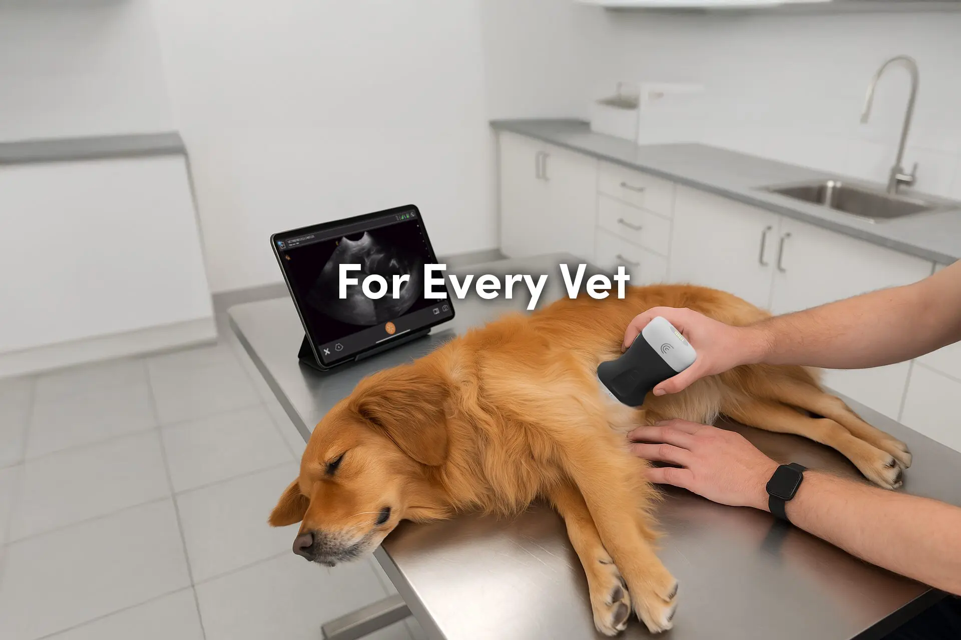
Dr. Edwards uses the Clarius C7 Vet HD3 scanner in her small animal practice. To learn more about how you can add wireless ultrasound to your practice, visit our Veterinary Specialty Page. There you’ll have access to additional webinars and classroom videos. Learn about the Clarius Membership which provides additional workflows for a wide variety of animal examinations.
Our new Clarius C7 HD3 Vet scanners are smaller and lighter. To find out which scanner is best for your practice, contact us today, or request a virtual ultrasound demo.
