


More than 4400 clinicians registered for a recent webinar — Ultrasound for Facial Aesthetics: A Beginner’s Guide to Scanning Like a Pro. A recording of the 1-hour webinar is available now to watch at your convenience.
The highly-rated webinar is presented by Shelley Guenther, an experienced sonographer and Clinical Manager at Clarius, with sonographer Janaye Smith, who performs live scanning using the Clarius L20 HD3. Scroll down for some key takeaways.
Introduction to Ultrasound
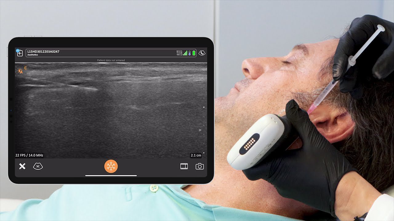
Medical ultrasound, also known as sonography, is a non-invasive and non-ionizing technique that uses sound waves to create pictures inside the body. Ultrasound has a variety of applications in facial aesthetics, including planning treatments, guiding injections, making diagnoses, and improving decision-making in clinical practice.
What is Ultrasound?
Ultrasound waves are sound waves with frequencies in the 2 to 20 MHz range, which is beyond the range of human hearing. The terms frequency and wavelength define an ultrasound wave. Frequency is the number of cycles per second, and wavelength is the length of a wave from one peak to the next.
How Does Ultrasound Work?
Ultrasound transducers, also known as probes or scanners, contain piezoelectric crystals that produce sound waves. When an electric current is applied to these crystals, they contract and expand, creating pressure waves that propagate into the body. The echoes that are reflected back to the crystals are then processed to create an ultrasound image.
Clarius handheld ultrasound scanners are miniature versions of traditional cart-based or laptop ultrasound systems. They use the same piezoelectric transducer technology to achieve high-definition imaging in an all-in-one system without wires.
Types of Ultrasound Transducers
Ultrasound probes come in different shapes and sizes and are named based on their frequency and array. The three main types of arrays are linear, curved, and phased.
- Linear array transducers are typically used for superficial imaging, such as skin, muscle, and breast tissue.
- Curved array transducers are used for deeper structures in the body, such as obstetrics and abdominal imaging.
- Phased array transducers are used for cardiac and some deeper abdominal imaging.
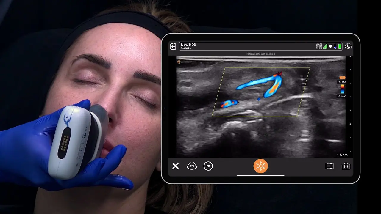
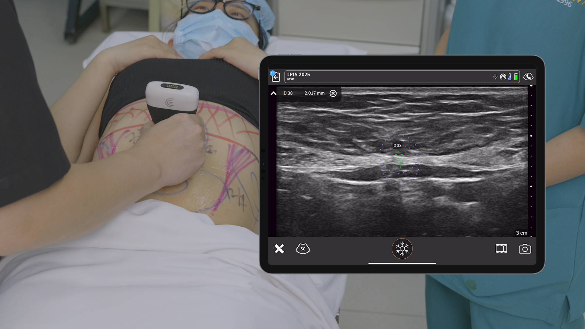
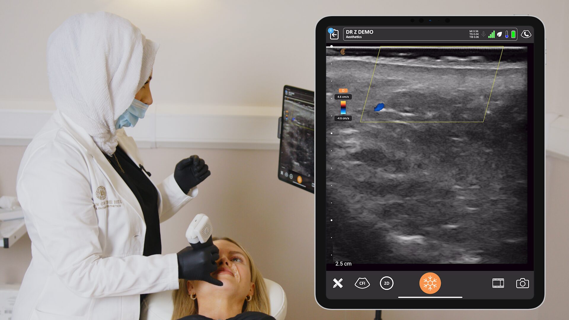
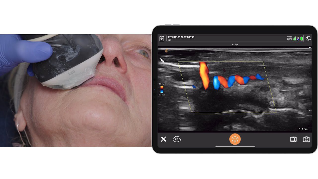
For facial aesthetics, a high-frequency linear array transducer is typically used. The Clarius L20 HD3 is a popular choice for facial aesthetics.
Ultrasound Scanning Techniques
Successful ultrasound scanning involves attention to patient positioning, probe grip, and gel application.
- Patient positioning should be comfortable for both the clinician and the patient, and should allow for a clear view of the imaging screen.
- Transducer grip should be firm but not too tight, and should allow for fine motor movements.
- Gel application is essential for ensuring good contact between the transducer and the patient, and for allowing sound to travel uninterrupted into the body.
Ultrasound Image Interpretation
Ultrasound images are composed of shades of gray, which indicate differences in tissues. The shades of gray are referred to as echogenicity.
- Hyperechoic or echogenic tissues are brighter, such as bone, collagen, and PMMA filler.
- Hypoechoic tissues are darker, such as muscle tissue.
- Anechoic tissues are black, such as fluid-filled structures.
- Isoechoic tissues are the same shade of gray as the surrounding tissues.
Ultrasound Artifacts
Ultrasound artifacts are visual features that appear on an image but do not correspond to actual structures in the body. They can be helpful or troublesome.
- Shadowing occurs when the sound beam encounters sound-absorbing materials, such as bone or calcifications.
- Posterior acoustic enhancement occurs when the speed of sound increases through fluid, causing brightness behind the fluid-filled structure.
Ultrasound Modes
The most common imaging mode is B-Mode, also known as 2D or grayscale imaging. Other modes include color Doppler, which is used to identify blood flow.
Practice Tips
Practice is essential for becoming proficient in ultrasound scanning.
- Practice on family, friends, or even tofu or a pork chop.
- Start with small and slow movements.
- Enroll in courses specific to your specialty.
Webinar Q&A
Q: Where can I purchase the gel that you like?
A: I purchased mine on Amazon, it’s called Wavelength CL. You can also try your hospital’s supplier.
Q: How high a frequency is high enough for facial aesthetics?
A: The higher the better, as long as you can still see what you need to see. If you only need to see 2-3 centimeters deep, 20 MHz is optimal.
Q: What do nerves look like on ultrasound?
A: Nerves are typically oval in cross-section and linear in long axis. They are bright with dark fascicles throughout them.
Q: How long does it take to get comfortable using ultrasound for aesthetics with no prior experience?
A: It varies, but it’s important to practice and get hands-on experience. There are many resources available to help you learn including hands-on workshops. Check out Clarius Classroom to watch dozens of video featuring aesthetic ultrasound experts and other Aesthetic webinars.
Q: Does the scanner pick up needle tips?
A: Yes, as long as the needle is at an angle that reflects the sound beam back to the scanner.
Q: How common is it for aesthetic physicians to use ultrasound in their practice?
A: It’s becoming more common, and patients are starting to demand it.
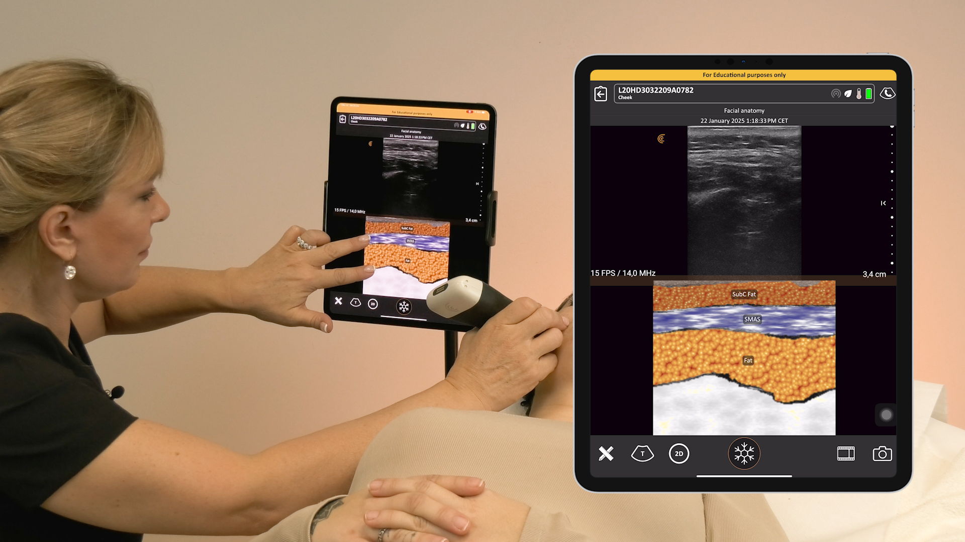
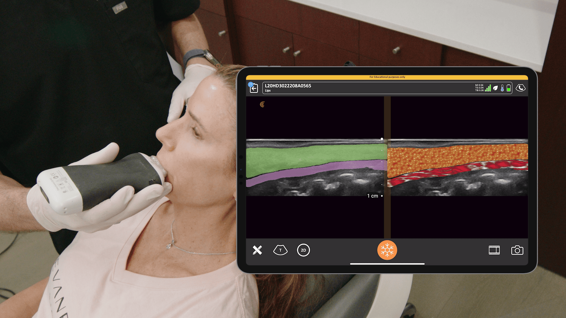
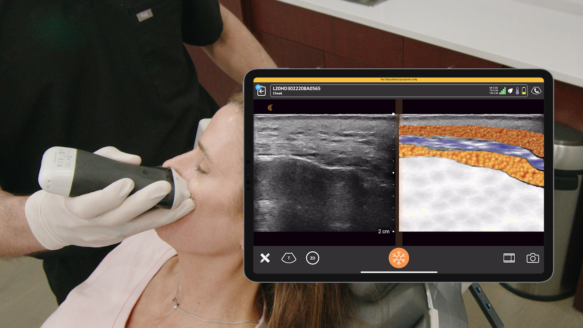
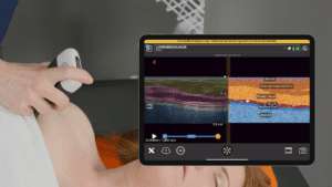
T-ModeTM: A New Way to Learn to Use Ultrasound
If you’re new to ultrasound, check out our new T-ModeTM, a groundbreaking educational technology to help new users to ultrasound advance their image interpretation skills using Clarius handheld scanners. It overlays distinctive colors, patterns, and labels to instantly identify and differentiate anatomical structures and tissue layers during aesthetic exams.
Watch this 2-minute demonstration with Shelley Guenther.
Curious About Clarius Ultrasound for Your Aesthetics Practice?
Check out our aesthetics specialty page for information about product options, including the Clarius L20 HD3 ultra-high frequency wireless handheld ultrasound scanner for facial aesthetic procedures. Designed to provide extremely high image quality in the near field, from the skin line to 4 cm, the Clarius L20 HD3 is ideal for a variety of clinical settings requiring superior superficial imaging. It is the only handheld ultrasound with ultra-high frequency of 20 MHz. Wireless and affordable, it delivers exceptional superficial imaging with an easy-to-use app for your iOS or Android device.
Schedule a personalized virtual demo tailored to your specialty. We’ll show you how easy it is to integrate handheld ultrasound into your workflow. We’ll customize the session to your needs and answer all your questions.
