
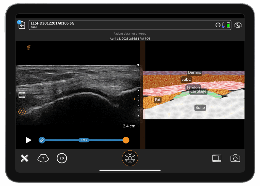
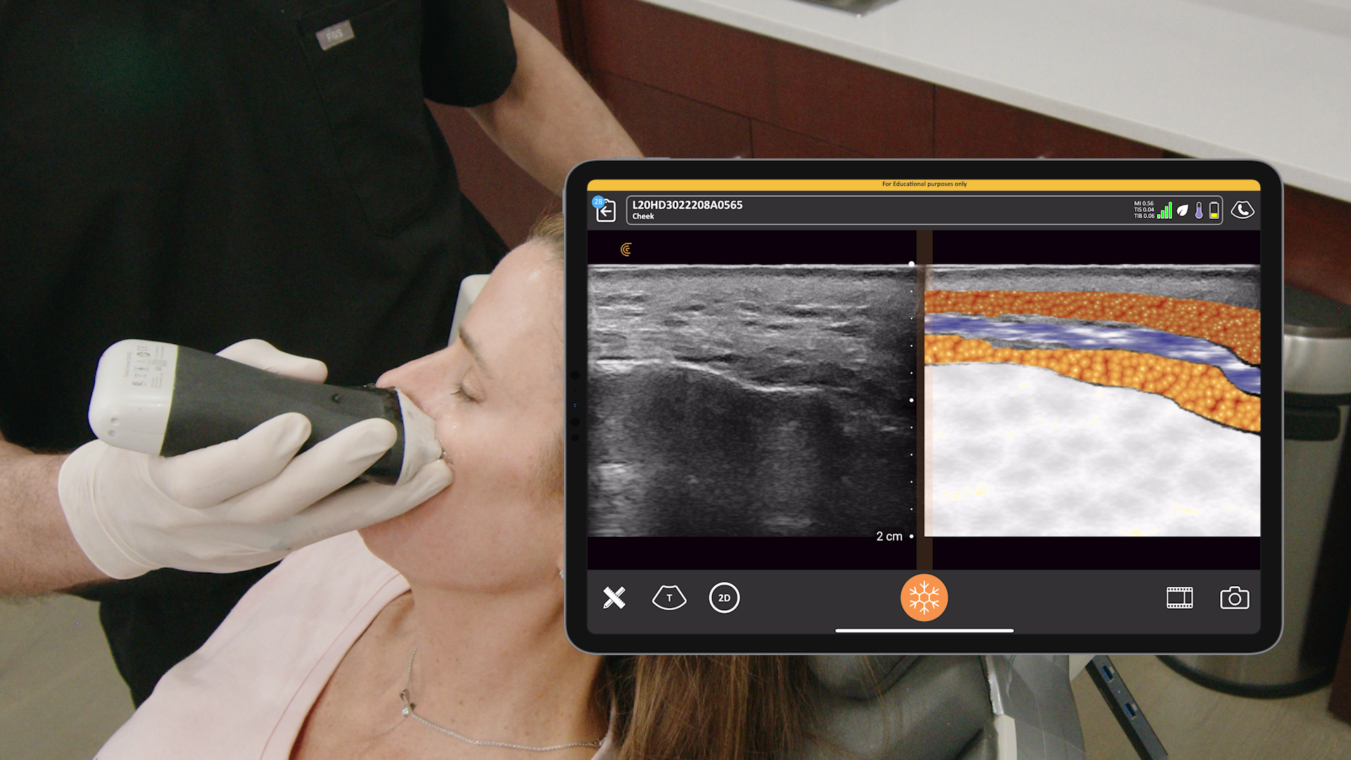
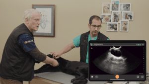
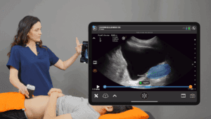
We’re excited to announce T-ModeTM Anterior Knee, the 11th AI model that Clarius has designed and clinically validated to make ultrasound easier to use for clinicians. This new feature, available now with the Clarius L15 HD3 and L7 HD3 scanners, transforms standard grayscale ultrasound images into vibrant, color-coded visuals that illuminate the subtle differences between tendons, bone, and other soft tissue in the anterior knee.
Chris Wolfe at Belmont University was one of the first to teach his students knee ultrasound anatomy with the help of new Clarius T-Mode Knee and says he saw them experience a “light bulb moment” during the demonstration.
T- Mode is providing a wonderful option in the curriculum right now. That adds so much benefit helping the student move and learning from a grayscale to seeing like, oh, this actually looks like something I’ve looked at in another manual,” says Chris. “We’re able to cover more structures in a timelier manner. When I’m trying to outline certain structures, it’s so helpful to have like a split screen to be able to look at grayscale and color images side by side.”
Watch this 2-minute T-Mode demonstration.
Ultrasound is frequently the first-line imaging modality for assessing knee pain, but learning to read and interpret the grayscale variations of ultrasound is known to be challenging for new users,” says Sarah Leverett, Vice President of Marketing at Clarius. “T-Mode is the expert assistant that helps novice MSK clinicians to quickly build imaging skills for confident assessments and in-clinic diagnoses. It’s also useful for patient conversations.”
A Complete MSK Ultrasound Solution
Shoulder and knee issues account for approximately 25% of all MSK complaints. Clarius offers an affordable and easy-to-use MSK ultrasound solution for in-clinic and MSK education use that includes:
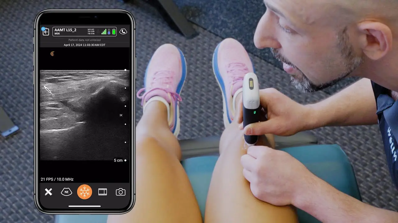
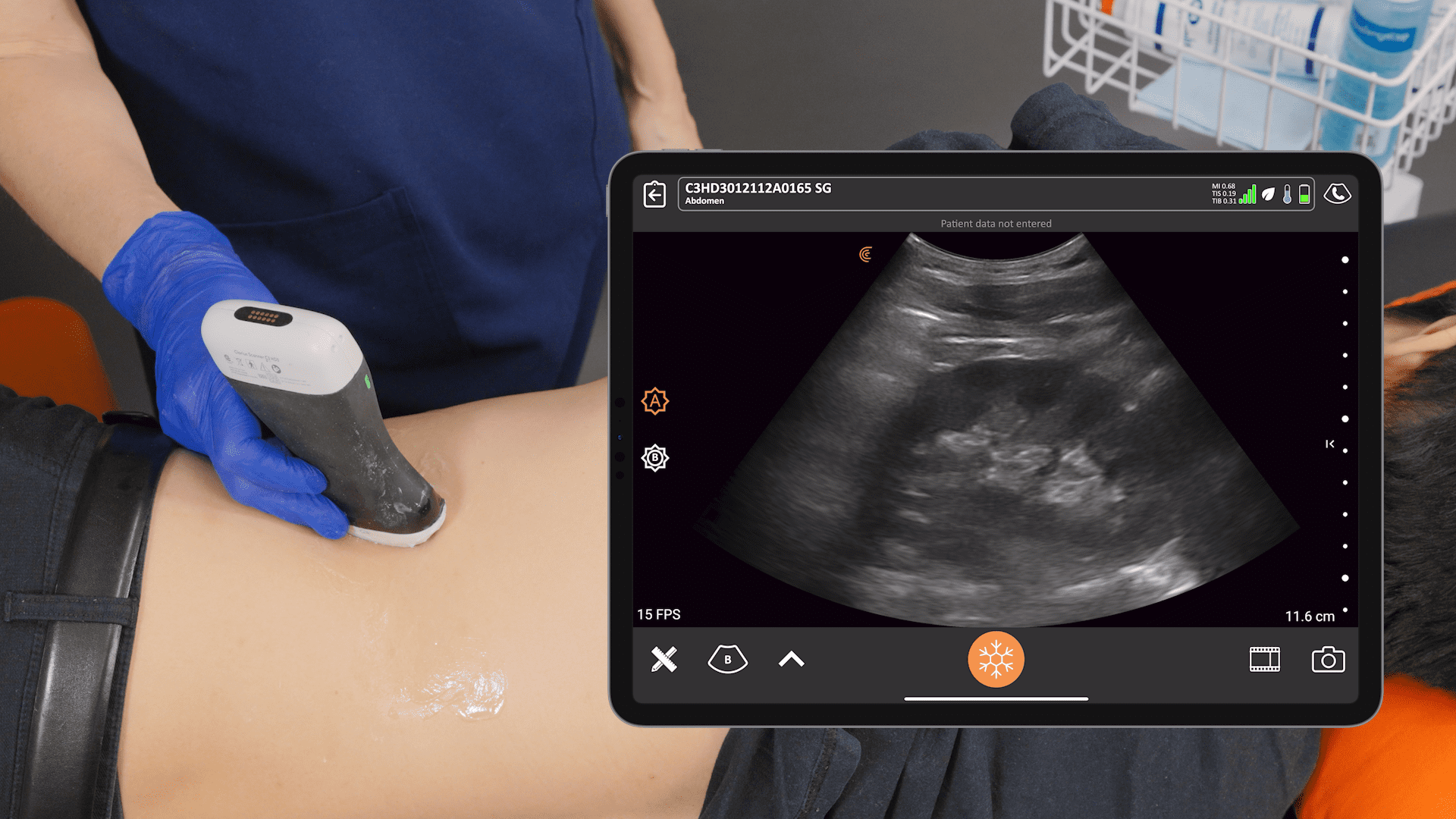
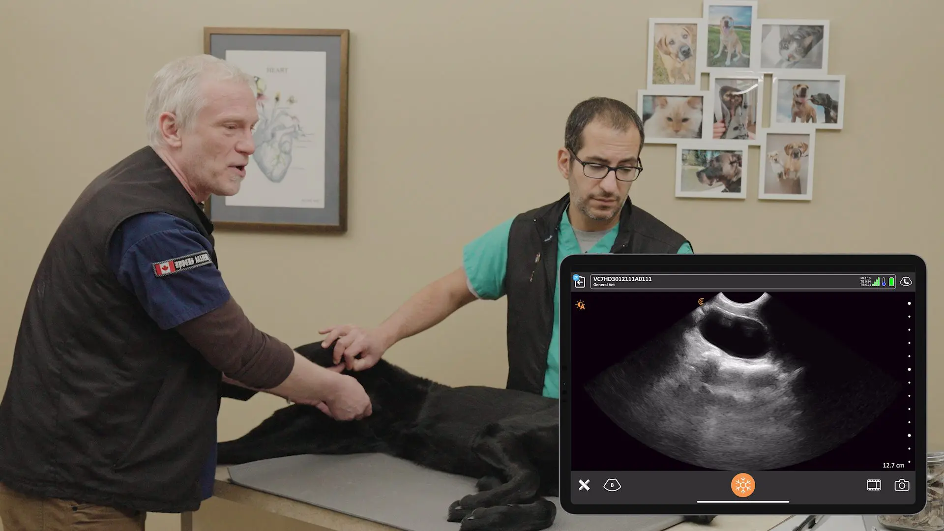
- Purpose-Built Wireless, Handheld Ultrasound Scanners: Engineered with best-in-class technology to deliver image quality comparable to traditional cart-based systems in an ultra-portable format for a small fraction of the cost.
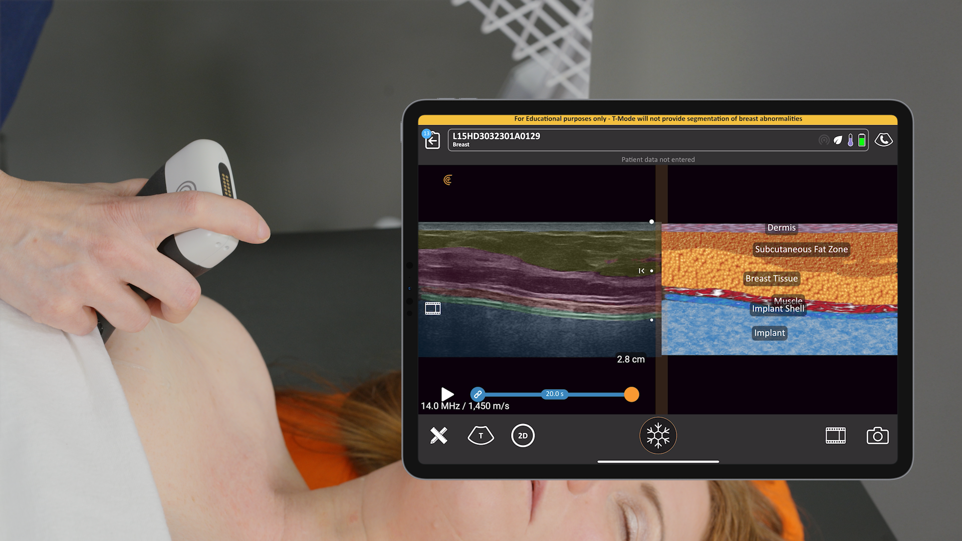
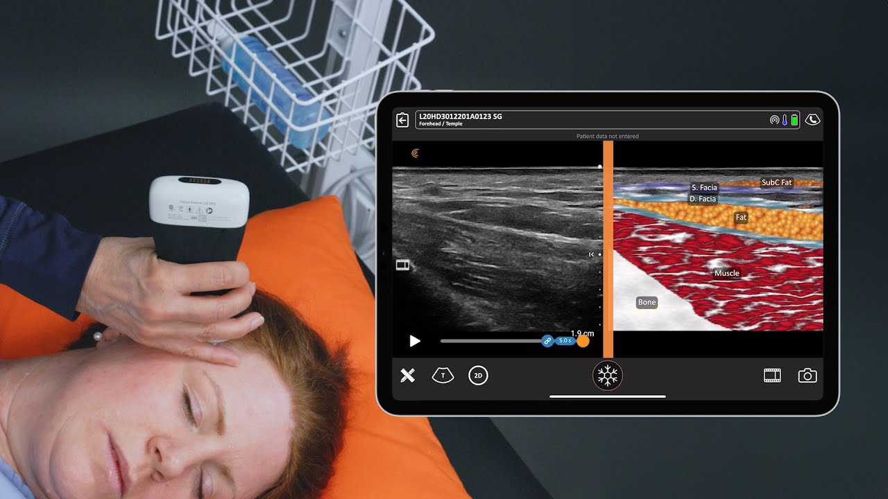
- Innovative T-Mode for Shoulder and Anterior Knee: Converts grayscale ultrasound images into vibrant, labeled visuals in real-time, simplifying anatomical understanding.
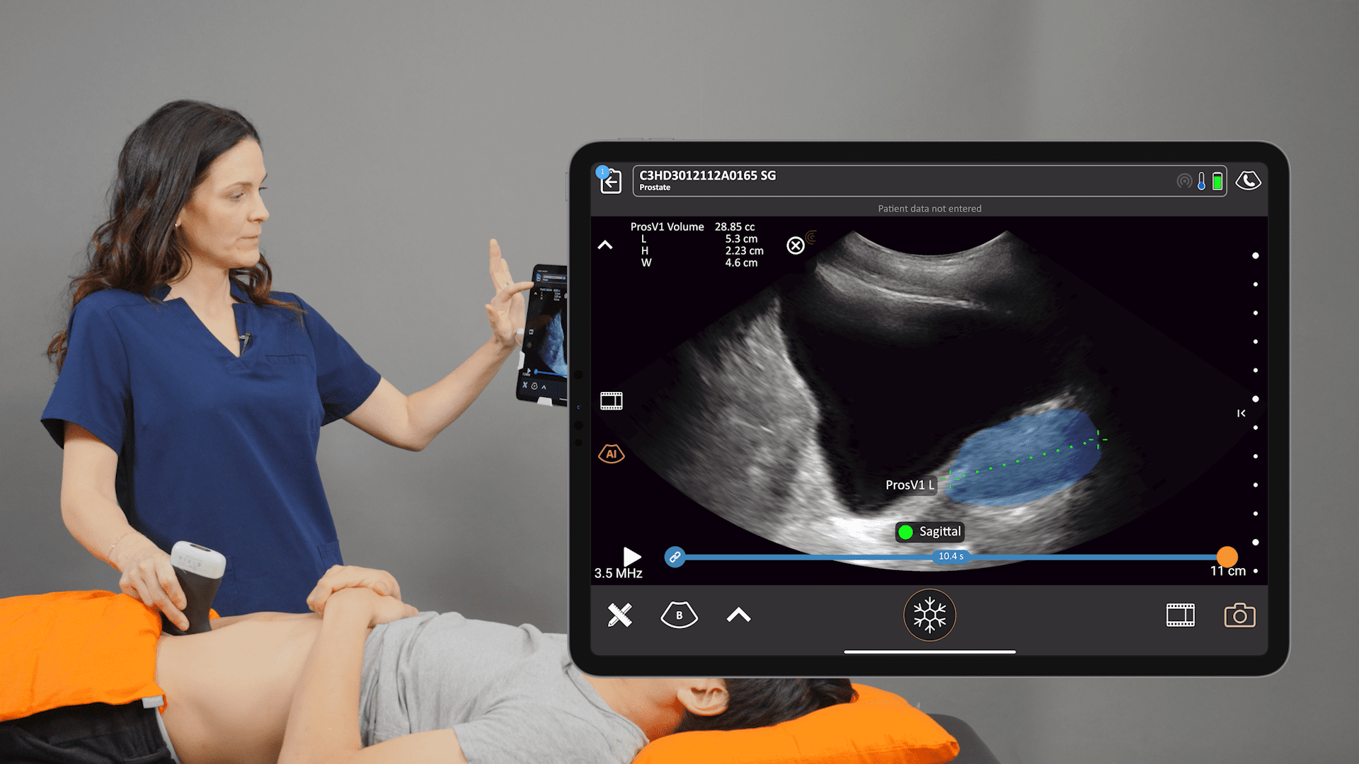
- MSK AI: Automatically segments, measures, and labels key structures like the patellar tendon, eliminating manual steps and allowing clinicians to focus more on patient care.
- Comprehensive Education Support: Providing resources and training to help clinicians effectively use ultrasound and the new T-Mode technology.
- Automatic Measurements and Calculations: Streamlining assessments for greater efficiency and accuracy.
- Integrated Patient Reports and Billing Codes: Facilitates documentation and billing; CPT codes are accessible on the Clarius App for clinicians in the United States.
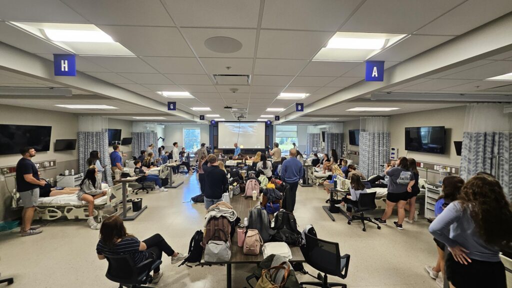
Clarius at Belmont University – What Students Say About T-Mode
Belmont University has been teaching with Clarius ultrasound scanners in its physical therapy program since September 2024. We recently had an opportunity to speak to the students about their experience with Clarius wireless ultrasound for physical therapy applications. Here’s what some of them had to say.
Hannah, a post-doctoral student, comments on T-Mode’s helpfulness with learning ultrasound anatomy
So today, having the T-Mode next to what we’re actually imaging was really helpful to just see it broken down – like this is muscle. This is the Bursa or fat. And just being able to see the multiple layers side by side was very helpful in learning.
I think having the ability to see side-by-side labeled images compared to what you’re actively seeing, like by pausing and seeing the image was super helpful.”
Izzy Gatey, a Second Year Student in the Physical Therapy Program, comments on the freedom of scanning without wires and Clarius Intelligence
I honestly love that it’s wireless. The cords usually get in the way when I’m using the transducer, so having the freedom to move between body parts without being restricted has been really nice. The wireless experience is amazing.
Today was my first time using T-Mode, and it was incredible. I already had a general sense of where the bone, muscles, and tendons were, but being able to actually confirm that on the screen really helped everything click. Now I’m starting to truly understand it.”
Jacob Storey is a physical therapist and an orthopedic resident and values clear image quality
The image quality of Clarius is a lot more clear than other units we’ve had before. Image quality is important for me just because for two reasons really, that you get to actually identify the anatomy that you’re looking at. But then also if you’re using it now on diagnosis, you want to actually be right about your diagnosis.”
Madison, a second-year student at Belmont University’s physical therapy program, appreciates Clarius image quality and plans to use ultrasound in her practice.
The image is incredibly clear, especially with diagnostic ultrasound and learning that process. It can be difficult to differentiate structures. So, the clearer the image and what this device can do has been very beneficial to the learning and to making that process a bit easier.
I see myself with patients being able to begin to give them answers a little bit quicker than maybe they have in the past. But more importantly, to confirm my own diagnostic assumptions or predictions that I think are going on, and to also kind of bring the patient along with me on the journey and to be able to show them like, hey, this is what’s going on in real time. It’s really exciting because I think patient education is something, especially in the field of physical therapy, that is vital to recovery and making them feel like they’re riding along in that process as well.”
Curious About Clarius MSK Ultrasound for Your Institution or Practice?
If we’ve piqued your interest in discovering whether Clarius ultrasound is right for your practice or teaching institution, we invite you to book a virtual demonstration. We’ll be delighted to show you why Clarius ultrasound is the leading choice of handheld ultrasound for discerning clinicians.
