

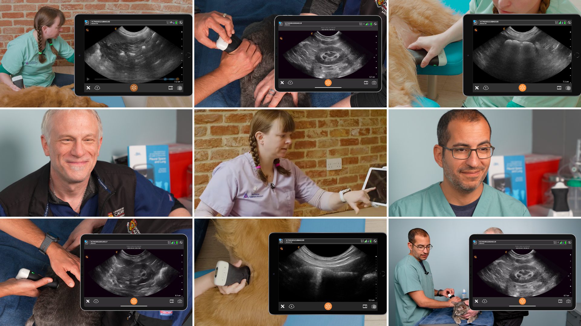
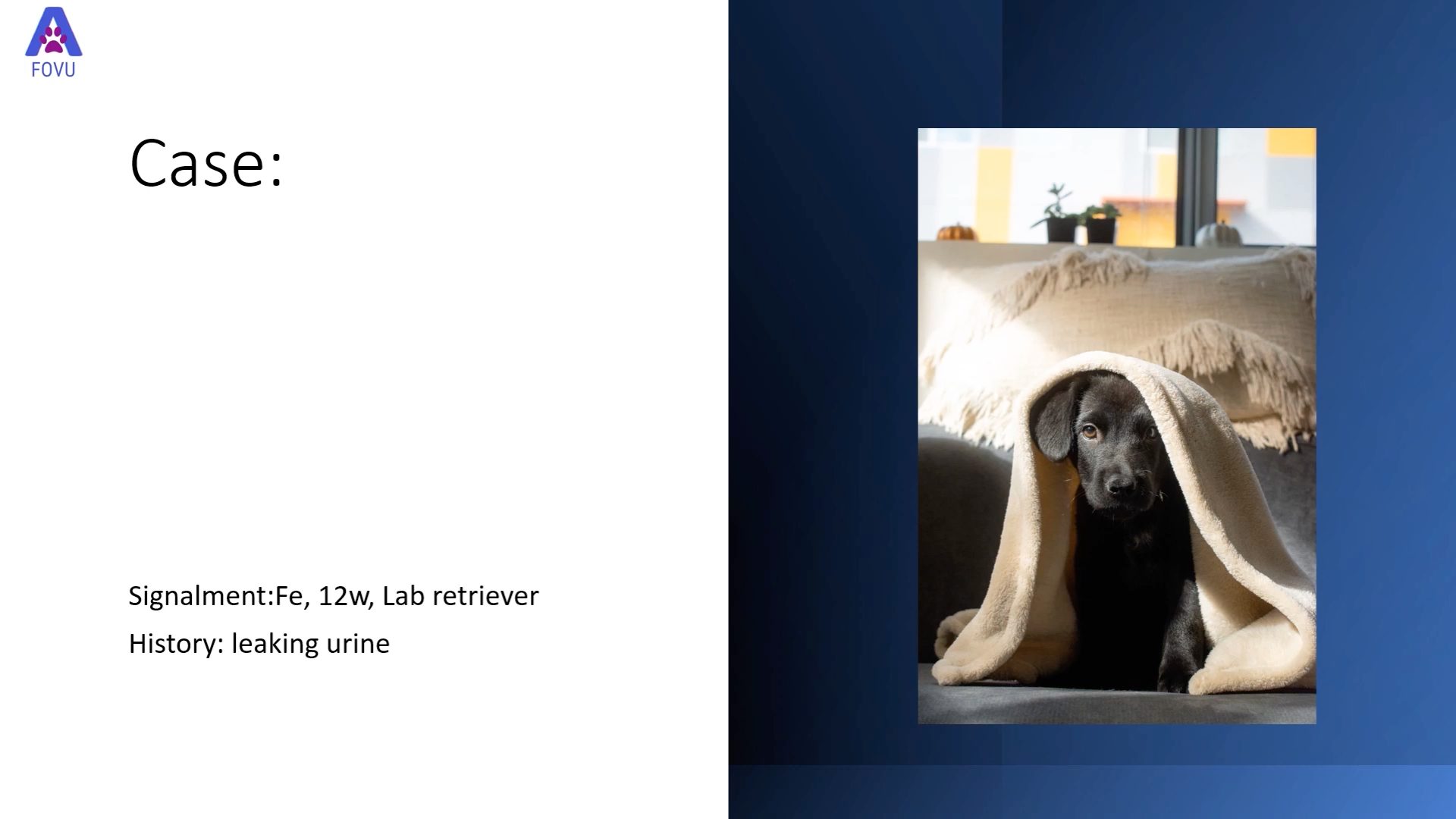
As more veterinarians join the Clarius ultrasound community, we’re producing more free educational tools to support point-of-care ultrasound training. This week we’re featuring practical tips from Dr. Camilla Edwards, DVIM, on how to diagnose pathology in the intestines of small animals, including cats and dogs.
An expert veterinary ultrasound educator, Dr. Edwards offered clear and methodical scanning techniques and pathology interpretation in a recent one-hour webinar: Practical Small Animal Ultrasound: Diagnosing Pathology with Intestinal, Gallbladder & Spleen Exams. You can watch the full webinar or read on for highlights.
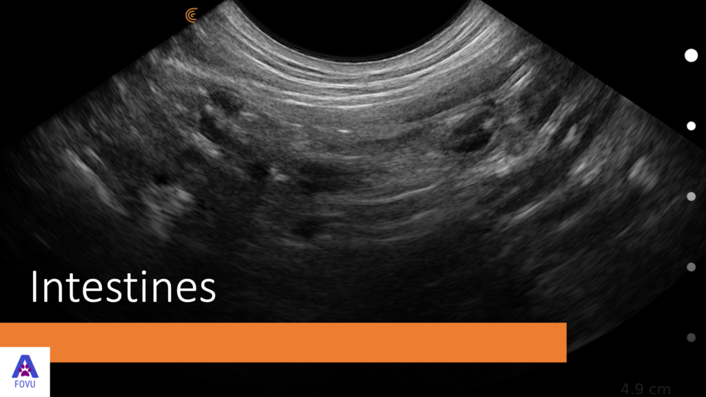
High Resolution Imaging Is Important for Identifying Differences from Normal Intestines
“For small animals, indications for scanning the intestines include vomiting, chronic diarrhea and abdominal pain. Before we see a demonstration on how to scan the intestines of a small dog, let’s cover some basics of what we’re looking for.
Our exam focuses on smaller areas and we look at the wall layering in the gastro intestinal tract, which can be quite important. Because we’re looking at layers of submillimeter structures, the resolution of your ultrasound system is important for differentiating normal from pathology.”
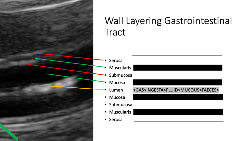
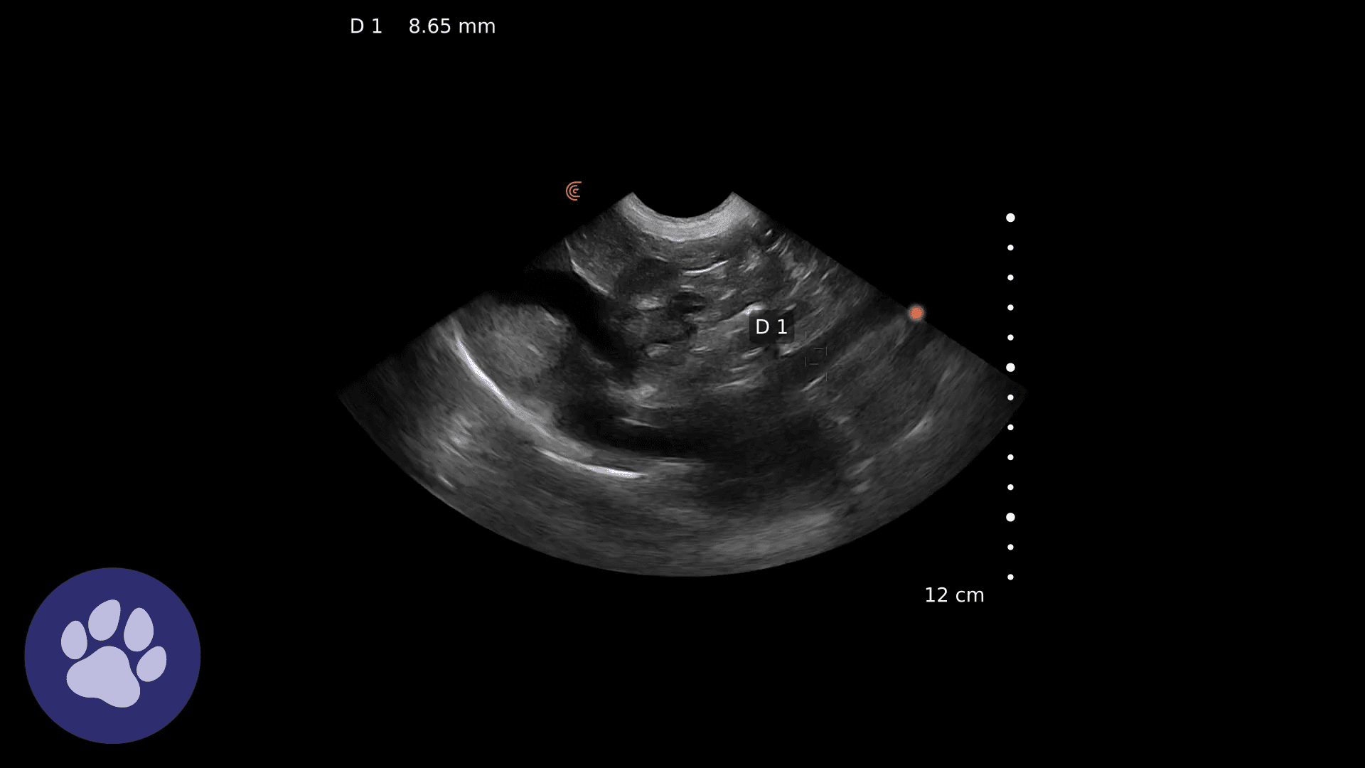
“In the image above we’re looking at one loop of the small intestine. The layers we see here and on the other side are as follows:
Serosa – is the first layer at the top shown as a thin white line
Muscularisis – is the next hypoechoic line
Submucosa – is the following hyperechoic line
Mucosa – is thicker and hypoechoic
Lumen – is in the middle and can be filled with gas, which will obstruct the view of the other side wall.
Note: It’s wise to know the animal’s diet whether they’re raw fed or if they’re kibble fed as you’ll get different images. You might find fluid or mucus as you see here. When we get down to the colon, you’ll find feces.”
Measuring Wall Layering in Intestines when Looking for Changes
“As you see below, the layering is different in various areas of the intestines. We’re looking for changes within the layers. For example, the muscularis layer might be thickened in cats with Inflammatory Bowel Disease (IBD).
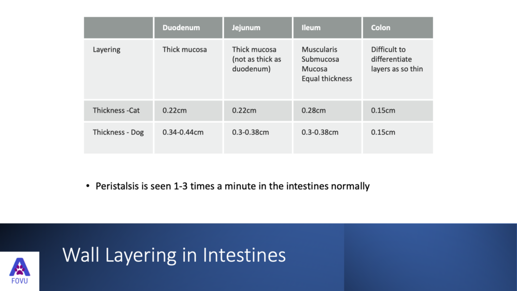
- Duodenum has a thicker mucosa than the rest.
- Jejunum has a reasonably thick mucosa, but not as thick as duodenum.
- Ileum has an equal thickness in the muscularis, submucosa, and mucosal layers.
- The colon can be difficult to differentiate as the layer is so thin: 0.15 centimeter in cats. In dogs, it does vary as well, but we’re expecting a bit of a thicker duodenum than the rest of the intestines.
Peristalsis – During the exam, we should see peristalsis one to three times a minute in the intestines, normally. It will be much less than that in animals that are sedated.
Normal Images of the Intestine
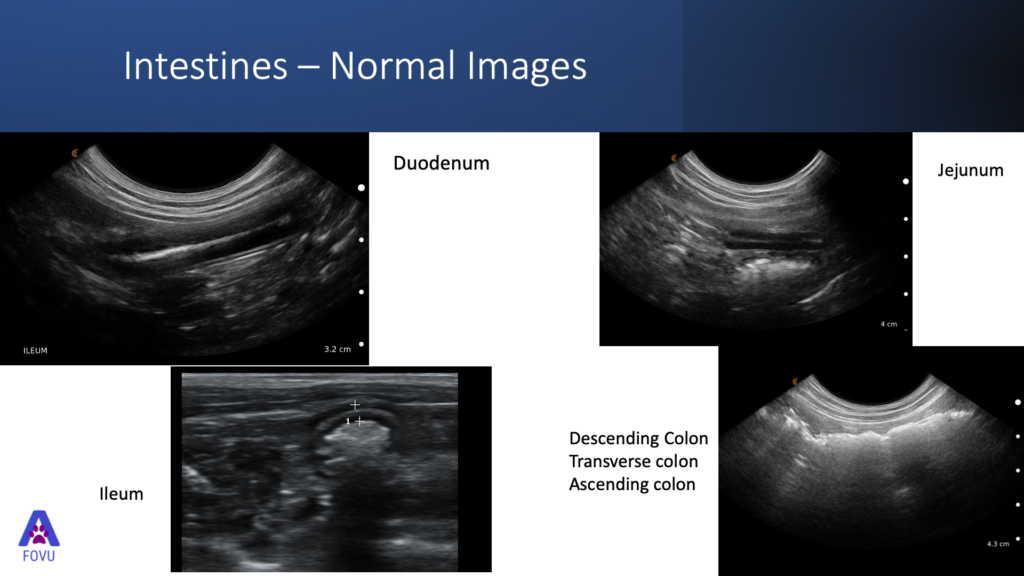
These are normal intestine images taken of my dog Pippi.
- We can see that this mucosal layer is slightly thinner than in the duodenum.
- In the image of the ileum, we can see the layers are much more evenly spread.
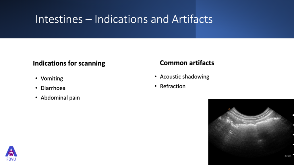
- Acoustic Shadowing as we see in this transverse image of the colon image is a common artifact. It’s the enemy when we’re scanning the intestines. We see the gas and the feces obstructing the view of the other side. That’s why we recommend scanning both sides of the animal.”
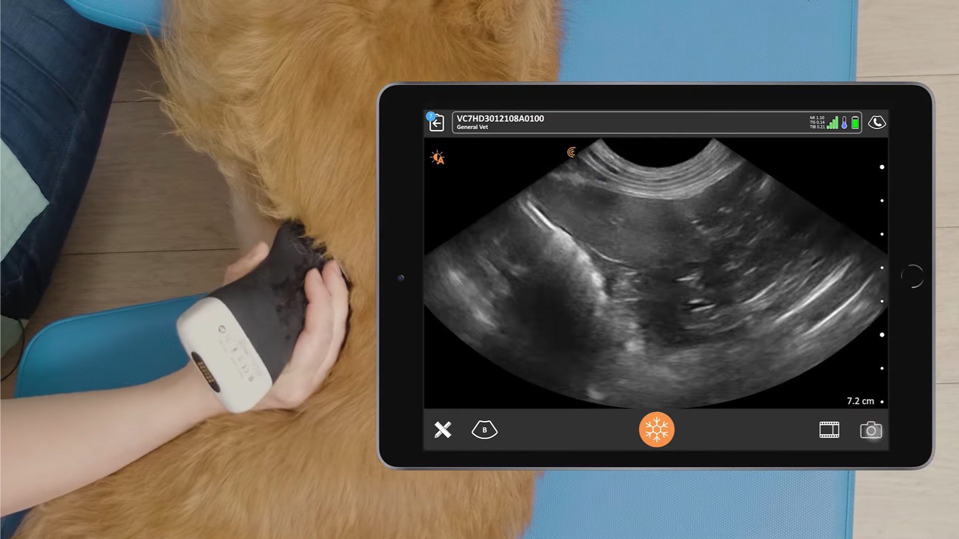
VIDEO: How to Scan the Intestines
Watch Dr. Edwards demonstrate her step-by-step process for scanning the intestine using a castle pattern in this five-minute video of her dog Pippa.
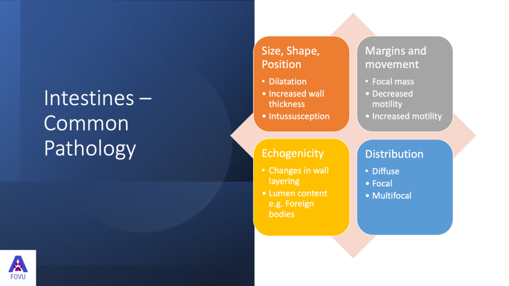
Common Pathology in Small Animal Intestines
“To detect pathology within the intestines, we need to systematically view the whole area from two different planes. We also need to look at the total thickness of the wall and the layering to detect if it’s appropriate for that part of the intestines.
Dilatation – can indicate an obstruction further along.
Increased wall thickness and interception – is easy to pick up during an ultrasound exam. You can make a diagnosis by seeing a loop and testing within another loop of intestine.
Focal masses and patterns of movement – is there decreased motility? If the animals are not sedated, then that might be for pathological reasons, or is there even increased motility?
Echogenicity – here we’re looking for changes in wall layering and we’re looking for lumen content.
Distribution – is it diffuse, focal, or multifocal?”
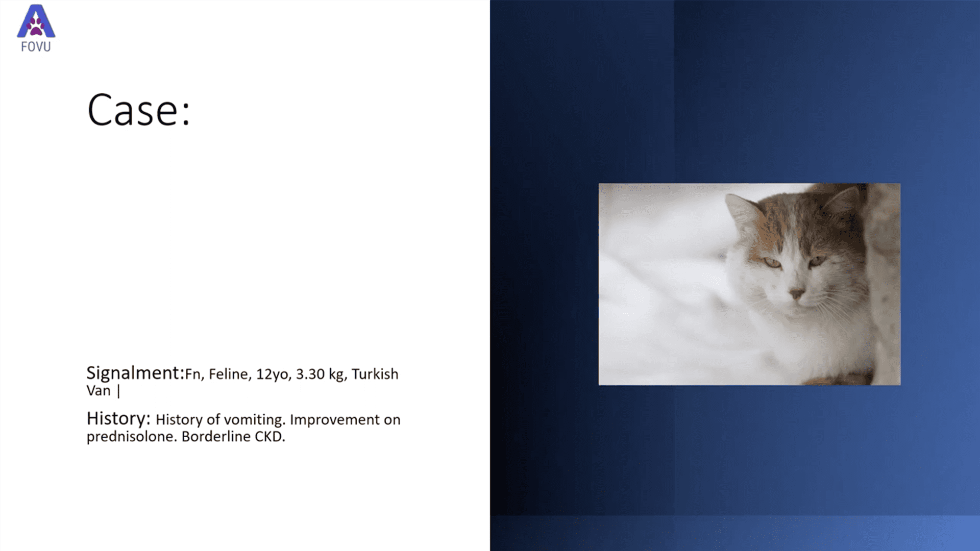
Intestinal Mass in a Feline
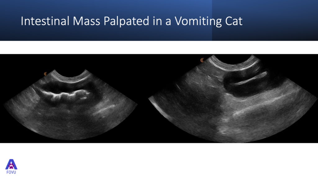
“Here we have images of abnormal intestines. There is an intestinal mass that we were able to palpate in a cat that was vomiting.
This is just a scan through one side of the mass whether the lumen comes into view. There is quite a focal lesion in this cat. And differentials for this presentation might include some localized inflammation and edema. Lymphoma is high on our list. It would be nice to check the local lymph nodes to see if they’re affected in any way.”
Ultrasound is Ideal for Foreign Body Identification
“Ultrasound is a great tool for detecting foreign bodies. It enables identifying and retrieving the foreign bodies quite readily. One of the things that gives foreign bodies away is that they are usually perfectly shaped. For example, you’re normally never going to find a perfect circle in the abdomen. If it’s a ball, they’re often hyper echoic and they often have an acoustic shadow as well.”
Sign Up for Next Webinar
If you’re interested in learning more about veterinary ultrasound, register for our free webinar on March 31: Veterinary Point-of-Care Pleural Space and Lung Ultrasound (PLUS) for Everyday Practice!
With high-quality imaging and an AI-driven app that is almost as easy to use as your smart phone, Clarius is the ideal ultrasound system for veterinarians who are new to ultrasound. To learn about how easy and affordable it is to add Clarius handheld ultrasound to your veterinary practice, visit our Clarius veterinary ultrasound page for a video demonstration and product details. Or contact us today to discuss which scanner is right for your veterinarian practice.
If you’re interested in alerts on new videos as they are published on Clarius Classroom, please subscribe to our Facebook, Twitter, LinkedIn, Instagram or YouTube channel.
You can also watch Dr. Edwards’ review of the Clarius C7 HD Vet on her website.


