


Facial fillers have become increasingly popular in aesthetic medicine, but complications can arise when they are misplaced or have migrated. Fortunately, ultrasound technology has revolutionized the way practitioners identify and treat these issues, offering a safer and more efficient approach to filler dissolution.
Dr. MJ Rowland-Warman, founder of Smileworks Hub and clinician director at Smileworks in Liverpool, considers ultrasound the most groundbreaking development in aesthetic medicine since the invention of filler. She practices non-surgical aesthetics with a special interest in the management of dermal filler complications using ultrasound.
We were delighted to have Dr. MJ share ultrasound-guided techniques for aesthetic procedures during a series of highly-rated webinars, which are now available on-demand. Watch Ultrasound-Guided Techniques to Rapidly Dissolve Fillers, Part 1: Misplaced Facial Fillers at your convenience. Scroll below for some highlights and two detailed video demonstrations.
The Power of Ultrasound in Aesthetic Medicine
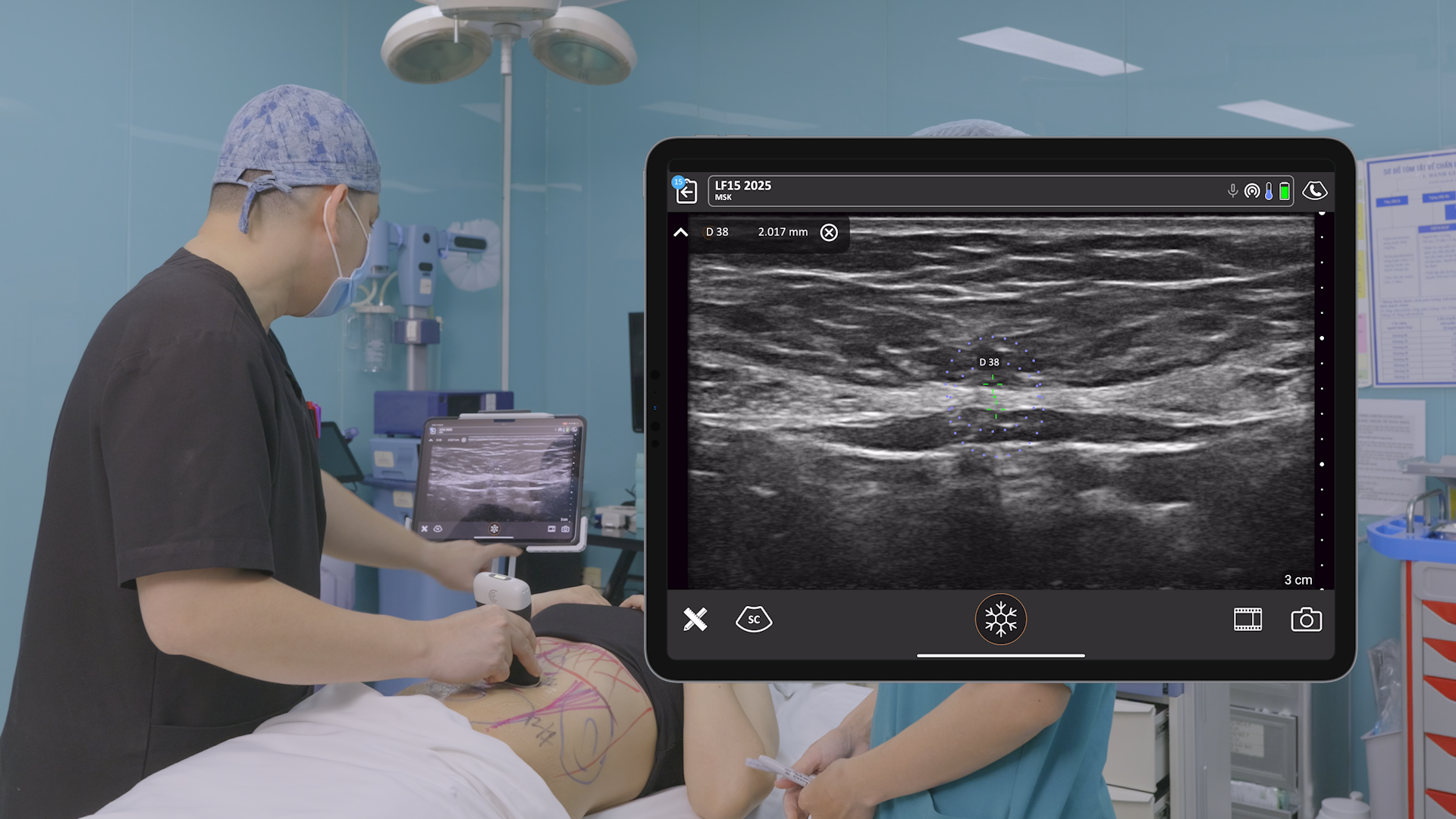
According to Dr. MJ, ultrasound has emerged as a game-changing tool in the field of aesthetic medicine. It allows practitioners to visualize facial anatomy beneath the skin, identify misplaced fillers, and guide treatment with unprecedented precision and improved safety. This technology is particularly valuable for addressing one of the most common complications in aesthetic procedures: misplaced facial fillers.
Understanding Misplaced Fillers
Misplaced fillers occur when the product is injected into an incorrect plane or location, or when it interferes with adjacent structures. This can happen immediately during treatment or develop over time due to tissue movement. While some practitioners may overlook minor misplacements, these issues can cause significant distress for patients and potentially damage the reputation of the aesthetic industry.
Benefits of Ultrasound-Guided Filler Dissolution
1. Precise Identification: Ultrasound allows practitioners to accurately locate misplaced filler deposits, eliminating guesswork.
2. Targeted Treatment: With ultrasound guidance, hyaluronidase can be injected directly into the filler deposit.
3. Reduced Hyaluronidase Usage: Ultrasound-guided techniques typically require less hyaluronidase compared to traditional „flooding“ methods, minimizing potential side effects.
4. Real-Time Visualization: Practitioners can observe the dissolution process in real-time, ensuring complete treatment.
5. Improved Patient Outcomes: Patients experience fewer interventions, faster resolution of complications, and better overall results.
The Dissolution Process
When performing ultrasound-guided filler dissolution, practitioners typically follow these steps:
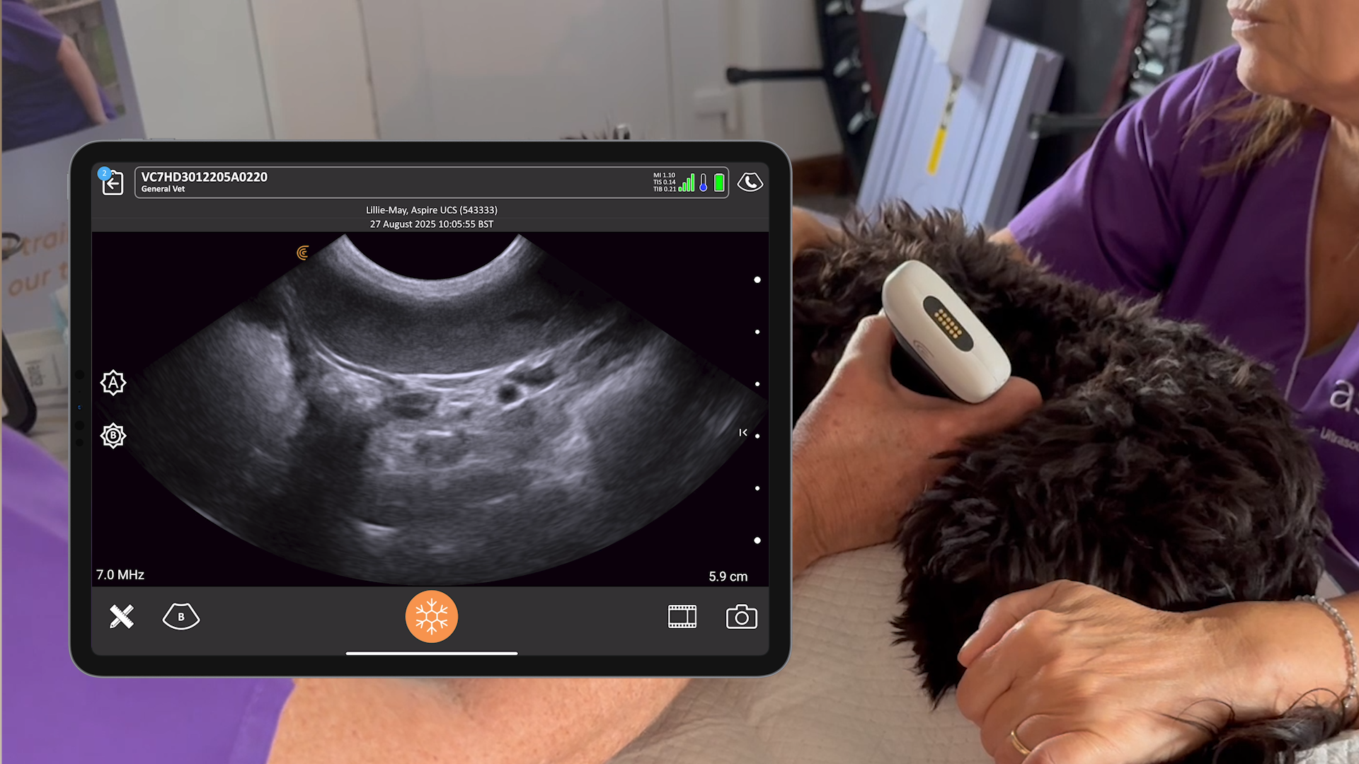
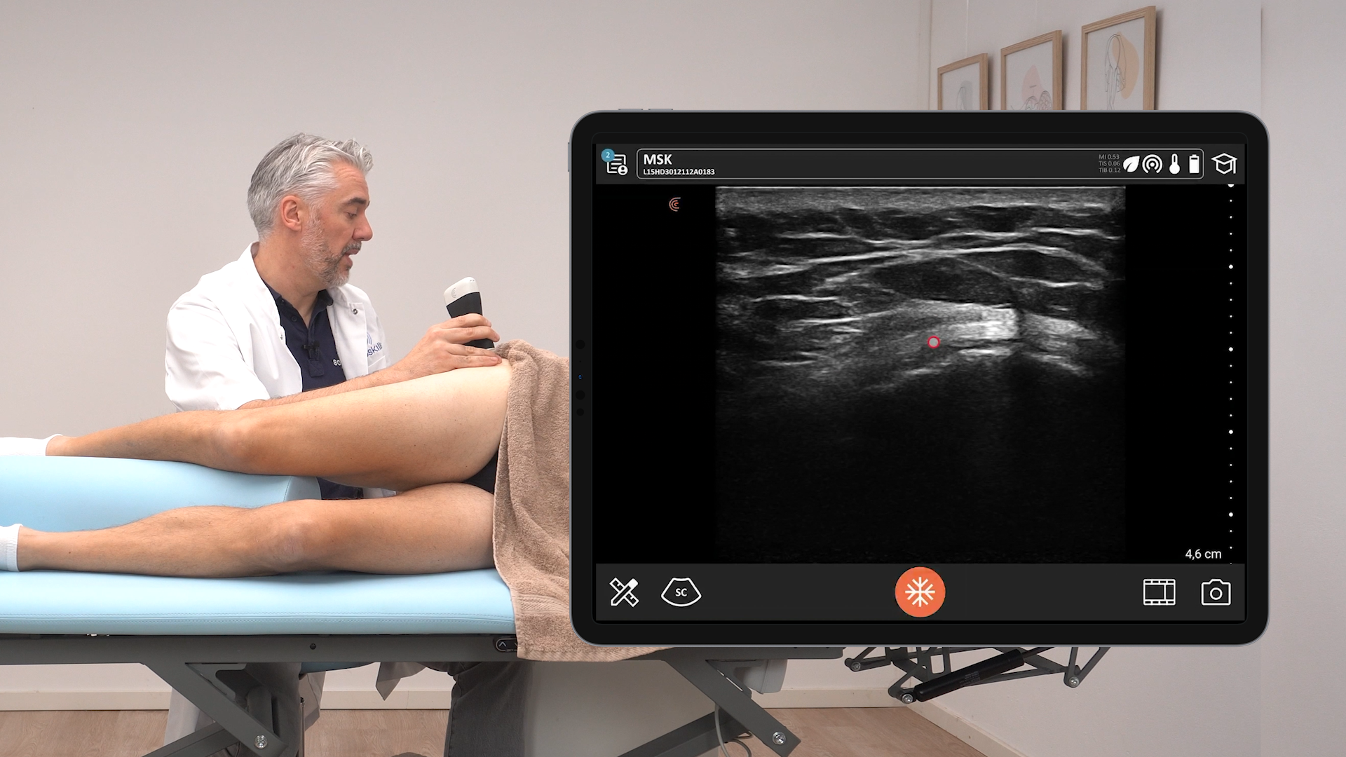
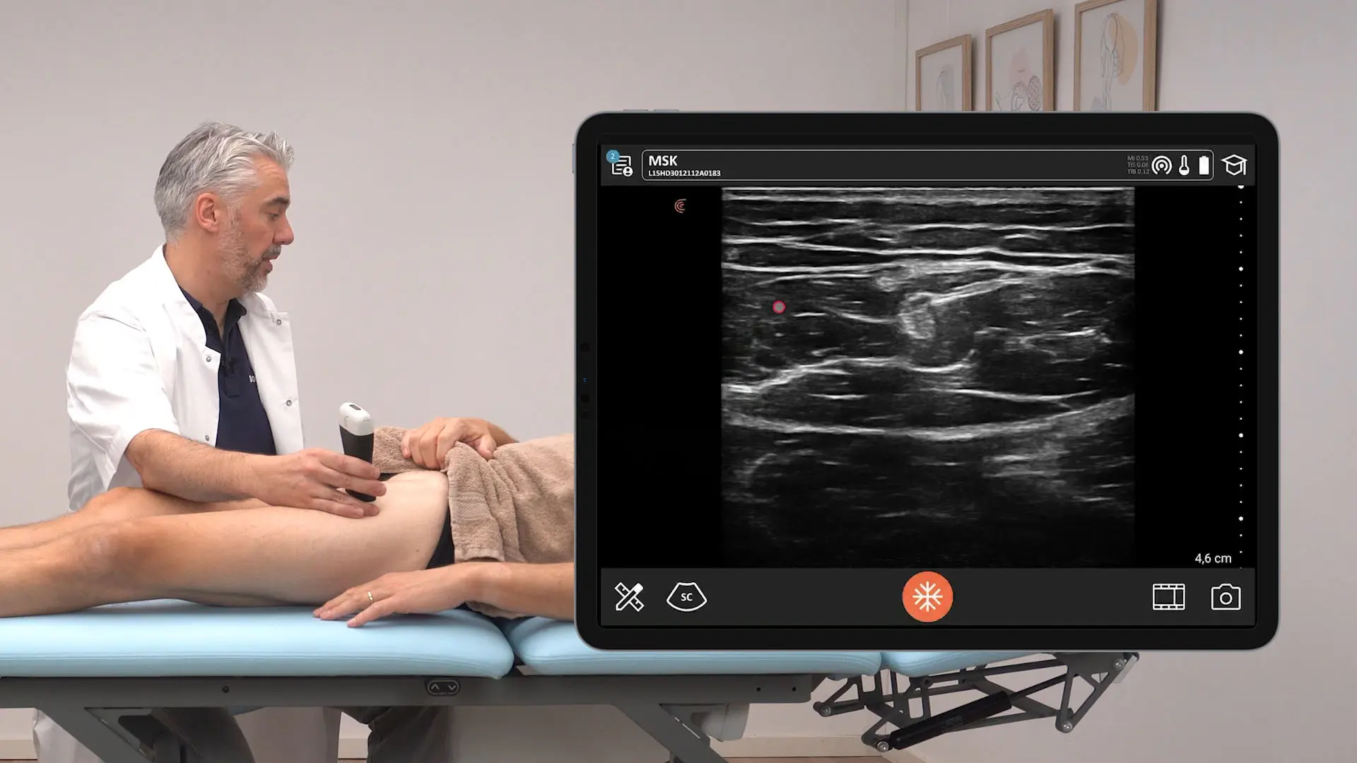
1. Scan the Area: Use the ultrasound to identify the misplaced filler and surrounding anatomy.
2. Prepare for Injection: Utilize an in-plane technique, aligning the needle with the ultrasound probe for optimal visibility.
3. Inject Hyaluronidase: Administer small, targeted doses of hyaluronidase directly into the filler deposit.
4. Monitor Dissolution: Observe the filler taking on a characteristic „cotton wool“ appearance as it dissolves.
5. Reassess and Repeat: If necessary, administer additional doses until the filler is completely dissolved.
Case Studies
Dr. MJ Rowland-Warmann, a renowned expert in aesthetic medicine, shared two compelling cases demonstrating the effectiveness of ultrasound-guided filler dissolution:
Tear Trough Complication
A patient presented with bilateral interorbital swelling 18 months after tear trough filler treatment. Ultrasound revealed misplaced filler and associated hypervascularity. Targeted dissolution provided swift resolution. Watch this video for her case review and demonstration.
Forehead Filler Complication
A patient experienced severe symptoms after receiving forehead filler from an unlicensed practitioner. Ultrasound-guided dissolution offered relief when previous attempts had failed. Watch this video for her case review and detailed demonstration.
Clarius Handheld Ultrasound: The Leading Choice for Aesthetic Professionals
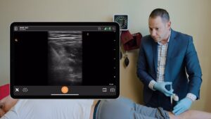
As Dr. MJ demonstrates during the webinar, ultrasound-guided techniques for dissolving misplaced facial fillers represent a significant advancement in aesthetic medicine. She has been using the Clarius L20 HD3 handheld ultrasound at her practice for three years and says using it for filler treatment “is a godsend, whether that be before treatment to vascular map, during treatment to place filler using ultrasound guidance, or after treatment to confirm placement.
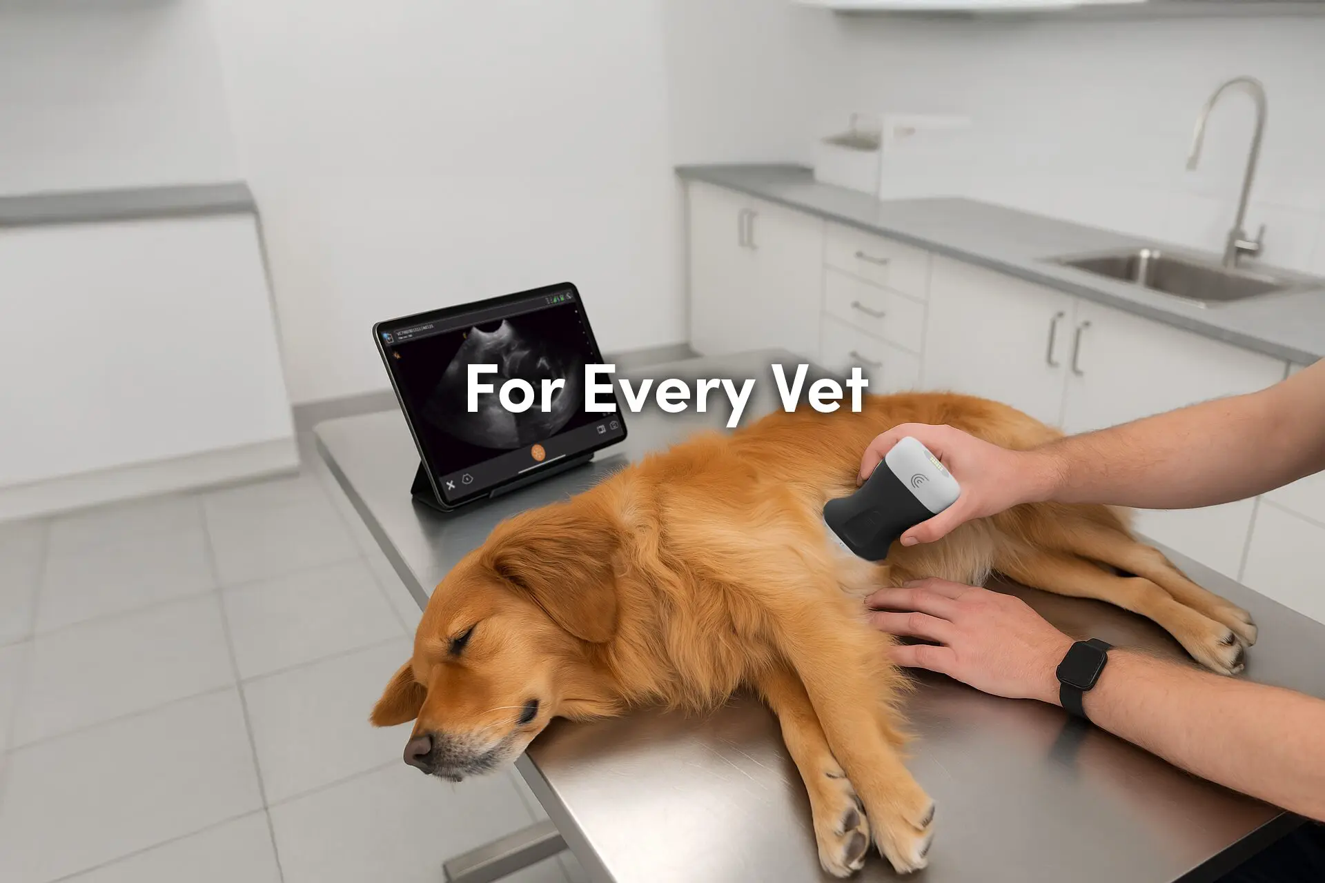
Wireless and pocket-sized, Clarius handheld ultrasound scanners deliver the high-definition imaging and performance of traditional ultrasound systems for a small fraction of the cost. They are the leading choice for plastic surgeons and aesthetics practitioners performing ultrasound-guided procedures to ensure patient safety.
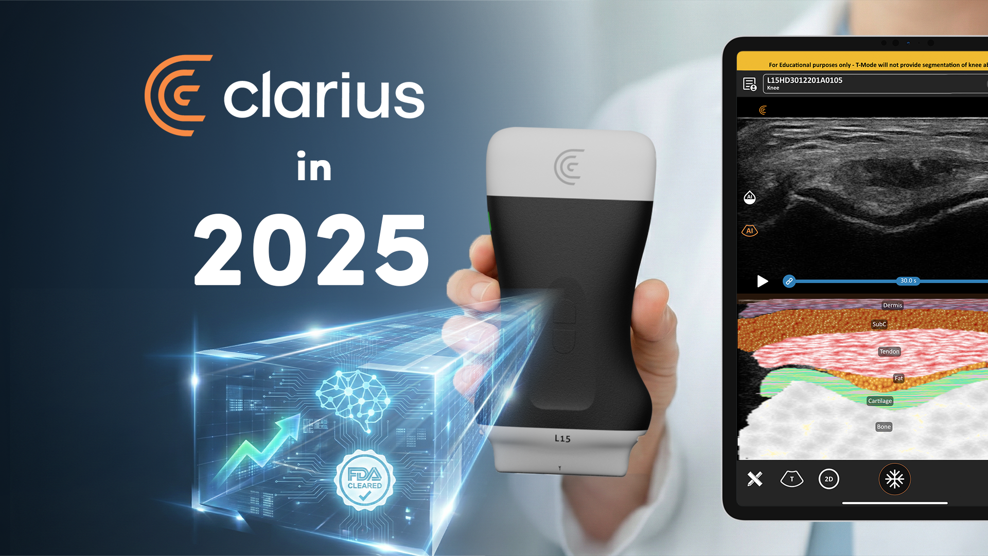
New T-Mode™AI by Clarius is a groundbreaking educational technology to help clinicians new to ultrasound advance their image interpretation skills using Clarius handheld scanners.
Visit our aesthetics page to learn more or request a virtual ultrasound demo today to learn how high-definition ultrasound imaging with voice controls can improve safety and deliver consistent patient outcomes at your aesthetic practice!


