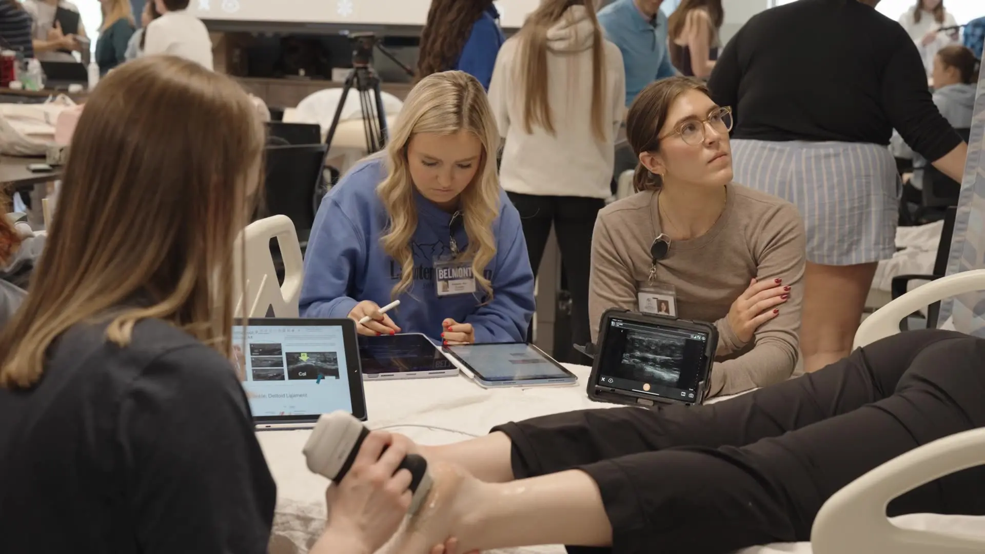
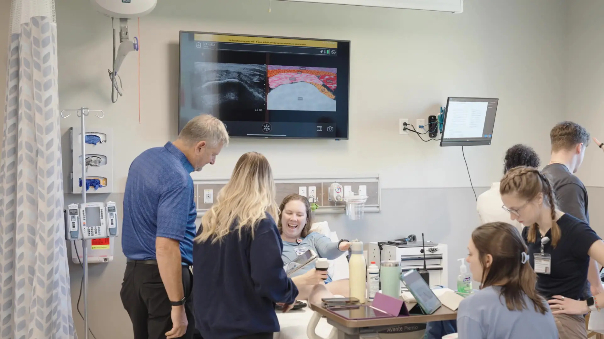
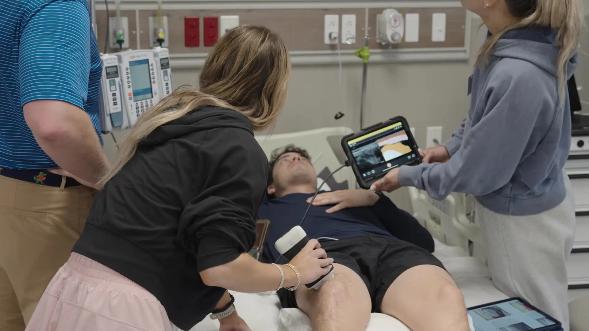
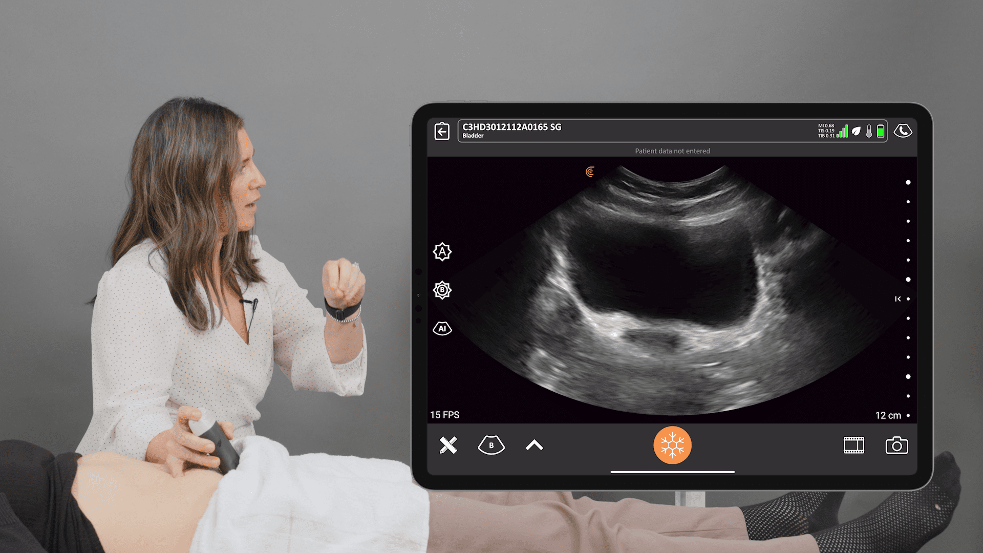
A 2023 paper published in the International Journal of Sports Physical Therapy notes that “Musculoskeletal (MSK) ultrasound (US) is quickly growing as a non-invasive and safe manner of assessing musculoskeletal structures (bones, muscles, tendons, ligaments) without the need for expensive or potentially harmful studies such as radiograph or MRI.”
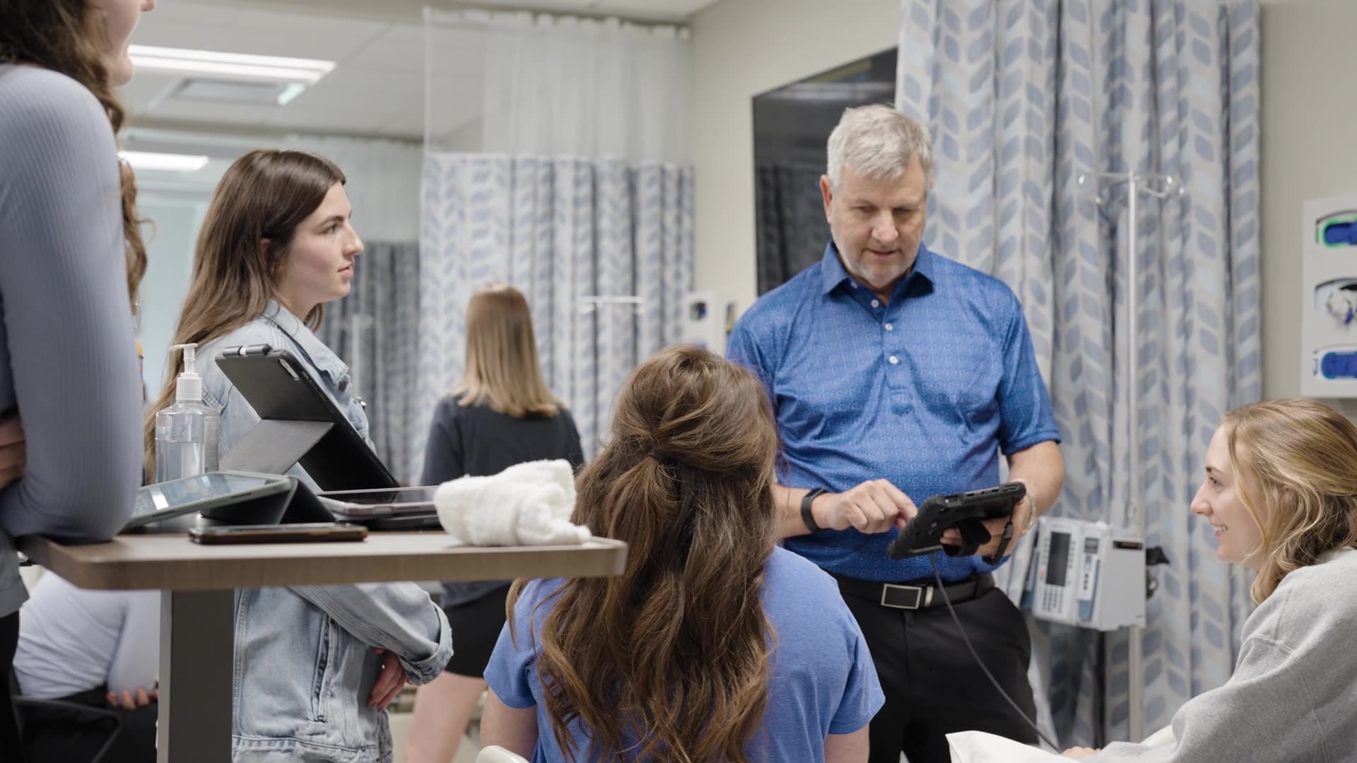
Dr. Matt Harkey, an assistant professor at Michigan State University conducting musculoskeletal research, wholeheartedly concurs. He switched from using blood biomarkers and MRI assessments to ultrasound about 10 years ago and in recent years has been using multiple Clarius scanners for his research studies.
Watch our 3-minute video to learn why Dr. Harkey uses Clarius for his research or scroll down for his five reasons for choosing Clarius.
1. Resolution of Ultrasound Imaging for MSK Measurements is Better than MRI
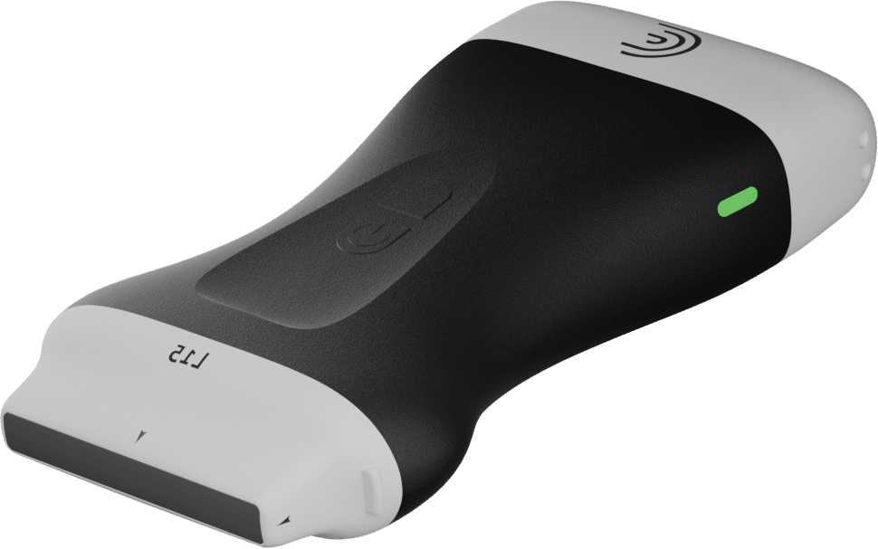
In the imaging world, ultrasound is still kind of the ugly stepchild that people still have these preconceived notions that ultrasound has poor resolution, you can’t standardize it, no one knows what they’re looking at. But I don’t think people realize that that’s not necessarily the case. That especially with the advancements in technology today, that you can get a scanner that is what a few thousand dollars versus a giant ultrasound machine that’s tens of thousands of dollars. You can get a tiny unit that hooks up to your phone, hooks up to an iPad and has better image quality than machines that were around five years ago. I don’t think people realize how good the technology of ultrasound and how much it’s progressing. »
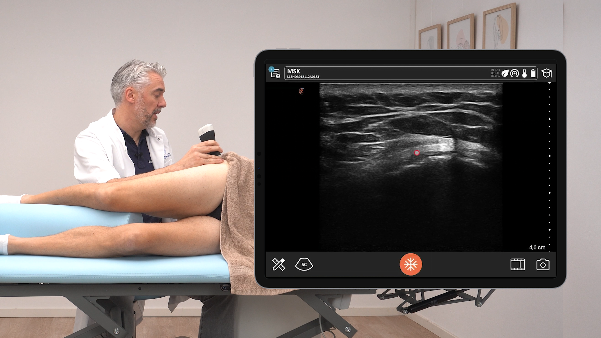
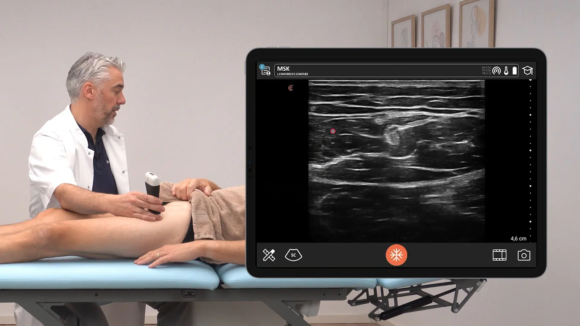
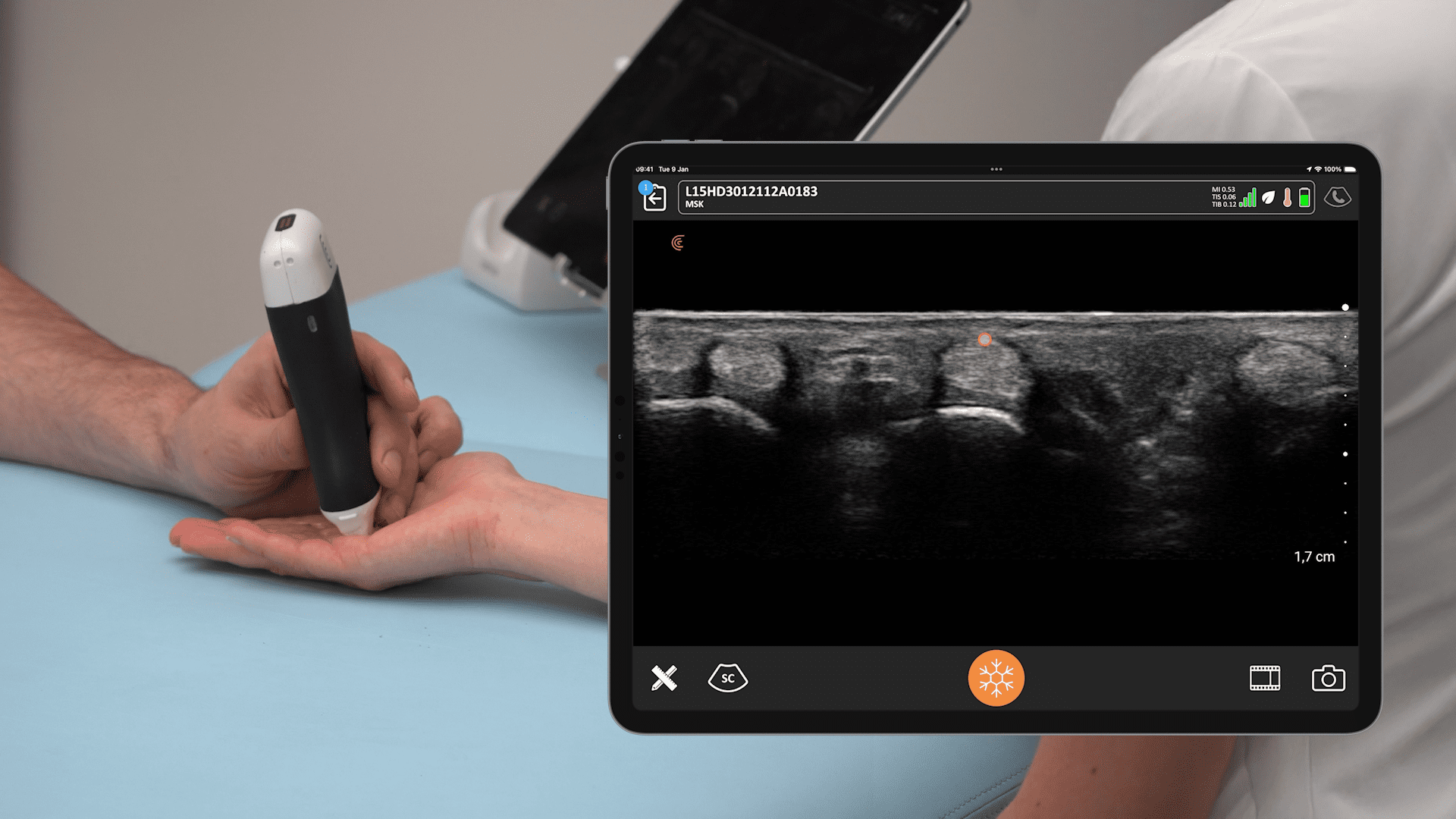
Yes, with MRI you can see the entire [knee] joint, but the resolution you’re getting on an MRI is much worse than what you’re getting on an ultrasound machine. Using an ultrasound probe, we’re getting better resolution and it allows for a more dynamic assessment in real time. Yes, it’s a 2D shot of one spot, but in real time you can move it around two different locations. You can have them flex their muscles, you can have them move their joint and assess what the structure looked like in various motions in real time and you’re having one-on-one patient interaction instead of sending someone off to an MRI machine.”
2. Clarius Ultrasound Is More Clinically Accessible in Different Settings than Traditional Machines
Clarius substantially surpasses a traditional machine because it’s much more clinically accessible and allows it to be applied in much different settings. It opens up so many more places that we can start to use the tool. There have been limitations in what we’ve been able to do with MSK research. But the fact that you could walk in and have everything you need in your pockets to do an ultrasound assessment is really going to change things.»
And I think another big piece that I didn’t necessarily realize with the Clarius scanner until recently is how dedicated Clarius is to develop their product and not just having this ultrasound wireless device that does a good job taking images, but finding ways to improve upon that device in ways that I didn’t think was possible. Some of the AI capabilities and the voice recognition capabilities really is something that you don’t get with a traditional ultrasound unit. Now I can actually say freeze and take an image to the iPad and it’ll do it for me.”
3. We’re Able to Measure and Quantify Structures with Clarius AI Tools
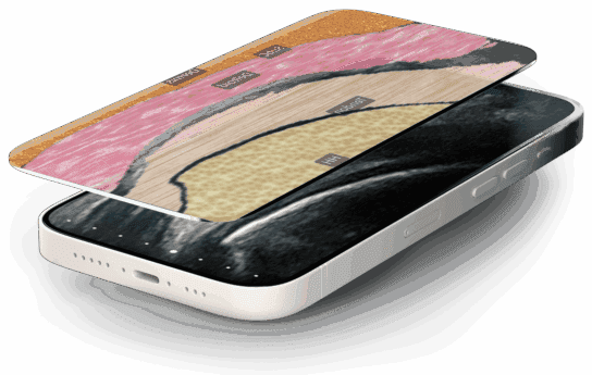
Clarius AI tools can actually help the reader scan a tissue and also quantify it and I’m not aware of other systems you can do that with in real time.»
A lot of my MSK research is quantifying various structures at the joint using Clarius ultrasound. With other systems, what I need to do is export it, put it into a different software, go in and fine tune and measure the structure. And then I get some numbers back way down the line. But now with these Clarius AI tools, all you have to do is put the probe on the structure, align it. And the probe does the work for you. That honestly, from what I’ve seen, can identify a patellar tendon as well as the human eye and then immediately provides you with a structural assessment of it.»
I’ve recently reached out to the Clarius AI group and I am starting to work with them on developing new ways and new structures to apply their AI capabilities to. »
4. Multiple Benefits of Wireless Ultrasound
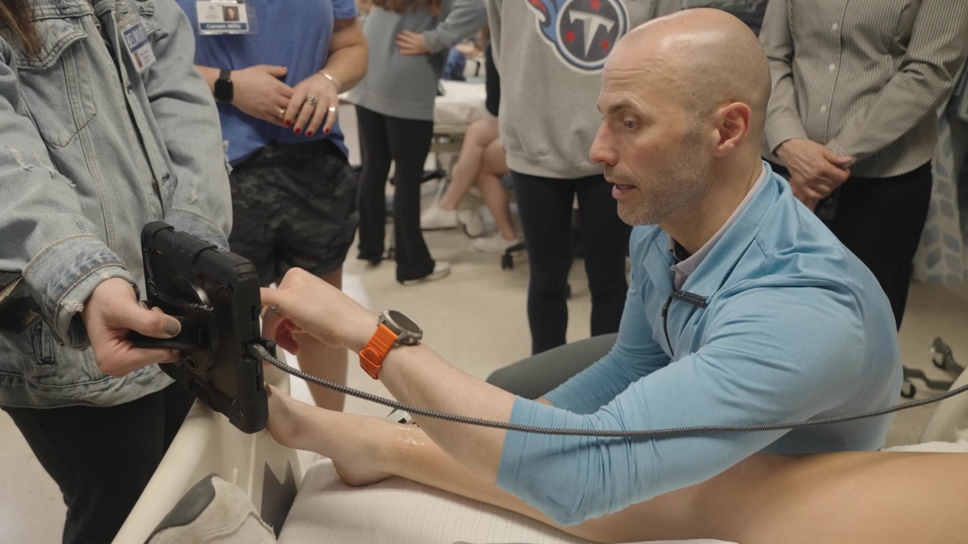
If you’ve done any ultrasound scanning, you’ll know that the cord is your enemy and the cord always wants to find the ultrasound gel and hit the ultrasound gel and then you’re just moving around and just you’re getting gel everywhere. It’s not that big of an inconvenience, but I honestly hated leaving the lab at the end of the day and they’re just being gel everywhere, and I wouldn’t even know where it’s coming from.»
But besides that, the wireless [scanner] opens up what you can assess and how you assess. Now that you don’t have that wire, you’re able to do assessments where they’re moving around. I wouldn’t do a dynamic scan on a patient if I was using a wired ultrasound machine, but the wireless piece really allows for that to be used.»
5. Dynamic Msk Assessments Are Now Easy When Clarius Ultrasound Is Paired with Usono’s Probefix
Dr. Harkey was an early user of a new wearable ultrasound solution that pairs Clarius HD3 high-definition wireless ultrasound scanner with research software and Usono’s ProbeFix Dynamic, enabling hands-free, stable, and reproducible ultrasound imaging during exercise and other activities where motion and continuous scanning is crucial. He recommends the combination to researchers performing dynamic analysis of muscle responding to activity.
I probably wouldn’t even have considered purchasing the Usono if I didn’t have a wireless scanner because it just allows for so much more opportunity of what you can scan, where you can scan, and the things that you’re able to do with the two products together. Combining the two together really allows for just a whole new realm of research that we can do in musculoskeletal imaging because we’re able to in real time see a dynamic assessment of how these structures are responding to load.”
Clarius for MSK Research
Clarius ultrasound tools for research provide access to raw data collected internally, gyroscope collection, a programable interface, and custom software for real-time analysis anywhere. The research package can be purchased with any scanner.
For details, visit our research page and book a virtual demonstration with a Clarius expert.
